

 PDF
PDF Citation
 Print
Print


Introduction
Pharmacokinetic principles for understanding volume kinetics
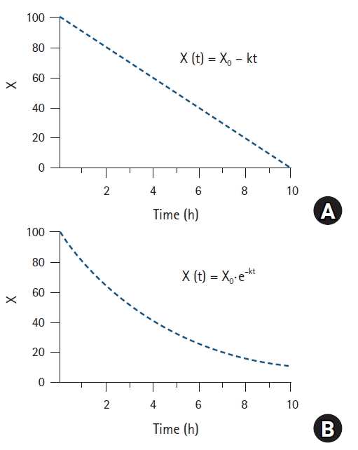
Kinetics of drugs
Mammillary compartmental model

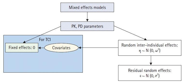
Population analysis
Calculation of plasma volume expansion caused by fluid administration using hemoglobin
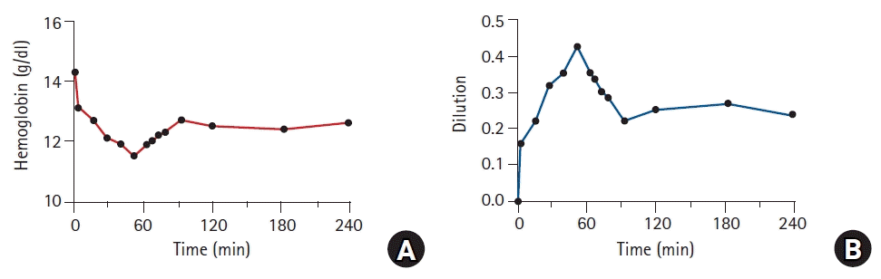 | Fig. 4.Changes in hemoglobin (A) and plasma dilution (B) caused by fluid administration. Male volunteer received 40 ml/kg Ringer’s lactate solution over 1 h. Dilution was calculated as follows:
where CHb(0) and Hct(0) are the hemoglobin concentration and hematocrit measured prior to the administration of Ringer’s lactate solution, respectively; CHb(t) is the hemoglobin concentration measured at any time t.
|
Structure model for volume kinetics
Table 1.
Dp(t): plasma dilution at any time t during intravenous infusion, CHb(0): hemoglobin concentration at t = 0 before intravenous infusion (g/dl), CHb(t): hemoglobin concentration at any time t after intravenous infusion (g/dl), Hct(0): hematocrit at t = 0 before intravenous infusion, Vp(0): plasma volume at t = 0 before intravenous infusion (ml), Vp(t): plasma volume at any time t after intravenous infusion (ml), Rate: infusion rate of the drug (mg/min), k10: elimination rate constant (1/min), A(t): drug amount at any time t (mg), V1: volume of distribution in the central compartment (ml), Cl: clearance (ml/min), V2: volume of distribution in the peripheral compartment (ml), Q: inter-compartmental clearance (ml/min), ki: infusion rate of fluid (ml/min), kb: basal elimination reflecting ongoing losses of water due to respiration, sweating, and basal renal filtration (ml/min), kr: renal clearance (ml/min), kt: distributional clearance (ml/min).
![]()
One-volume model
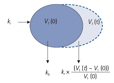 | Fig. 5.One-volume model. ki: infusion rate of the fluid (ml/min), kb: basal elimination reflecting ongoing losses of water due to respiration, sweating, and basal renal filtration (ml/min), kr: renal clearance (ml/min), V1(0): plasma volume at t = 0 before intravenous infusion (ml), V1(t): plasma volume at any time t after intravenous infusion (ml).
|
Two-volume model
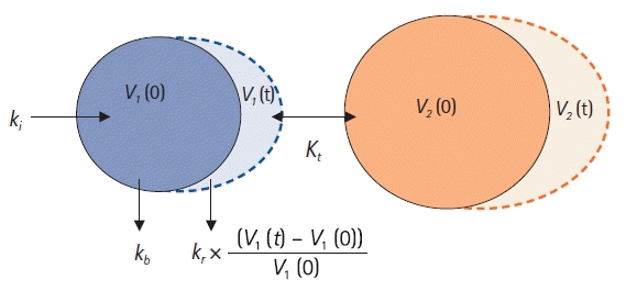 | Fig. 6.Two-volume model. ki: infusion rate of the fluid (ml/min), kb: basal elimination reflecting ongoing losses of water due to respiration, sweating, and basal renal filtration (ml/min), kr: renal clearance (ml/min), V1(0): plasma volume at t = 0 before intravenous infusion (ml), V1(t): plasma volume at any time t after intravenous infusion (ml), V2(0): peripheral volume at t = 0 before intravenous infusion (ml), V2(t): peripheral volume at any time t after intravenous infusion (ml), kt: distributional clearance (ml/min).
|
Calculation of renal clearance using urine volume
Covariates describing inter-individual variabilities of volume kinetic parameters
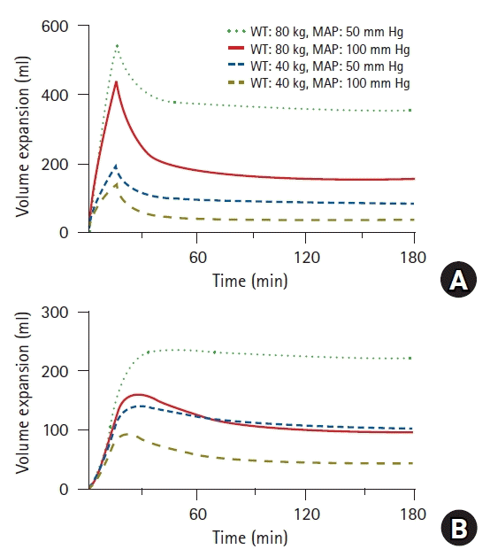 | Fig. 7.Simulated volume expansions of central (A) and peripheral (B) compartments in hypothetical patient receiving 10 ml/kg of Ringer’s lactate solution over 15 min followed by a rate of 8 ml/kg/h for 165 min. Four different cases were simulated based on weight (40 vs. 80 kg) and mean arterial pressure (50 vs. 100 mmHg). WT: weight, MAP: mean arterial pressure.
|
Clinical application of volume kinetics
Limitations of fluid kinetics
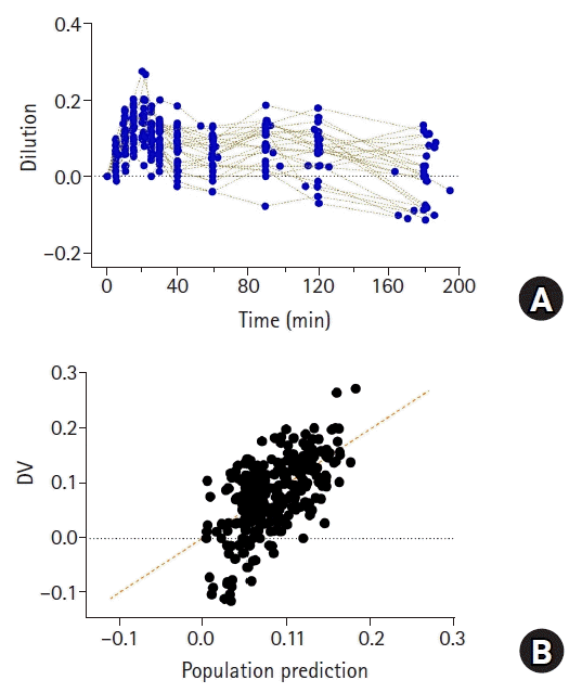 | Fig. 9.Plasma dilution (A) in 27 patients undergoing open gastrectomy. Patients were administered with 1,000 ml of Ringer’s lactate solution for 20 min, followed by continuous infusion at 6 ml/kg/h until time of last blood collection for volume kinetic analysis. Observed values (plasma dilutions) and population predicted values by volume kinetic model (B). Orange dashed line represents line of identity.
|
 XML Download