Abstract
This case report involves tracheal intubation using i-gel® in combination with a lightwand in a patient with a difficult airway, classified as Cormack-Lehane grade 3. I-gel® was used during anesthesia induction to properly maintain ventilation. The authors have previously reported successful tracheal intubation on a patient with a difficult airway through the use of i-gel® and a fiberoptic bronchoscope. However, if the use of a fiberoptic bronchoscope is not immediately available in a patient with a difficult airway, tracheal intubation may be performed by using i-gel® and a lightwand in a patient with difficult airway, allowing the safe induction of anesthesia.
To ensure proper ventilation and the supply of inhalation anesthetics during general anesthesia, tracheal intubation is the most commonly used method in maintaining the airway. When it is difficult to perform a tracheal intubation, it is possible to maintain the airway by using a larygeal mask airway (LMA) or supraglottic airway device [1]. However, during a lengthy surgery or when a patient is in a non-supine position during surgery, one can expect that maintaining an LMA or supraglottic airway device will be difficult. Therefore, there should be a change in the tracheal intubation procedure.
I-gel® (Intersurgical Ltd., Wokingham, Berkshire, UK) is a supraglottic airway device which has a soft, gel-like, non-inflatable cuff, designed to provide an anatomical, impression fit over the laryngeal inlet. The shape, softness and contours accurately mirror the perilaryngeal anatomy [2].
The authors have reported having experience with tracheal intubation on a patient with a difficult airway in whom i-gel® and a fiberoptic bronchoscope were used for tracheal intubation [3]. However, in emergency situations, it is not always possible to use a fiberoptic bronchoscope. The authors also performed tracheal intubation on a patient with a difficult airway, Cormack-Lehane classification grade 3, by inserting a lightwand into the i-gel® while maintaining adequate ventilation of the patient. Following the removal of the i-gel® and lightwand, surgery commenced without any complications.
A 59-year-old, 168 cm, 69 kg male with a diagnosis of rotator cuff syndrome was scheduled for arthroscopy. The patient, American Society of Anesthesiologists Physical Status II, had a past medical history of diabetes but no other unusual conditions. Preoperative assessment of the patient revealed a thyromental distance of greater than 6 cm, Mallampati classification Class 2, mouth opening of greater than 3 fingers, and no neck movement disorder.
Prior to administration of anesthesia, the patient was administered intramuscular injection of glycopyrrolate 0.2 mg and midazolam 2.5 mg as premedication. While closely monitoring the patient's electrocardiogram, pulse oximetry, non-invasive arterial pressure, and bispectral index score (BIS), the patient was administered 100% oxygen via face mask and intravenously injected with propofol 140 mg and lidocaine 40 mg. After confirming the loss of spontaneous breathing and BIS of less than 60, the patient was administered rocuronium 40 mg by intravenous injection. After waiting two minutes, tracheal intubation was attempted. However, tracheal intubation using a laryngoscope failed because the patient's glottic opening was not visible and the epiglottis was only slightly visible, revealing a Cormack-Lehane classification grade 3. At this point, since the patient was equipped with a face mask, two-handed ventilation was possible. The patient's blood pressure was 112/63 mmHg, heart rate 68 beats/min, and oxygen saturation was 100%. However, the ventilation did not proceed well. The peak airway pressure was over 30 mmHg and there was a possibility of introducing air into the stomach. It could also be expected that pulse oximeter saturation (SpO2) could decrease due to the fact that the end tidal CO2 (ETCO2) was in a decreased state.
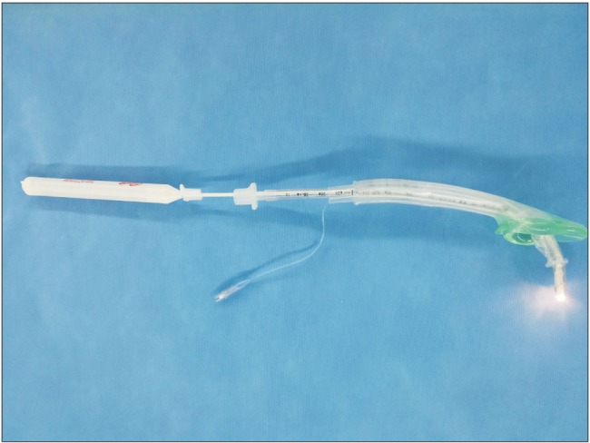
In order to properly ventilate the patient, a size 4 i-gel® was inserted. Proper ventilation was confirmed by auscultation and capnography. Audible air leak pressure was 30 mmHg, and appropriate tidal volume was measured. Because during the surgery, the patient would be required to maintain the lateral decubitus position, and the duration of the surgery could be three hours or longer, it was decided to insert an endotracheal tube in the patient. First, through the i-gel®, a 7.0 mm inner diameter endotracheal tube was inserted using blind intubation, but the endotracheal tube went into the esophagus. Although the authors have had previous successful experience in tracheal intubation using a fiberoptic bronchoscope, that was not possible in this case because the necessary equipment was not available. Therefore, the authors decided to use a lightwand (Surch-Lite™, Aaron Medical Industries, St. Petersburg, FL, USA) instead of a fiberoptic bronchoscope. The lightwand was inserted into a 7.0 mm inner diameter endotracheal tube, then the endotracheal tube was passed through the i-gel® (Fig. 1). The tracheal tube was inserted after dimming the lights in the operating room and the light source was visible at the end of the probe through the transparent skin. Successful tracheal intubation was confirmed by auscultation and capnography. The authors, concerned about the possibility of accidental extubation while removing the i-gel ®, removed both the 7.0 mm endotracheal tube and the i-gel® using a tube exchanger and inserted an 8.0 mm endotracheal tube. After the insertion of the 8.0 mm endotracheal tube, surgery proceeded.
After the surgery, the patient was extubated after regaining full consciousness and breathing. No complications or side effects were observed due to the tracheal intubation.
The safest method of maintaining the airway during general anesthesia is by tracheal intubation to supply the patient with inhalation anesthetics, adequate ventilation, and oxygenation. However, tracheal intubation is not possible in 0.1-0.4% of patients [4]. Failure to properly maintain an airway can lead to anesthesia-related complications such as tracheotomy, brain damage, and cardiopulmonary arrest, and can possibly result in mortality [5].
The LMA, intubating laryngeal mask airway (ILMA), supraglottic airway device, fiberoptic bronchoscope, Glidescope® (video-larynoscope), and lightwand are all used to secure the airway. When tracheal intubation is difficult, especially in cases of difficult ventilation using a face mask, the LMA or supraglottic airway device are useful in maintaining the airway [678].
The advantages of the i-gel® insertion technique are that it is easier and does not require higher skill compared to other supraglottic airway devices. In a study comparing i-gel® and LMA insertion, the first attempt success rate was 90.0% for i-gel® and 48.3% for the LMA by novice physicians. In addition, i-gel® use provided almost equal success rates for experienced and novice physicians (91 vs. 90%), whereas LMA use resulted in significantly lower success rates for novices (48.3 vs. 80.4%) [9]. Furthermore, for the novice physicians, the median insertion times on actual patients were as follows: i-gel®, 17.5 seconds; LMA, 32 seconds; and ProSeal® LMA, 53 seconds [1011]. A study was conducted to evaluate the success rate of blind intubations using the ILMA and i-gel®. A lower percentage of intubations, 40%, were achieved with i-gel®, versus the 70% achieved with the ILMA [12]. Thus, blind tracheal intubation using i-gel® is likely to be unsuccessful. To compensate for the above, after inserting the i-gel®, if tracheal intubation is required, intubation using a fiberoptic bronchoscope has been reported to be successful. However, in cases where a fiberoptic bronchoscope is not available, a different approach is required.
Lightwand tracheal intubation is a technique in which an illuminated stylet is inserted into the endotracheal tube, and the tip of the tube is directed into the trachea guided by transillumination of the neck tissue. Before intubation, if the end of the lightwand is bent in a form similar to a hockey stick, the lightwand can aid easier tracheal intubation in patients with limited mouth opening and limited cervical spine motion [1314]. We believe bending the tip of the lightwand upward makes it easier to enter the vocal cords. The benefit of the lightwand is that even during unexpected intubation failure, it is a simple and useful device for performing tracheal intubation. Most anesthesiologists can relatively easily handle a lightwand, and the success rate of the lightwand for skilled anesthesiologists in patients with difficult airways has been reported to be 96.8 to 100% [15]. Furthermore, since most hospitals are equipped with lightwands, it is readily available for use. However, since the lightwand is a type of blind technique which can cause injury to the oral cavity, pharynx, and larynx, care and attention are required to prevent injury. Nevertheless, trauma to the airway after lightwand intubation is generally minor, and may include bleeding, sore throat, hoarseness, and dysphagia.
In the case described here, during anesthesia induction in a patient with a difficult airway, the authors maintained adequate ventilation for the safety of the patient by first using the i-gel®. Thereafter, it was determined that the patient required tracheal intubation, considering the duration of the operation and the patient's position during the surgery. After failed blind tracheal intubation using the i-gel®, tracheal intubation using a lightwand instead of a fiberoptic bronchoscope was successful. The I-gel® and lightwand were removed using a tube exchanger and the surgery was completed without any particular complications. We were concerned about the possibility of accidental extubation while removing the i-gel®. A tube exchanger can be used to facilitate the exchange from a single lumen tube to a double lumen tube, or vice versa. This device has a center hollow channel and a universal fit adapter through which oxygen insufflation or jet ventilation can be administered to allow more time for the tube exchanging process. Although there was the possibility of airway damage, we thought that this was safer than re-intubation.
There are no previous reported cases dealing with tracheal intubation using the i-gel® and a lightwand. Through this particular case, it was confirmed that a lightwand can replace a fiberoptic bronchoscope when performing tracheal intubation using i-gel®.
References
1. Eindhoven GB, Dercksen B, Regtien JG, Borg PA, Wierda JM. A practical clinical approach to management of the difficult airway. Eur J Anaesthesiol Suppl. 2001; 23:60–65. PMID: 11766249.

2. Levitan RM, Kinkle WC. Initial anatomic investigations of the I-gel airway: a novel supraglottic airway without inflatable cuff. Anaesthesia. 2005; 60:1022–1026. PMID: 16179048.
3. Lim HK, Chun CK, Shinn HK, Lee CS, Hwang SI, Lee SM, et al. Endotracheal intubation using i-gel and a flexible fiber optic bronchoscope: A case report. Anesth Pain Med. 2012; 7:147–150.
4. Benumof JL. Management of the difficult adult airway. With special emphasis on awake tracheal intubation. Anesthesiology. 1991; 75:1087–1110. PMID: 1824555.
5. Loh KS, Irish JC. Traumatic complications of intubation and other airway management procedures. Anesthesiol Clin North America. 2002; 20:953–969. PMID: 12512271.

6. Brain AI. The laryngeal mask--a new concept in airway management. Br J Anaesth. 1983; 55:801–805. PMID: 6349667.

7. Kapila A, Addy EV, Verghese C, Brain AI. The intubating laryngeal mask airway: an initial assessment of performance. Br J Anaesth. 1997; 79:710–713. PMID: 9496200.

8. Brain AI, Verghese C, Addy EV, Kapila A. The intubating laryngeal mask. I: Development of a new device for intubation of the trachea. Br J Anaesth. 1997; 79:699–703. PMID: 9496198.

9. Stroumpoulis K, Isaia C, Bassiakou E, Pantazopoulos I, Troupis G, Mazarakis A, et al. A comparison of the i-gel and classic LMA insertion in manikins by experienced and novice physicians. Eur J Emerg Med. 2012; 19:24–27. PMID: 21593672.

10. Wharton NM, Gibbison B, Gabbott DA, Haslam GM, Muchatuta N, Cook TM. I-gel insertion by novices in manikins and patients. Anaesthesia. 2008; 63:991–995. PMID: 18557971.

11. Klaver NS, Kuizenga K, Ballast A, Fidler V. A comparison of the clinical use of the Laryngeal Tube S and the ProSeal Laryngeal Mask Airway by first-month anaesthesia residents in anaesthetised patients. Anaesthesia. 2007; 62:723–727. PMID: 17567350.
12. Sastre JA, Lopez T, Garzon JC. Blind tracheal intubation through two supraglottic devices: i-gel versus Fastrach intubating laryngeal mask airway (ILMA). Rev Esp Anestesiol Reanim. 2012; 59:71–76. PMID: 22480552.
13. Davis L, Cook-Sather SD, Schreiner MS. Lighted stylet tracheal intubation: a review. Anesth Analg. 2000; 90:745–756. PMID: 10702469.

14. Weis FR Jr. Light-wand intubation for cervical spine injuries. Anesth Analg. 1992; 74:622. PMID: 1554139.

15. Apfelbaum JL, Hagberg CA, Caplan RA, Blitt CD, Connis RT, Nickinovich DG, et al. Practice guidelines for management of the difficult airway: an updated report by the American Society of Anesthesiologists Task Force on Management of the Difficult Airway. Anesthesiology. 2013; 118:251–270. PMID: 23364566.


 PDF
PDF Citation
 Print
Print


 XML Download