Journal List > Cancer Res Treat > v.54(1) > 1154878

Lee, Kim, Kim, Kim, Kim, Im, Jeon, Keam, Ku, and Heo: Acquired Resistance Mechanism of EGFR Kinase Domain Duplication to EGFR TKIs in Non–Small Cell Lung Cancer

Abstract

Purpose

Epidermal growth factor receptor kinase domain duplication (EGFR-KDD) is a rare and poorly understood oncogenic mutation in non–small cell lung cancer (NSCLC). We aimed to investigate the acquired resistance mechanism of EGFR-KDD against EGFR-TKIs.

Materials and Methods

We identified EGFR-KDD in tumor tissue obtained from a patient with stage IV lung adenocarcinoma and established the patient-derived cell line SNU-4784. We also established several EGFR-KDD Ba/F3 cell lines: EGFR-KDD wild type (EGFR-KDDWT), EGFR-KDD domain 1 T790M (EGFR-KDDD1T), EGFR-KDD domain 2 T790M (EGFR-KDDD2T), and EGFR-KDD both domain T790M (EGFR-KDDBDT). We treated the cells with EGFR tyrosine kinase inhibitors (TKIs) and performed cell viability assays, immunoblot assays, and ENU (N-ethyl-N-nitrosourea) mutagenesis screening.

Results

In cell viability assays, SNU-4784 cells and EGFR-KDDWT Ba/F3 cells were sensitive to 2nd generation and 3rd generation EGFR TKIs. In contrast, the T790M-positive EGFR-KDD Ba/F3 cell lines (EGFR-KDDT790M) were only sensitive to 3rd generation EGFR TKIs. In ENU mutagenesis screening, we identified the C797S mutation in kinase domain 2 of EGFR-KDDBDT Ba/F3 cells. Based on this finding, we established an EGFR-KDD domain 1 T790M/domain 2 cis-T790M+C797S (EGFR-KDDT/T+C) Ba/F3 model, which was resistant to EGFR TKIs and anti-EGFR monoclonal antibody combined with EGFR TKIs.

Conclusion

Our study reveals that the T790M mutation in EGFR-KDD confers resistance to 1st and 2nd generation EGFR TKIs, but is sensitive to 3rd generation EGFR TKIs. In addition, we identified that the C797S mutation in kinase domain 2 of EGFR-KDDT790M mediates a resistance mechanism against 3rd generation EGFR TKIs.

Introduction

Lung cancer has become a remarkable example of precision medicine among solid tumors, because of the progress in understanding genomic landscape and the development of targeted therapies [1]. Epidermal growth factor receptor (EGFR) mutations are prevalent oncogenic drivers in lung adenocarcinoma that confer susceptibility to tyrosine kinase inhibitors (TKIs) [24]. EGFR exon 19 deletion and the L858R mutation are the most frequent sensitizing mutations in EGFR [5].
EGFR kinase domain duplication (EGFR-KDD) is a rare mutation that represents 0.2% of EGFR-mutated lung adenocarcinoma [6]. In contrast to ligand-dependent EGFR activation in typical conditions, EGFR-KDD forms ligand-independent intra-molecular dimers and amplifies downstream signal with ligand-dependent inter-molecular dimers and multimers [79]. Notably, discovery of EGFR-KDD as a novel oncogenic driver has led to few functional studies [8,10] and acquired resistance mechanism of EGFR-KDD against EGFR TKIs is not fully understood.
The EGFR T790M mutation is the most frequent driver of acquired resistance in approximately 60% of patients with sensitizing EGFR mutations who experience progression after treatment with 1st and 2nd generation EGFR TKIs [11,12]. Other mechanisms involve on-target mutations, bypass signaling cascades, and histological transformation [13].
In this study, we established patient-derived cell lines and developed EGFR-KDDT790M Ba/F3 models to characterize and evaluate drug sensitivity of T790M-positive EGFR-KDD mutated cells. The T790M-positive EGFR-KDD mutation were identified from a patient whose tumor acquired the T790M mutation as a resistance mechanism to erlotinib. We also suggest that the EGFR C797S mutation, in combination with the EGFR-KDDT790M mutation, mediates a potential resistance mechanism to osimertinib, based on N-ethyl-N-nitrosourea (ENU) mutagenesis screening results.

Materials and Methods

1. Targeted next-generation sequencing and fluorescence in situ hybridization

The DNA extracted from formalin-fixed paraffin-embedded tumor tissues and patient malignant pleural effusion were sequenced using an next-generation sequencing (NGS) panel named Seoul National University Hospital (SNUH) FIRST Lung Cancer Panel (LCP) [14]. The sequencing coverage and quality statistics for each sample are summarized in S1 Table.
EGFR fluorescence in situ hybridization (FISH) was performed as previously described [15]. We calculated the EGFR copy number with an LSI EGFR Spectrum Orange/CEP7 Spectrum Green probe kit (Abbott Molecular) and performed analysis using Colorado scoring criteria. FISH probe signals were detected with the Olympus BX51TRF microscope (Olympus, Tokyo, Japan).

2. Patient-derived cell line

The SNU-4784 cell line was derived from a malignant pleural effusion in a patient with pulmonary adenocarcinoma who was diagnosed with the EGFR-KDD mutation by targeted NGS. The SNU-4784 cell line was provided and authenticated with short tandem repeat profiling by the Korean Cell Line Bank (KCLB, Seoul, Korea) (S2 Table). The cells were grown in RPMI-1640 media supplemented with 10% fetal bovine serum (FBS), 1% penicillin/streptomycin, and 2 mM L-glutamine.

3. Droplet digital polymerase chain reaction

Genomic DNA from the patient’s malignant pleural effusion was isolated using Exgene Cell SV mini kit (GeneAll, Lisbon, Portugal). DNA fragments were packaged into droplets using a QX200 droplet generator (Bio-Rad, Hercules, CA) and Thr 790 sites were amplified with specific probes. Droplets with amplified DNA fragments were analyzed using a QX200 droplet reader (Bio-Rad) according to the manufacturer’s guidelines. Data and graphs were created with QuantaSoft Analysis Pro software.

4. Cell lines and reagents

The 293FT (ATCC PTA-5077, discontinued) cell line was purchased from ATCC (Manassas, VA). The Ba/F3 cell line was purchased from DSMZ (Braunschweig, Germany). Ba/F3 cells were grown in RPMI-1640 media supplemented with 10% FBS, 1% penicillin/streptomycin, 2 mM L-glutamine, and 4 ng/mL interleukin (IL)-3 (ProSpec, Rehovot, Israel). The 293FT cells were grown in DMEM supplemented with 10% FBS and 2 mM L-glutamine. Gefitinib, erlotinib, afatinib, dacomitinib, osimertinib, and lazertinib were purchased from Selleck Chemicals (Boston, MA).

5. Cloning and construction of EGFR-KDD Ba/F3 cell lines

The pBabe EGFRWT was kindly provided by Matthew Meyerson (Dana-Farber Cancer Research Institute, Boston, MA; Addgene plasmids #11011). The pBabe EGFRT790M was purchased from Addgene (Addgene, Watertown, MA; Addgene plasmid #32070). The pBabe EGFRC797S was generated by site-directed mutagenesis (Agilent Technologies, Santa Clara, CA) of the pBabe EGFRWT [16]. The EGFR-KDDWT (pMSCV EGFR-KDD) vector was kindly provided by Dr. Christine M. Lovly (Vanderbilt-Ingram Cancer Center, Nashville, TN). The pBabe EGFR-KDDD1T, EGFR-KDDD2T, EGFR-KDDBDT, EGFR-KDDT/T+C were generated with an In-Fusion HD Cloning Kit (Takara Bio USA, Inc., Mountain View, CA) and specific primers (S3 Table). Each of EGFR-KDD constructs were transduced into Ba/F3 cells and cells were selected with puromycin (2–16 μg/mL).

6. PCR and sequencing

Genomic DNA and mRNA were isolated from the SNU-4784 cell line using ALL-prep DNA/RNA micro kit (Qiagen, Venlo, Netherlands). The DNA of the EGFR-KDD Ba/F3 cell lines was isolated using an Exgene Cell SV mini kit (GeneAll, Lisbon, Portugal). For use in cloning, the EGFR exon 18–25 of pBabe EGFRWT, pBabe EGFRT790M, and pBabe EGFRC797S were amplified with specific primers (S3 Table) using CloneAmp HiFi PCR Premix (Takara Bio USA, Inc.). DNA sequences were analyzed by Sanger sequencing.

7. Cell viability assay

The EGFR-KDD Ba/F3 cell lines were cultured in 96-well plates with EGFR TKIs in RPMI-1640 media for 72 hours. EGFR TKIs were diluted from 10 mM to 0.01 nM. Cell viability was measured using Chromo-CK (Chromogen, Korea) and absorbance at 450 nm was detected using microplate reader (BioTek, Winooski, VT). SNU-4784 cells were cultured as described above. Cell viability was analyzed using a Cell Titer Glo-Luminescent cell viability assay (Promega, Madison, WI). We measured luminescent signal with a PerkinElmer Victor Light 1420 Luminescence Counter (PerkinElmer, Waltham, MA). The IC50 values were calculated with Sigmaplot 10.0 software (Systat Software Inc., San Jose, CA) and the graphs were created using GraphPad Prism 7 software (GraphPad Software, San Diego, CA). Experiments were independently repeated three times.

8. Immunoblot assay

The EGFR-KDD Ba/F3 cell lines and SNU-4784 cells were cultured on 6-well plates and treated with EGFR TKIs for 4 hours. Cells were lysed with 10× cell lysis buffer (Cell Signaling Technology, Danvers, MA), phenylmethylsulfonyl fluoride (Sigma, St. Louis, MO), PhosSTOP (Roche, Basel, Switzerland), and a proteinase inhibitor cocktail (Merck, Kenilworth, NJ). Protein was quantified using a protein assay dye reagent concentrate (Bio-Rad). Samples were loaded and separated using NuPAGE Bis-Tris Gels (Invitrogen). Separated samples were transferred to polyvinylidene difluoride membranes (Bio-Rad), and signal was detected with ECL Prime Western Blotting Detection Reagent (GE Healthcare, Chicago, IL). Total EGFR (#4267s), phospho-EGFR (#3777s), total Akt (#4685), phospho-Akt (#4060s), total Erk p42/p44 (#9102), phospho-Erk (#9106), and GAPDH (#5174) antibodies were purchased from Cell Signaling Technology. Experiments were independently repeated three times.

9. ENU mutagenesis screening

The EGFR-KDDBDT Ba/F3 cells were seeded at 5×10⁶ cells/ml in 6-well plates and exposed to 50 μg/mL ENU for 24 hours. After that, cells were washed with RPMI-1640 media three times and then were cultured for 3 days. The cultured cells were seeded in 96-well plates with 2 μM osimertinib in RPMI-1640. We observed cells by light microscopy and periodically changed the osimertinib-containing media. DNA extracts from the drug-resistant clone were amplified with PCR and analyzed by Sanger sequencing.

Results

1. Patient and clinical history

A 56-year-old man, who had never smoked, was diagnosed with stage IV lung adenocarcinoma with metastasis to the brain. After his disease progressed following third-line chemotherapy, tumor tissue was newly obtained from the primary lung tumor. NGS-based multigene panel testing detected the EGFR-KDD mutation, whereas other common EGFR mutations were absent (S4A Fig.). The presence of EGFR-KDD was confirmed with further DNA sequencing (Fig. 1B). Based on this finding, erlotinib treatment was initiated and the patient achieved a partial response. After 8 months of erlotinib therapy, the cancer progressed with increased pleural effusion, bone metastases, and peritoneal seeding. Upon disease progression, malignant cells derived from pleural effusion were collected for genetic profiling. Then, both EGFR-KDD and the EGFR T790M mutation were detected in erlotinib-resistant tumor cells and confirmed with droplet digital polymerase chain reaction (Fig. 1C, S4B and S5 Figs.). However, we could not confirm whether T790M occurred in one domain or both of EGFR-KDD. FISH analysis showed no evidence of EGFR amplification (S6 Fig.). The patient received osimertinib and achieved a partial response for 7 months. Clinical history is summarized in Fig. 1A.

2. Characteristics of the patient-derived cell line

We confirmed the EGFR-KDD mutation in the patient-derived cell line SNU-4784 by Sanger sequencing; unexpectedly, the T790M mutation was not found in this cell line (S7A and S7B Fig.). We performed cell viability and immunoblot assays to determine the efficacy of EGFR TKIs in SNU-4784 cell line. We found that afatinib (IC50, 0.70±0.16nM) and osimertinib (IC50, 8.98±1.71 nM) inhibited cell viability (Fig. 2A, S8 Table). Furthermore, afatinib inhibited expression of phospo-EGFR, phospho-Akt, and phospho-Erk signals that are driven by activated EGFR (Fig. 2B) [17]. These results indicate that the EGFR-KDD mutation is sensitive to EGFR TKIs, especially afatinib, and this was demonstrated in a previous study [8].

3. Characteristics of T790M-positive EGFR-KDD Ba/F3 models

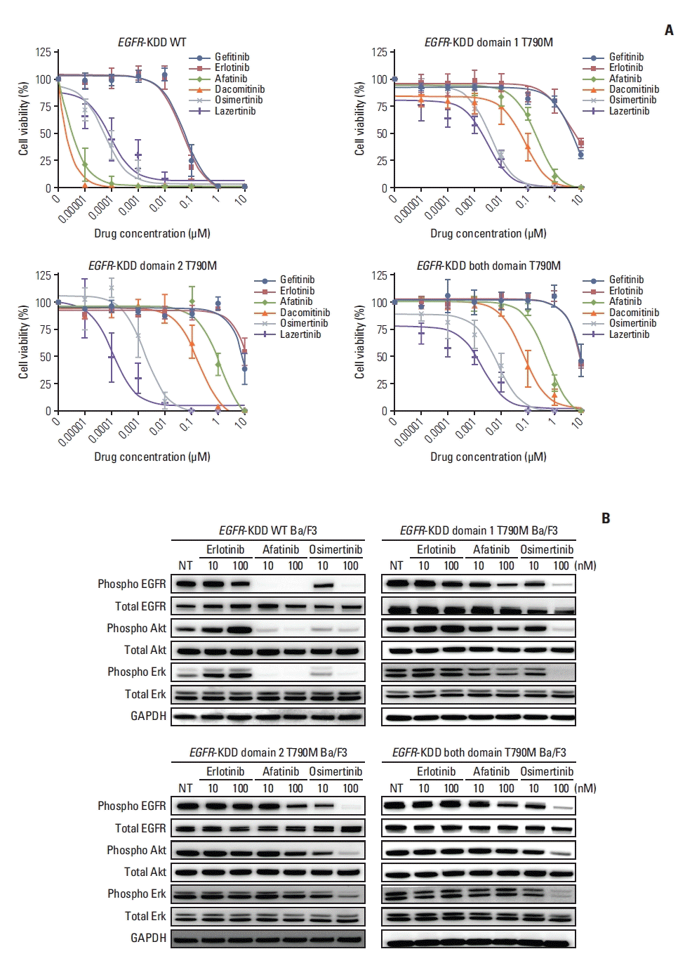
To characterize the T790M-positive EGFR-KDD mutation, we established several EGFR-KDDT790M Ba/F3 models. These included EGFR-KDD domain 1 T790M (EGFR-KDDD1T), EGFR-KDD domain 2 T790M (EGFR-KDDD2T), and EGFR-KDD both domain T790M (EGFR-KDDBDT) (S9 Fig.), and we performed cell viability assays and immunoblot assays with these Ba/F3 cell lines. We found that the EGFR-KDDWT Ba/F3 cells were sensitive to 2nd generation EGFR TKIs afatinib (IC50, < 0.01 nM) and dacomitinib (IC50, < 0.01 nM), in addition to 3rd generation EGFR TKIs osimertinib (IC50, 0.07±0.04nM) and lazertinib (IC50, 0.21±0.2 nM) (Fig. 3A, S10 Table). These results were consistent with our findings in a SNU-4784 cell line with an identical EGFR-KDDWT mutation (Fig. 2A). However, the EGFR-KDDT790M Ba/F3 cell lines were only sensitive to 3rd generation EGFR TKIs, osimertinib and lazertinib. Notably, lazertinib, an irreversible 3rd generation EGFR TKI, strongly inhibited the EGFR-KDDT790M mutation (Fig. 3A, S10 Table) [18]. Also, osimertinib inhibited phospho-EGFR, phospho-Akt, and phospo-Erk protein expression in EGFR-KDDT790M Ba/F3 cell lines. In contrast, 1st and 2nd generation EGFR TKIs did not effectively inhibit those signals (Fig. 3B). Thus, the EGFR T790M mutation, in combination with EGFR-KDD, induced resistance against 1st and 2nd generation EGFR TKIs.

4. ENU mutagenesis screening to investigate a resistance mechanism of EGFR-KDDT790M against osimertinib

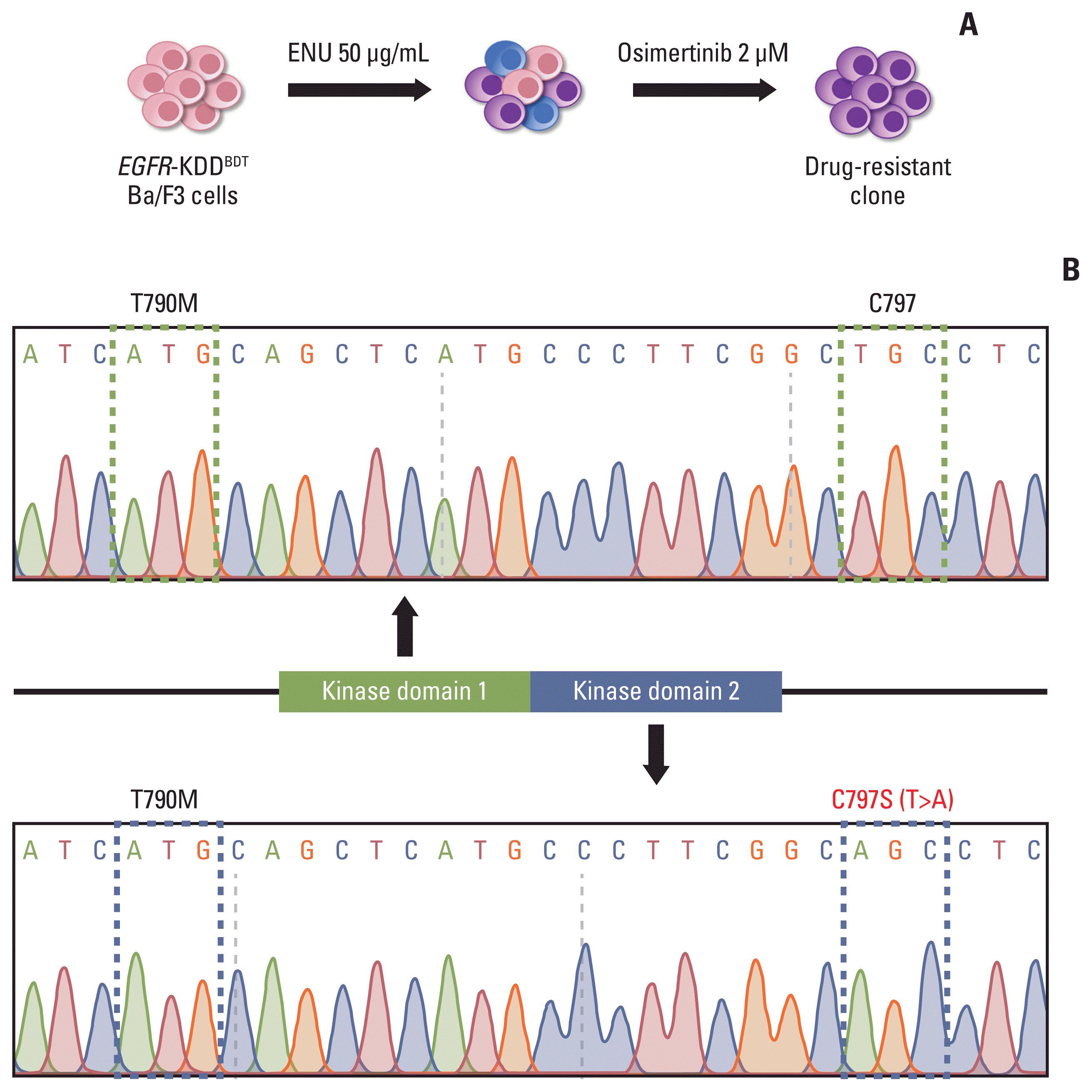
To identify the resistance mechanism against 3rd generation EGFR TKIs, we performed ENU mutagenesis screening in EGFR-KDDBDT Ba/F3 cells treated with osimertinib. We mutagenized cells via exposure to 50 μg/mL ENU and selected resistant cells with 2 μM/mL osimertinib (Fig. 4A). We sequenced DNA of the emergent resistant cells by Sanger sequencing and found the C797S mutation in kinase domain 2 of osimertinib-resistant EGFR-KDDBDT cells (Fig. 4B). Importantly, all osimertinib-resistant clones had the identical C797S mutation (Fig. 4B, S11 Fig.). These results suggest that kinase domain 2 of EGFR-KDD is more vulnerable than domain 1, and that the C797S mutation underlies a potential mechanism for EGFR-KDDT790M resistance against osimertinib.

5. The EGFR-KDDT/T+C Ba/F3 model indicating an acquired resistance mechanism

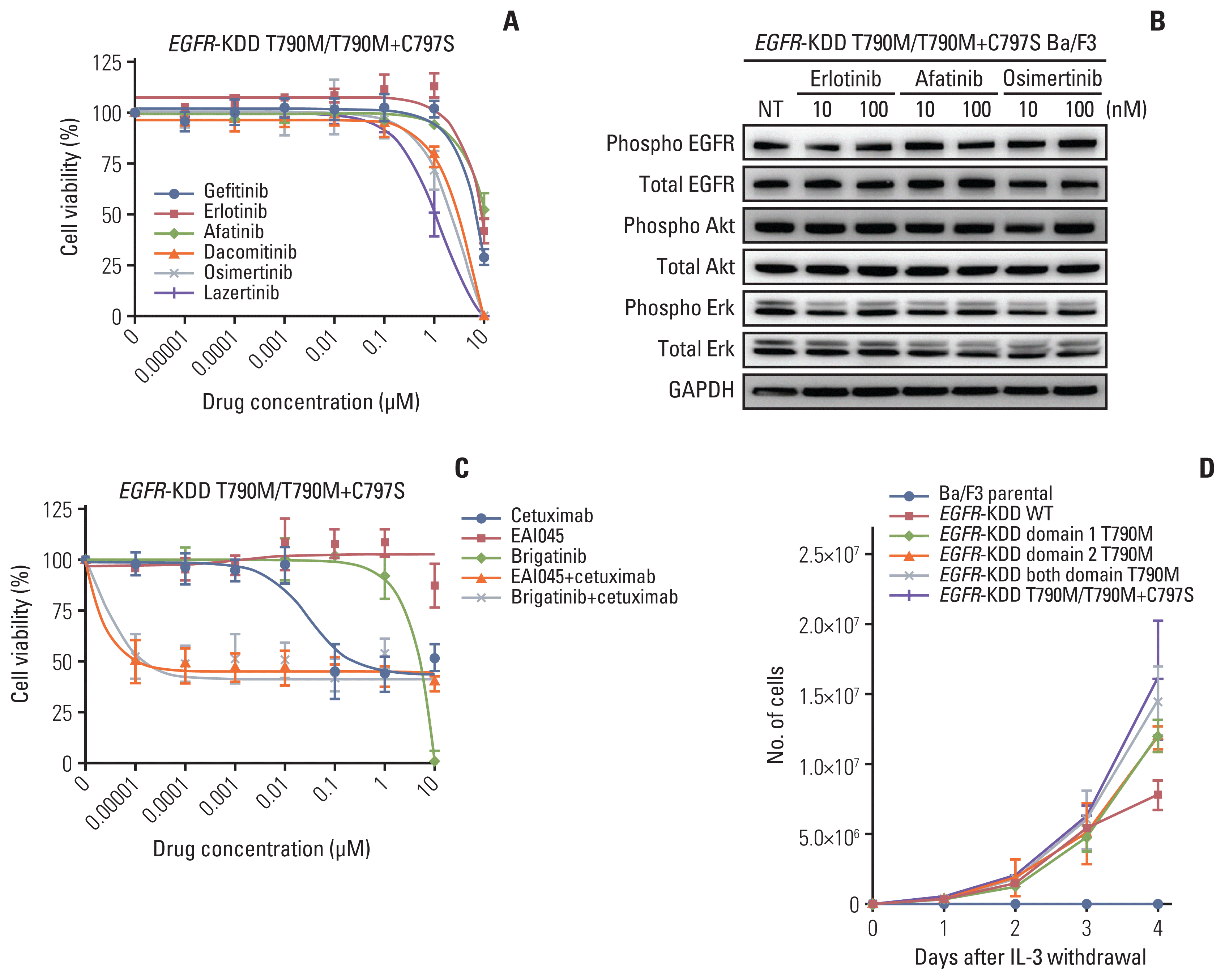
To characterize the EGFR-KDD domain 1 T790M/domain 2 cis-T790M+C797S mutation, we established an EGFR- KDDT/T+C Ba/F3 model. We performed cell viability assays and immunoblot assays to investigate the efficacy of EGFR TKIs in EGFR-KDDT/T+C Ba/F3 cells. Cells were resistant to 1st and 2nd generation EGFR TKIs, and also 3rd generation EGFR TKIs were not effective at inhibiting cell growth. (Fig. 5A). Furthermore, EGFR TKIs did not inhibit phospho-EGFR, phospho-Akt, or phospho-Erk protein expression (Fig. 5B). We then performed additional cell viability assays and immunoblot assays to identify the effect of a cetuximab combination treatment, a known therapeutic strategy for overcoming the C797S positive mutation [19,20]. Cells were exposed to EAI045 (an allosteric EGFR inhibitor) or brigatinib, in combination with cetuximab. We found that the cetuximab combination treatment did not inhibit the proliferation of the EGFR-KDDT/T+C Ba/F3 cells (Fig. 5C). In addition, the growth of EGFR-KDDT/T+C Ba/F3 cells in IL-3–free media was the most aggressive among the EGFR-KDD Ba/F3 cell lines (Fig. 5D). Taken together, our findings indicate that the cis-C797S mutation in kinase domain 2 of EGFR-KDDT790M induced drug resistance and rapid cell proliferation.

Discussion

Although EGFR-KDD was first reported in a glioblastoma, the mutation also exists in lung cancers and is a therapeutic target of EGFR TKIs [8,10,21,22]. The in-tandem and in-frame duplication of exons 18–25 encodes a permanently activated kinase with in vitro oncogenic potential [7,8].
Up to 10% of EGFR wild type patients still respond to EGFR TKIs, although the molecular basis for the response is unknown [23,24]. This gap in knowledge may be due to conventional PCR-based exon sequencing that can fail to detect rare EGFR structural variants, including EGFR-KDD and EGFR fusions, due to large intragenic repeats and/or intronic breakpoints. Thus, comprehensive molecular profiling is important to identify all genomic alterations that may lead to new therapeutic targets.
Since EGFR-KDD was discovered in a patient with metastatic lung adenocarcinoma, the duplication has been reported in more than 10 non–small cell lung cancer (NSCLC) patients, many of whom have never smoked. Patients with EGFR-KDD, including the patient in our study, are responsive to EGFR TKI. However, a recent study showed that two of the five NSCLC patients with EGFR-KDD did not respond to EGFR TKI therapies, suggesting the existence of a separate uncharacterized primary resistance mechanism [8,25].
Here we studied a patient with lung adenocarcinoma with an EGFR-KDD mutation, which was identified with NGS-based multigene panel testing. Consistent with previous reports, the patient was responsive to erlotinib as an EGFR TKI therapy [6]. Although patient-derived cell line lacks of EGFR T790M mutation (EGFR-KDDWT), that might be caused by clonal selection during establishment of patient-derived cell line due to low EGFR T790M proportion of patient’s tumor (S2 Fig.), we reaffirmed the finding of EGFR-TKI sensitivity against EGFR-KDDWT mutation with patient-derived cell line.
Due to the rarity of EGFR-KDD cases, there are few studies on the acquired resistance mechanism of EGFR-KDD tumors. The amplification of EGFR was previously demonstrated as one of acquired resistance mechanisms of EGFR-KDD tumors [8,25,26]. Also, the existence of EGFR T790M mutations in the post-TKI biopsy tumors, which might be related to developing resistant clones, was reported in previous studies [22,25].
Our results demonstrated that T790M contributed to the acquired resistance of EGFR-KDD tumors to erlotinib, which was further supported by the response to osimertinib, and this was also demonstrated with EGFR-KDDT790M Ba/F3 models. Our study provides a new report to indicate a drug can overcome the resistance mechanism in a patient with NSCLC harboring EGFR-KDD.
Additionally, our results indicated that the cis-C797S mutation underlies a resistance mechanism that acts with the EGFR-KDDT790M mutation against 3rd generation TKIs. The C797S mutation is known to cause acquired resistance to 3rd generation EGFR TKIs, and there is no targeted therapy for the cis-T790M/C797S mutation [2729]. Cetuximab combination treatments were studied as a strategy to overcome the C797S mutation, but the EGFR-KDDT/T+C mutation was resistant to those strategies [19,20].
In summary, we report a patient with lung adenocarcinoma harboring EGFR-KDD who experienced an anti-tumor response to erlotinib, as well as acquired resistance caused by T790M. Tumor progression in the patient was inhibited by osimertinib, and these findings were supported by our in vitro studies. Also, our results suggest that the EGFR-KDDT/T+C mutation mediates resistance mechanism against 3rd generation EGFR TKIs and cetuximab combination strategies. Further studies should target this type of mutation to benefit EGFR-KDD patients who develop acquired resistance.

Notes

Ethical Statement

This study was conducted after the review and approval of the institutional review board (IRB approval number: H-1708-158-880) and was done in compliance with the Declaration of Helsinki. Detailed data regarding patient’s characteristics, treatments, response, and survival were collected. Written informed consent was obtained from the patient.

Author Contributions

Conceived and designed the analysis: Lee C, Kim M, Kim DW, Kim TM.

Collected the data: Lee C, Kim M.

Contributed data or analysis tools: Lee C, Kim M, Kim S, Im SW, Jeon YK, Ku JL.

Performed the analysis: Lee C, Kim M, Im SW, Jeon YK.

Wrote the paper: Lee C, Kim M, Kim DW, Kim TM, Keam B, Heo DS.

Conflicts of Interest

Dr. D-W Kim’s Institution (Seoul National University Hospital) received grants from Alpha Biopharma, Amgen, Astrazeneca/Medimmune, Boehringer-Ingelheim, Daiichi-Sankyo, Hanmi, Janssen, Merus, Mirati Therapeutics, MSD, Novartis, ONO Pharmaceutical, Pfizer, Roche/Genentech, Takeda, TP Therapeutics, Xcovery and Yuhan. In addition, Dr. D-W Kim received travel and accommodation support for advisory board meeting attendance from Amgen and Daiichi-Sankyo. All these COI’s are outside of the submitted work. The other authors declare no potential conflicts of interest.

ACKNOWLEDGMENTS

The EGFR-KDD wild type vector (pMSCV EGFR-KDD) was kindly provided by Dr. Christine M. Lovly (Vanderbilt-Ingram Cancer Center, Nashville, Tennessee).
This study was supported by a grant of the Korea Health Technology R&D Project through the Korea Health Industry Development Institute (KHIDI), funded by the Ministry of Health & Welfare, Republic of Korea (grant number: HI14C1277).

References

1. Herbst RS, Morgensztern D, Boshoff C. The biology and management of non-small cell lung cancer. Nature. 2018; 553:446–54.
crossref
2. Lynch TJ, Bell DW, Sordella R, Gurubhagavatula S, Okimoto RA, Brannigan BW, et al. Activating mutations in the epidermal growth factor receptor underlying responsiveness of non-small-cell lung cancer to gefitinib. N Engl J Med. 2004; 350:2129–39.
crossref
3. Paez JG, Janne PA, Lee JC, Tracy S, Greulich H, Gabriel S, et al. EGFR mutations in lung cancer: correlation with clinical response to gefitinib therapy. Science. 2004; 304:1497–500.
4. Seo JS, Ju YS, Lee WC, Shin JY, Lee JK, Bleazard T, et al. The transcriptional landscape and mutational profile of lung adenocarcinoma. Genome Res. 2012; 22:2109–19.
crossref
5. Gazdar AF. Activating and resistance mutations of EGFR in non-small-cell lung cancer: role in clinical response to EGFR tyrosine kinase inhibitors. Oncogene. 2009; 28(Suppl 1):S24–31.
crossref
6. Costa DB. Kinase inhibitor-responsive genotypes in EGFR mutated lung adenocarcinomas: moving past common point mutations or indels into uncommon kinase domain duplications and rearrangements. Transl Lung Cancer Res. 2016; 5:331–7.
crossref
7. Du Z, Lovly CM. Mechanisms of receptor tyrosine kinase activation in cancer. Mol Cancer. 2018; 17:58.
crossref
8. Gallant JN, Sheehan JH, Shaver TM, Bailey M, Lipson D, Chandramohan R, et al. EGFR kinase domain duplication (EGFR-KDD) is a novel oncogenic driver in lung cancer that is clinically responsive to afatinib. Cancer Discov. 2015; 5:1155–63.
crossref
9. Du Z, Brown BP, Kim S, Ferguson D, Pavlick DC, Jayakumaran G, et al. Structure-function analysis of oncogenic EGFR Kinase Domain Duplication reveals insights into activation and a potential approach for therapeutic targeting. Nat Commun. 2021; 12:1382.
crossref
10. Zhu YC, Wang WX, Xu CW, Tan QH, Li JY, Zhuang W, et al. Lung adenocarcinoma patient with an EGFR kinase domain duplication (KDD) and the response to icotinib. J Thorac Dis. 2018; 10:E359–63.
crossref
11. Oxnard GR, Arcila ME, Sima CS, Riely GJ, Chmielecki J, Kris MG, et al. Acquired resistance to EGFR tyrosine kinase inhibitors in EGFR-mutant lung cancer: distinct natural history of patients with tumors harboring the T790M mutation. Clin Cancer Res. 2011; 17:1616–22.
crossref
12. Yu HA, Arcila ME, Rekhtman N, Sima CS, Zakowski MF, Pao W, et al. Analysis of tumor specimens at the time of acquired resistance to EGFR-TKI therapy in 155 patients with EGFR-mutant lung cancers. Clin Cancer Res. 2013; 19:2240–7.
crossref
13. Sequist LV, Waltman BA, Dias-Santagata D, Digumarthy S, Turke AB, Fidias P, et al. Genotypic and histological evolution of lung cancers acquiring resistance to EGFR inhibitors. Sci Transl Med. 2011; 3:75ra26.
crossref
14. Im SW, Chae J, Jang SS, Choi J, Yun J, Cha S, et al. A newly developed capture-based sequencing panel for genomic assay of lung cancer. Genes Genomics. 2020; 42:751–9.
crossref
15. Kim TM, Song A, Kim DW, Kim S, Ahn YO, Keam B, et al. Mechanisms of acquired resistance to AZD9291: a mutation-selective, irreversible EGFR inhibitor. J Thorac Oncol. 2015; 10:1736–44.
16. Lee Y, Kim TM, Kim DW, Kim S, Kim M, Keam B, et al. Preclinical modeling of osimertinib for NSCLC with EGFR exon 20 insertion mutations. J Thorac Oncol. 2019; 14:1556–66.
crossref
17. Normanno N, De Luca A, Bianco C, Strizzi L, Mancino M, Maiello MR, et al. Epidermal growth factor receptor (EGFR) signaling in cancer. Gene. 2006; 366:2–16.
crossref
18. Ahn MJ, Han JY, Lee KH, Kim SW, Kim DW, Lee YG, et al. Lazertinib in patients with EGFR mutation-positive advanced non-small-cell lung cancer: results from the dose escalation and dose expansion parts of a first-in-human, open-label, multicentre, phase 1–2 study. Lancet Oncol. 2019; 20:1681–90.
crossref
19. Jia Y, Yun CH, Park E, Ercan D, Manuia M, Juarez J, et al. Overcoming EGFR(T790M) and EGFR(C797S) resistance with mutant-selective allosteric inhibitors. Nature. 2016; 534:129–32.
crossref
20. Uchibori K, Inase N, Araki M, Kamada M, Sato S, Okuno Y, et al. Brigatinib combined with anti-EGFR antibody overcomes osimertinib resistance in EGFR-mutated non-small-cell lung cancer. Nat Commun. 2017; 8:14768.
crossref
21. Ozer BH, Wiepz GJ, Bertics PJ. Activity and cellular localization of an oncogenic glioblastoma multiforme-associated EGF receptor mutant possessing a duplicated kinase domain. Oncogene. 2010; 29:855–64.
crossref
22. Baik CS, Wu D, Smith C, Martins RG, Pritchard CC. Durable response to tyrosine kinase inhibitor therapy in a lung cancer patient harboring epidermal growth factor receptor tandem kinase domain duplication. J Thorac Oncol. 2015; 10:e97–9.
crossref
23. Garassino MC, Martelli O, Broggini M, Farina G, Veronese S, Rulli E, et al. Erlotinib versus docetaxel as second-line treatment of patients with advanced non-small-cell lung cancer and wild-type EGFR tumours (TAILOR): a randomised controlled trial. Lancet Oncol. 2013; 14:981–8.
crossref
24. Kawaguchi T, Ando M, Asami K, Okano Y, Fukuda M, Nakagawa H, et al. Randomized phase III trial of erlotinib versus docetaxel as second- or third-line therapy in patients with advanced non-small-cell lung cancer: Docetaxel and Erlotinib Lung Cancer Trial (DELTA). J Clin Oncol. 2014; 32:1902–8.
crossref
25. Wang J, Li X, Xue X, Ou Q, Wu X, Liang Y, et al. Clinical outcomes of EGFR kinase domain duplication to targeted therapies in NSCLC. Int J Cancer. 2019; 144:2677–82.
crossref
26. Kim JT, Zhang W, Lopategui J, Vail E, Balmanoukian A. Patient with stage IV NSCLC and CNS metastasis with EGFR exon 18–25 kinase domain duplication with response to osimertinib as a first-line therapy. JCO Precision Oncol. 2021; 5:88–92.
27. Thress KS, Paweletz CP, Felip E, Cho BC, Stetson D, Dougherty B, et al. Acquired EGFR C797S mutation mediates resistance to AZD9291 in non-small cell lung cancer harboring EGFR T790M. Nat Med. 2015; 21:560–2.
crossref
28. Niederst MJ, Hu H, Mulvey HE, Lockerman EL, Garcia AR, Piotrowska Z, et al. The allelic context of the C797S mutation acquired upon treatment with third-generation EGFR inhibitors impacts sensitivity to subsequent treatment strategies. Clin Cancer Res. 2015; 21:3924–33.
crossref
29. Park S, Ku BM, Jung HA, Sun JM, Ahn JS, Lee SH, et al. EGFR C797S as a resistance mechanism of lazertinib in non-small cell lung cancer with EGFR T790M mutation. Cancer Res Treat. 2020; 52:1288–90.
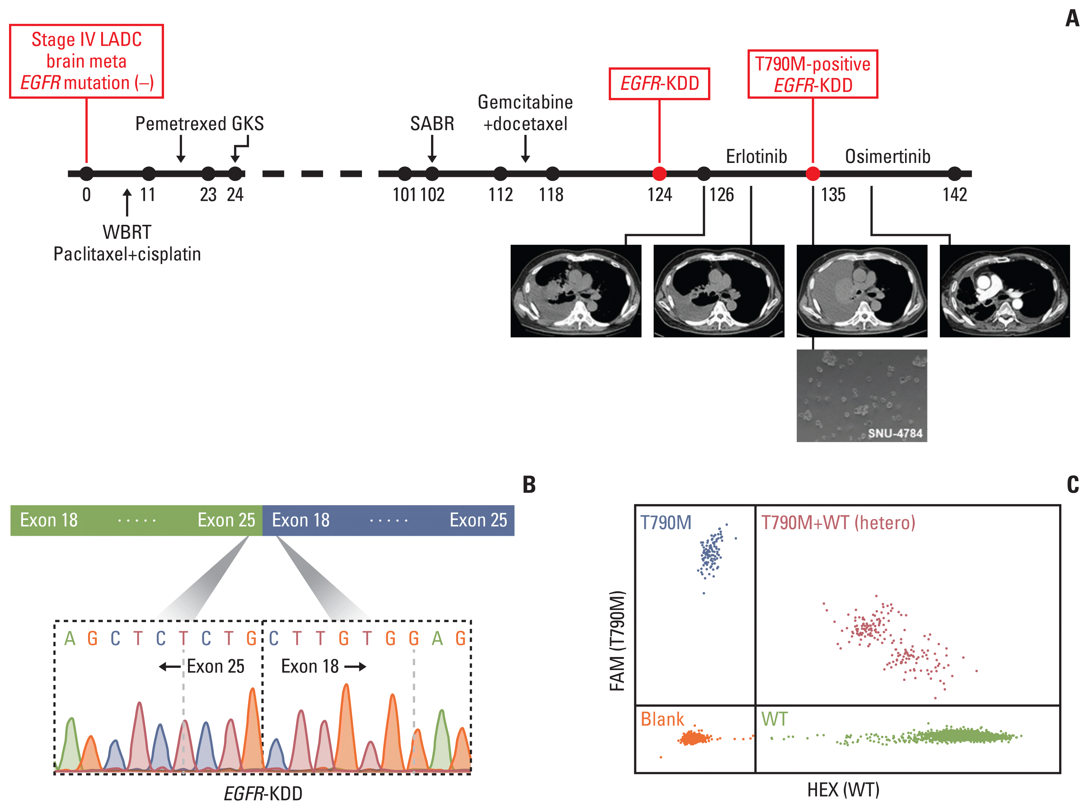
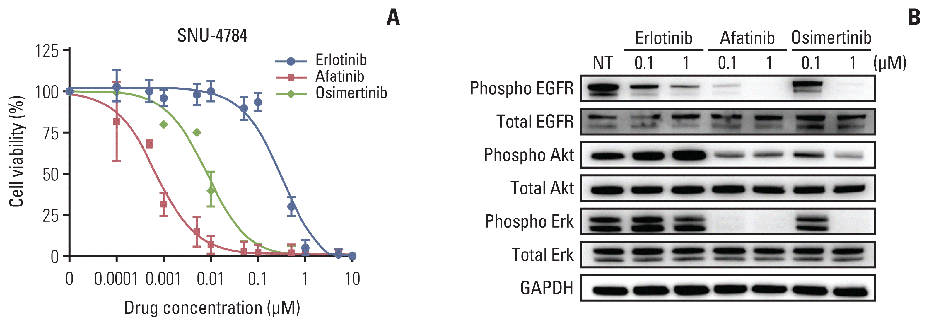
Fig. 1
Clinical history and genomic features for a patient with EGFR KDD. (A) Clinical history of a patient with EGFR-KDD. Numbers beneath the line represent months after diagnosis. Patient’s computerized tomography images indicated tumor masses pre-erlotinib treatment (first), post-erlotinib with partial response (second), post-erlotinib with progressive disease (third), post-osimertinib with partial response (fourth). A patient-derived cell line SNU-4784 was established with pleural effusion upon emerging erlotinib resistance, but without the EGFR T790M mutation. (B) EGFR-KDD breakpoint in a patient cDNA from pleural effusion. (C) The EGFR T790M mutation confirmed by droplet digital polymerase chain reaction (orange dots, blank; green dots, wild type; blue dots, T790M; red dots, T790M plus WT). EGFR-KDD, epidermal growth factor receptor kinase domain duplication.
crt-2021-385f1.gif
Fig. 2
Characteristics of the patient-derived EGFR-KDD cell line SNU-4784. (A) Cell viability assay in SNU-4784 cell line. Cells were exposed to erlotinib, afatinib, and osimertinib for 72 hours. (B) Immunoblot assay of SNU-4784 cell line. Cells were exposed to erlotinib, afatinib, or osimertinib for 4 hours. Cell viability assay and the immunoblot assay were independently repeated three times. EGFR-KDD, epidermal growth factor receptor kinase domain duplication; GAPDH, glyceraldehyde 3-phosphate dehydrogenase.
crt-2021-385f2.gif
Fig. 3
Characteristics of the EGFR-KDDT790M Ba/F3 cell lines. (A) Cell viability assays in EGFR-KDDWT and EGFR-KDDT790M Ba/F3 cell lines. Cells were exposed to EGFR TKIs for 72 hours. Graphs represent mean±SD values. (B) Immunoblot assays of EGFR-KDDWT and EGFR-KDDT790M Ba/F3 cell lines. Cells were exposed to erlotinib, afatinib, or osimertinib for 4 hours. Cell viability assays and immunoblot assays were independently repeated three times. EGFR-KDD, epidermal growth factor receptor kinase domain duplication; GAPDH, glyceraldehyde 3-phosphate dehydrogenase.
crt-2021-385f3.gif
Fig. 4
ENU mutagenesis screening to identify the potential resistance mechanism underlying the EGFR-KDDBDT mutation. (A) Graphical scheme of ENU mutagenesis screening. EGFR-KDDBDT Ba/F3 cells were exposed to 50 μg/mL ENU and selected with 2 μM osimertinib. (B) Sanger sequencing of osimertinib-resistant EGFR-KDDBDT Ba/F3 cells. EGFR C797S mutation in kinase domain 2. EGFR-KDD, epidermal growth factor receptor kinase domain duplication; ENU, N-ethyl-N-nitrosourea.
crt-2021-385f4.gif
Fig. 5
Characteristics of the EGFR-KDDT/T+C Ba/F3 cell line. (A) Cell viability assay of the EGFR-KDDT/T+C Ba/F3 cell line. Cells were exposed to EGFR TKIs for 72 hours. (B) Immunoblot assays of the EGFR-KDDT/T+C Ba/F3 cell line. Cells were exposed to EGFR TKIs for 4 hours. (C) Cell viability assay in the EGFR-KDDT/T+C Ba/F3 cell line. Cells were exposed to EGFR TKIs and cetuximab for 72 hours. For combination treatment, 10 μg/mL cetuximab was added. (D) Growth curve of the EGFR-KDD Ba/F3 cell lines. Cells were grown in interleukin-3 free media and the Ba/F3 parental cells were used as controls. EGFR-KDD, epidermal growth factor receptor kinase domain duplication; TKI, tyrosine kinase inhibitor.
crt-2021-385f5.gif
TOOLS
Similar articles