Journal List > Cancer Res Treat > v.54(1) > 1154871

Kim, Shin, Yoon, Choi, Namgoong, Kim, Koh, Lee, Yoon, Lyu, Han, Hahn, and Cho: Children’s Hepatic Tumors International Collaboration-Hepatoblastoma Stratification (CHIC-HS) System for Pediatric Patients with Hepatoblastoma: A Retrospective, Hospital-Based Cohort Study in South Korea

Abstract

Purpose

In 2017, the Children’s Hepatic Tumors International Collaboration-Hepatoblastoma Stratification (CHIC-HS) system was introduced. We aimed to evaluate the accuracy of CHIC-HS System for the prediction of event-free survival (EFS) in Korean pediatric patients with hepatoblastoma.

Materials and Methods

This two-center retrospective study included consecutive Korean pediatric patients with histopathologically confirmed hepatoblastoma from March 1988 through September 2019. We compared EFS among four risk groups according to the CHIC-HS system. Discriminatory ability of CHIC-HS system was also evaluated using optimism-corrected C-statistics. Factors associated with EFS were explored using multivariable Cox regression analysis.

Results

We included 129 patients (mean age, 2.6±3.3 years; female:male, 63:66). The 5-year EFS rates in the very low, low, intermediate, and high-risk groups, according to the CHIC-HS system were 90.0%, 82.8%, 73.5%, and 51.3%, respectively. The CHIC-HS system aligned significantly well with EFS outcomes (p=0.004). The optimism-corrected C index of CHIC-HS was 0.644 (95% confidence interval [CI], 0.561 to 0.727). Age ≥ 8 (vs. age ≤ 2; hazard ratio [HR], 2.781; 95% CI, 1.187 to 6.512; p=0.018), PRE-Treatment EXTent of tumor (PRETEXT) stage IV (vs. PRETEXT I or II; HR, 2.774; 95% CI, 1.228 to 5.974; p=0.009), and presence of metastasis (HR, 2.886; 95% CI, 1.457 to 5.719; p=0.002), which are incorporated as the first three nodes in the CHIC-HS system, were independently associated with EFS.

Conclusion

The CHIC-HS system aligned significantly well with EFS outcomes in Korean pediatric patients with hepatoblastoma. Age group, PRETEXT stage, and presence of metastasis were independently associated with EFS.

Introduction

Hepatoblastoma is the most common malignant liver tumor in children, with an estimated annual incidence to be 1.5 cases per million [1]. Recently, there has been remarkable progress in terms of survival among hepatoblastoma patients due to advances in chemotherapy strategies and surgical techniques, including liver transplantation [2], with the 3-year event-free survival (EFS) reaching approximately 80%–90% [3,4]. However, there are still considerable differences in outcomes between low-risk and high-risk hepatoblastoma [5,6].
Thus, four major international liver groups—the Childhood Liver Tumors Strategy Group (SIOPEL), Children’s Oncology Group (COG), German Society for Paediatric Oncology and Haematology (GPOH), and Japanese Study Group for Pediatric Liver Tumors (JPLT)—have introduced their own risk-stratified multimodal treatment strategies [3,7,8]. However, it is difficult to compare study results from the different groups. Therefore, these four groups developed a risk stratification system based on the Children’s Hepatic tumors International Collaboration (CHIC) database, called the CHIC-Hepatoblastoma Stratification (CHIC-HS) system [9], for use in the Pediatric Hepatic International Tumor Trial (PHITT). However, so far, there is a paucity of data evaluating the risk stratification accuracy of CHIC-HS system.
Therefore, we aimed to validate how well the CHIC-HS system predicts EFS in a hospital-based retrospective cohort comprising Korean pediatric patients with hepatoblastoma.

Materials and Methods

1. Patients

Consecutive hepatoblastoma patients who were managed at the study hospitals from March 1988 through September 2019 were retrospectively reviewed. For the robustness of the statistical analysis, Severance hospital was participated upon request in addition to Asan Medical Center. The inclusion criteria were as follows: (1) age at diagnosis < 18 years; (2) histopathologically confirmed hepatoblastoma; (3) abdominal computed tomography (CT) or magnetic resonance imaging (MRI) performed before treatment and initial imaging data available for analysis; and (4) available electronic medical records, including laboratory and follow-up data.

2. Data collection and imaging analysis

The CHIC-HS system stratifies patients into four risk groups depending on the following factors: age (classified into three groups, i.e., ≤ 2 vs. 3–7 vs. ≥ 8 years), serum α-fetoprotein (AFP; classified into four groups, i.e., < 100 vs. 100–999 vs. 103–106 vs. > 106 ng/mL), and PRETEXT stage and its annotation factors. Therefore, those data were recorded and assessed.
Imaging analysis for the PRETEXT staging and annotation factors were conducted by three experienced pediatric radiologists (H.M.Y., H.J.S., and Y.A.C., with 6, 6, and 20 years of experience in pediatric body imaging, respectively), based on pretreatment CT or MRI images. The PRETEXT group indicates overall tumor extent and depends on the number of hepatic sections that are free from tumor as follows: PRETEXT I, three contiguous hepatic sections free from tumor; PRETEXT II, two contiguous sections free from tumor; PRETEXT III, one section free from tumor; and PRETEXT IV, no tumor-free sections. Annotation factors include vascular involvement (V, hepatic vein/inferior vena cava; P, portal vein), extrahepatic tumor extension (E), multifocality (F), tumor rupture (R), caudate lobe involvement (C), lymph node metastases (N), and distant metastases (M). All abdominal CT scans were performed with contrast enhancement protocols including the portal venous phase. In all patients, chest CT scans were performed to evaluate possible lung metastases. To determine the annotation factor M, all available imaging studies (positron emission tomography/CT scans, whole-body MRIs, or bone scans) were thoroughly reviewed, if available. Surgical findings were also reviewed to check for possible misinterpretation of imaging studies and details such as diaphragm involvement or peritoneal seeding.
Because of the small number of patients, the four PRETEXT stages, five annotation factors, and four AFP groups were integrated into three simplified PRETEXT stages (I or II vs. III vs. IV), one aggregated VPEFR factor (positive for at least one of V, P, E, F, or R factors), and two AFP categories (< 1,000 vs. ≥ 1,000 ng/mL). Patients were classified into four risk groups according to the CHIC-HS system [9].

3. Statistical analysis

The primary study outcome was EFS, defined as the time from enrollment (the date of initial abdominal CT scan) until the occurrence of one of the following events: first relapse, disease progression, development of a second malignancy, or death from any cause [9]. The study was conducted according to Strengthening the Reporting of Observational Studies in Epidemiology [STROBE] guidelines [10]. Relapse and disease progression were evaluated based on the Response Evaluation Criteria in Solid Tumors (RECIST) ver. 1.1 criteria [11]. To evaluate the discriminatory performance of the CHIC-HS system, the cumulative EFS was calculated and compared between four risk groups according to the CHIC-HS system (very low vs. low vs. intermediate vs. high) using the Kaplan-Meier (KM) method and the log-rank test.
We also calculated C-statistics with 95% confidence intervals (CIs) of the CHIC-HS system to evaluate the discriminatory ability of each system using Z tests [12]. To avoid overestimation of the C-statistics, optimism-corrected C-statistics were calculated by subtracting optimism from the original value using the bootstrap method. C-statistics were interpreted as follows: > 0.8 indicated excellent discrimination, 0.7–0.8 indicated good discrimination, 0.6–0.7 indicated some clinical value, and < 0.6 indicated no clinical value [13].
In addition, multivariable Cox regression to explore factors associated with EFS. The following variables were included in the analysis: sex, age group, PRETEXT stage, annotation factors (V, P, E, F, R, C, N, and M). Additionally, subgroups of the simplified classification using the merged PRETEXT stages, aggregated VPEFR, and dichotomized AFP categories were also included in the analysis. Variables were selected via backward elimination.
All statistical analyses were conducted using R software ver. 3.6.1 (R Foundation for Statistical Computing, Vienna, Austria) with “tableone” and “survival” packages. All tests were two-sided, with the significance level at 0.05.

Results

1. Patients

A total of 204 potentially eligible patients who underwent initial abdominal CT or MRI for suspected hepatoblastoma were retrospectively identified by a systematic, computerized search of a tertiary referral center database. Among these patients, 75 were excluded for the following reasons: (1) confirmed diagnosis other than hepatoblastoma (n=40); (2) insufficient imaging data for PRETEXT staging, e.g., unavailable initial chest CT or abdominal CT/MRI (n=24); (3) immediate follow-up loss after PRETEXT staging (n=8); and (4) no histologic confirmation (n=3). Consequently, a total of 129 patients (mean age±standard deviation [SD], 2.6±3.3 years; female:male, 63:66) were included and analyzed (Table 1).
The majority of patients were 2 years old or younger (66.7%; 86/129) and had serum AFP levels between 1,000 and 106 ng/mL (87.2%; 109/125). The proportions of patients classified as PRETEXT I, II, III, and IV were 7.8% (10/129), 39.5% (51/129), 30.2% (39/129), and 22.5% (29/129), respectively. Distant metastases (annotation factor M+) were present in 26.4% (34/129) of the patients at the time of diagnosis. Additionally, the proportions of patients with positive V, P, E, F, and R factors were 34.9% (45/129), 18.6% (24/129), 8.5% (11/129), 35.7% (46/129), and 7.8% (10/129), respectively; 54.3% (70/129) of the patients were positive in terms of at least one of V, P, E, F, or R. The proportions of the patients who were positive for C and N were 24.0% (31/129) and 8.5% (11/129), respectively. There were four patients whose AFP levels were not available; among those, two could be classified as high-risk regardless of their serum AFP levels (a 3-year-old male with PRETEXT IV and metastasis and a 10-year-old female with PRETEXT IV without metastasis). In the other two patients, we assumed serum AFP to be higher than 1,000 ng/mL because 96% of the study population had AFP ≥ 1,000 ng/mL. These two patients were 9 months and 1 year of age, and their PRETEXT stages were III and IV, respectively. Additionally, the 9-month-old male with a PRETEXT stage of III was V+. Therefore, those patients could be classified into the intermediate group (the 9-month-old male with PRETEXT III and V+ without metastasis and the 1-year-old male with PRETEXT IV without metastasis). Consequently, patients were classified as very low risk in 8.5% (11/129) of cases, low risk in 31.0% (40/129) of cases, intermediate risk in 23.3% (30/129) of cases, and high risk in 37.2% (48/129) of cases.

2. EFS based on the CHIC-HS system

During the median follow-up of 36.6 months (interquartile range, 11.3 to 81.8 months), 36 patients (27.9%) experienced a clinical event (i.e., first relapse, disease progression, development of a second malignancy, or death from any cause). Indeed, the 6-month, 1-, 2-, 3-, and 5-year EFS rates were 88.1%, 83.1%, 74.5%, 69.4%, and 69.4%, respectively.
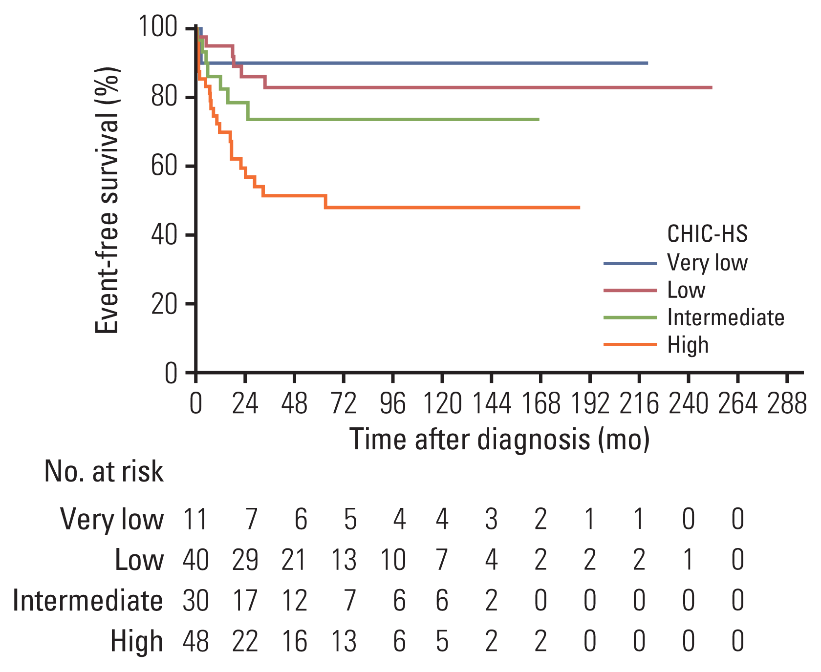
When stratified according to the CHIC-HS system, the 6-month, 1-, 2-, 3-, and 5-year EFS rates were 90.0%, 90.0%, 90.0%, 90.0%, and 90.0% in very low-risk group; 94.9%, 94.9%, 86.0%, 82.8%, and 82.8% in the low-risk group; 86.2%, 86.2%, 78.4%, 73.5%, and 73.5% in intermediate-risk group; and 83.3%, 69.8%, 59.4%, 51.3%, and 51.3% in the high-risk group, respectively (Fig. 1).
Univariable Cox regression analysis revealed hazard ratios (HRs) (with the very low-risk group set as the reference category) of 1.48 (95% CI, 0.18 to 12.27), 2.65 (95% CI, 0.33 to 21.57), and 5.82 (95% CI, 0.78 to 43.21) for the low-, intermediate-, and high-risk groups, respectively. Consequently, the CHIC-HS system aligned significantly well with EFS outcomes (p=0.004). The optimism-corrected C indices of the CHIC-HS was 0.644 (95% CI, 0.561 to 0.727), indicating some clinical value.

3. Factors associated with EFS

Results of univariable and multivariable Cox regression analysis to explore factors associated with EFS are presented in Table 2. Multivariable analyses showed that age ≥ 8 (vs. age ≤ 2; HR, 2.781; 95% CI, 1.187 to 6.512; p=0.018), PRE-Treatment EXTent of tumor (PRETEXT) stage IV (vs. PRETEXT I or II; HR, 2.774; 95% CI, 1.228 to 5.974; p=0.009), and presence of metastasis (HR, 2.886; 95% CI, 1.457 to 5.719; p=0.002) were independently associated with EFS.

Discussion

Our retrospective study showed that the CHIC-HS system was a significant predictor of EFS (p=0.004), with an optimism-corrected C index of 0.644 (95% CI, 0.561 to 0.727). The 5-year EFS rates in the very low-, low-, intermediate-, and high-risk group were 90.0%, 82.8%, 73.5%, and 51.3%, respectively. Multivariable Cox regression analysis identified age group, PRETEXT stage, and the presence of metastasis as independent predictors of EFS.
The CHIC-HS system, created based on 1,605 children treated during eight multicenter hepatoblastoma trials over 25 years, incorporates the presence of metastasis, age group, serum AFP level, aggregated VPEFR factor status, and resectability [9]. To date, there is a paucity of data evaluating the accuracy CHIC-HS system to predict prognosis. Our study demonstrated a respectable level of discrimination achieved by the CHIC-HS. Multivariable Cox regression analysis identified age group, PRETEXT stage, and metastasis status as independent factors associated with EFS, which are incorporated as the first three nodes in the CHIC-HS system. This at least partly confirms the external validity of the CHIC-HS system.
However, it should be emphasized that the optimism-corrected C index did not reach the level of good discrimination (< 0.7). In the KM curve for the very low risk group (Fig. 1), there was an early descent at 3 months after diagnosis. Notably, one female patient classified as very low risk according to the CHIC-HS system died 77 days after her diagnosis. The patient was a preterm baby born at 27 weeks’ gestation with extremely low birth weight (910 g) and diagnosed with hepatoblastoma 5 months of age. The patient died due to acute respiratory distress syndrome combined with heart and renal failure. For this patient, the association between hepatoblastoma and death is unclear, and a previous study did not find a significant association between prematurity and EFS [14]. If the death was not clinically associated with hepatoblastoma, there might be a chance to underestimate discriminatory performance of CHIC-HS system. However, since this is a rare but possible scenario in real practice, this should be further investigated.
This study had several other limitations. First, there might be a potential bias derived from the study’s retrospective design. In particular, as already mentioned, serum AFP levels at the diagnosis were not available, thereby limiting the analysis. Second, since the study was based on a hospital-based cohort, the generalizability of our results might be limited. Of note, our study included a local cohort over a long period. This might impose a risk of bias due to different treatment modalities applied at different times during the study period. Notably, liver transplantation has been widely performed at the institutions since 2006. Besides this, patients in the late period had relatively shorter follow-up periods. However, considering that most events generally occur within 3 years of diagnosis, the impact of this limitation might not be significant.
In conclusion, the CHIC-HS system aligned significantly well with EFS outcomes in Korean pediatric patients with hepatoblastoma. Age group, PRETEXT stage, and presence of metastasis were independently associated with EFS. Considering the retrospective nature of this study and its long study period implying temporal shifts in treatment strategies, further large-scale prospective studies are warranted to evaluate the CHIC-HS system.

Notes

Ethical Statement

This bi-center retrospective study was approved by the relevant institutional review boards (IRB number, 2019-1403 for Asan Medical Center; 4-2020-0052 for Severance Hospital), and the requirement for informed consent was waived due to the study’s retrospective design. The study was conducted according to Strengthening the Reporting of Observational Studies in Epidemiology [STROBE] guidelines.

Author Contributions

Conceived and designed the analysis: Shin HJ, Yoon HM.

Collected the data: Kim PH, Shin HJ, Yoon HM.

Contributed data or analysis tools: Kim PH, Shin HJ, Yoon HM, Choi YH, Namgoong JM, Kim DY, Koh KN, Lee MJ, Yoon H, Lyu CJ, Han JW, Hahn SM, Cho YA.

Performed the analysis: Kim PH, Shin HJ, Yoon HM.

Wrote the paper: Kim PH.

Supervision: Yoon HM, Cho YA.

Conflicts of Interest

Conflict of interest relevant to this article was not reported.

References

1. Ranganathan S, Lopez-Terrada D, Alaggio R. Hepatoblastoma and pediatric hepatocellular carcinoma: an update. Pediatr Dev Pathol. 2020; 23:79–95.
crossref
2. Carceller A, Blanchard H, Champagne J, St-Vil D, Bensoussan AL. Surgical resection and chemotherapy improve survival rate for patients with hepatoblastoma. J Pediatr Surg. 2001; 36:755–9.
crossref
3. Czauderna P, Lopez-Terrada D, Hiyama E, Haberle B, Malogolowkin MH, Meyers RL. Hepatoblastoma state of the art: pathology, genetics, risk stratification, and chemotherapy. Curr Opin Pediatr. 2014; 26:19–28.
4. Perilongo G, Maibach R, Shafford E, Brugieres L, Brock P, Morland B, et al. Cisplatin versus cisplatin plus doxorubicin for standard-risk hepatoblastoma. N Engl J Med. 2009; 361:1662–70.
crossref
5. Hiyama E, Hishiki T, Watanabe K, Ida K, Ueda Y, Kurihara S, et al. Outcome and late complications of hepatoblastomas treated using the Japanese Study Group for Pediatric Liver Tumor 2 Protocol. J Clin Oncol. 2020; 38:2488–98.
crossref
6. Haberle B, Maxwell R, Schweinitz DV, Schmid I. High dose chemotherapy with autologous stem cell transplantation in hepatoblastoma does not improve outcome: results of the GPOH Study HB99. Klin Padiatr. 2019; 231:283–90.
crossref
7. Meyers RL, Tiao G, de Ville de Goyet J, Superina R, Aronson DC. Hepatoblastoma state of the art: pre-treatment extent of disease, surgical resection guidelines and the role of liver transplantation. Curr Opin Pediatr. 2014; 26:29–36.
8. Perilongo G, Malogolowkin M, Feusner J. Hepatoblastoma clinical research: lessons learned and future challenges. Pediatr Blood Cancer. 2012; 59:818–21.
crossref
9. Meyers RL, Maibach R, Hiyama E, Haberle B, Krailo M, Rangaswami A, et al. Risk-stratified staging in paediatric hepatoblastoma: a unified analysis from the Children’s Hepatic tumors International Collaboration. Lancet Oncol. 2017; 18:122–31.
crossref
10. von Elm E, Altman DG, Egger M, Pocock SJ, Gotzsche PC, Vandenbroucke JP, et al. Strengthening the Reporting of Observational Studies in Epidemiology (STROBE) statement: guidelines for reporting observational studies. BMJ. 2007; 335:806–8.
crossref
11. Eisenhauer EA, Therasse P, Bogaerts J, Schwartz LH, Sargent D, Ford R, et al. New response evaluation criteria in solid tumours: revised RECIST guideline (version 1.1). Eur J Cancer. 2009; 45:228–47.
crossref
12. Kang L, Chen W, Petrick NA, Gallas BD. Comparing two correlated C indices with right-censored survival outcome: a one-shot nonparametric approach. Stat Med. 2015; 34:685–703.
13. Wong MC, Ching JY, Ng S, Lam TY, Luk AK, Wong SH, et al. The discriminatory capability of existing scores to predict advanced colorectal neoplasia: a prospective colonoscopy study of 5,899 screening participants. Sci Rep. 2016; 6:20080.
crossref
14. Czauderna P, Haeberle B, Hiyama E, Rangaswami A, Krailo M, Maibach R, et al. The Children’s Hepatic tumors International Collaboration (CHIC): Novel global rare tumor database yields new prognostic factors in hepatoblastoma and becomes a research model. Eur J Cancer. 2016; 52:92–101.
crossref

Fig. 1
Event-free survival rates based on the 2017 Children’s Hepatic Tumors International Collaboration-Hepatoblastoma Stratification (CHIC-HS) system.
crt-2021-265f1.gif
Table 1
Baseline characteristics of the 129 patients
Characteristic No. (%)
Age at initial diagnosis, mean±SD (yr) 2.6±3.3
 ≤ 2 86 (66.7)
 3–7 27 (20.9)
 ≥ 8 16 (12.4)
Sex
 Female 63 (48.8)
 Male 66 (51.2)
Serum AFP concentration (ng/mL) (n=125)
 < 100 1 (0.8)
 100–999 4 (3.2)
 1,000–106 109 (87.2)
 > 106 11 (8.8)
PRETEXT stage
 I 10 (7.8)
 II 51 (39.5)
 III 39 (30.2)
 IV 29 (22.5)
Annotation factors
 V (HV or IVC involvement) 45 (34.9)
 P (PV involvement) 24 (18.6)
 E (extrahepatic tumor extension) 11 (8.5)
 F (multifocality) 46 (35.7)
 R (tumor rupture) 10 (7.8)
 C (caudate involvement) 31 (24.0)
 N (lymph node metastasis) 11 (8.5)
 M (distant metastasis) 34 (26.4)
 One or more of V, P, E, F, or R 70 (54.3)
CHIC-HS risk
 Very low 11 (8.5)
 Low 40 (31.0)
 Intermediate 30 (23.3)
 High 48 (37.2)
Follow-up period, median (IQR, mo) 36.6 (11.3–81.8)
No. of patients with an event 36 (27.9)
No. of deaths 22 (17.8)
Table 2
Summary of Cox regression analysis
Variable Univariable Multivariable


Unadjusted HR (95% CI) p-value Adjusted HR (95% CI) p-value
Sex

 Female Reference

 Male 1.542 (0.795–2.994) 0.200

Age group (yr)

 ≤ 2 Reference Reference

 3–7 2.206 (1.033–4.714) 0.041 1.809 (0.839–3.901) 0.130

 ≥ 8 2.788 (1.202–6.467) 0.017 2.781 (1.187–6.512) 0.018

PRETEXT

 I Reference

 II 0.930 (0.206–4.200) 0.925

 III 0.983 (0.208–4.630) 0.982

 IV 2.983 (0.681–13.066) 0.147

PRETEXT (simplified)

 I or II Reference Reference

 III 1.045 (0.432–2.524) 0.923 1.233 (0.497–3.059) 0.652

 IV 3.172 (1.504–6.689) 0.002 2.774 (1.228–5.974) 0.009

V

 Yes 2.463 (1.277–4.749) 0.007 Eliminated

P

 Yes 2.371 (1.136–4.947) 0.021 Eliminated

E

 Yes 1.685 (0.593–4.790) 0.327

F

 Yes 2.553 (1.324–4.926) 0.005 Eliminated

R

 Yes 2.179 (0.846–5.610) 0.107

C

 Yes 1.348 (0.650–2.799) 0.423

N

 Yes 3.017 (1.317–6.909) 0.009 Eliminated

M

 Yes 3.513 (1.816–6.795) < 0.001 2.886 (1.457–5.719) 0.002

V, P, E, F, or R

 Yes 2.339 (1.149–4.761) 0.019 Eliminated

AFP

 < 1,000 0.865 (0.078–9.547) 0.906

 ≥ 1,000 1.683 (0.403–7.033) 0.476

Variables with p < 0.05 in the univariable analyses were entered into the multivariable analysis. AFP, α-fetoprotein; C, caudate lobe involvement; CI, confidence interval; E, extrahepatic tumor extension; F, multifocality; HR, hazard ratio; M, distant metastases; N, lymph node metastases; P, portal vein; PRETEXT, 2017 PRE-Treatment EXTent of tumor staging system; R, tumor rupture; REF, reference category; V, hepatic vein/inferior vena cava.

TOOLS
Similar articles