INTRODUCTION
CASE REPORT
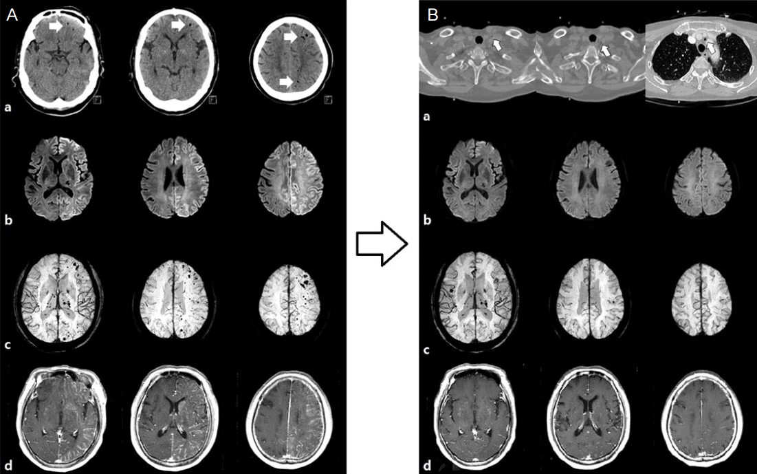
 | Figure 1.(A) Brain computed tomography (CT) and magnetic resonance image (MRI) demonstrate acute cerebral ischemia with cerebral air embolism. (a) Unenhanced CT shows several hypodense lesions in left cerebral hemisphere, suggesting multiple scattered small air bubbles (white arrows). (b) In diffusion weighted image (DWI), typical diffuse high signal intensity lesions in left entire cerebral hemisphere and basal ganglia are seen. (c) Susceptibility weighted image (SWI) shows multiple scattered air bubbles in mainly left frontal subcortex. (d) Diffuse parenchymal and leptomeningeal enhancement are seen in left cerebral hemisphere suggesting early blood-brain barrier (BBB) disruption. (B) Neck CT reveals small air collection and follow-up magnetic resonance imaging (MRI) at 8 days later demonstrate resolution of cerebral air embolism after treatment. (a) In neck CT, small amount of air collection in left lower lateral neck and superior mediastinum are noted (white arrows). (b, c, d) Brain MRI shows resolution of diffuse ischemic lesions, air embolism and BBB disruption in left entire cerebral hemisphere in DWI, SWI and enhanced T1 images in contrast to previous images. |


 PDF
PDF Citation
 Print
Print


 XML Download