

 PDF
PDF Citation
 Print
Print


서 론
연수에는 무의식적인 호흡기능에 중추적인 역할을 하는 세포핵들이 있으므로, 외측연수경색의 증상으로 중추수면무호흡이 발생할 수 있는 것으로 알려져 있다. 본 저자들은 최근 일측성 외측연수경색에 의한 중추수면무호흡 증례를 경험하여 이를 뇌간의 자율호흡조절 네트워크의 최신 지견과 함께 보고하고자 한다.
Go to : 
증 례
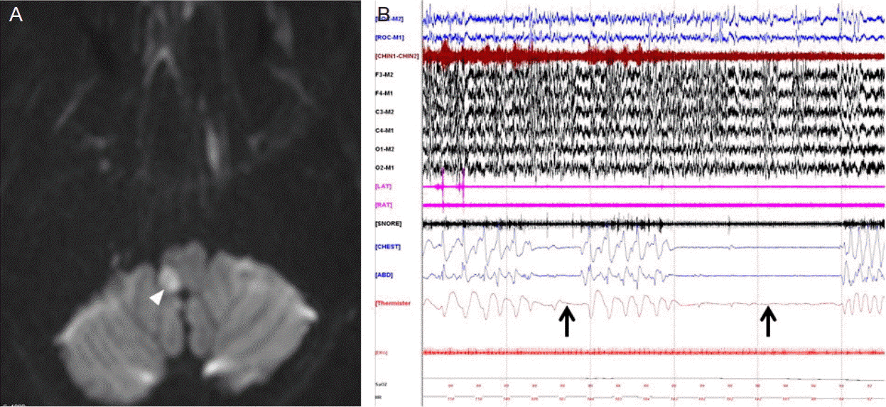
특이 과거력이 없던 49세 여환이 내원 1시간 전 갑자기 발생한 어지러움, 체위불균형, 구음장애와 삼킴곤란을 주소로 내원하였다. 내원시 다량의 객담 증상을 호소하였으나 신체검사상 발열이나 청진상 이상소견은 관찰되지 않았다. 신경학적진찰상에서는 좌측 방향으로의 수평 안진(horizontal nystagmus), 구개수의 좌측 편위 및 우측 연구개 저하, 우측 상하지 실조, 우측 쏠림, 우측 안면 및 좌측 반신의 통각 및 온도감각 저하, 우측의 호너 증후군(Horner’s syndrome)이 관찰되었다. 확산강조영상에서도 우측 외측 연수에 고신호강도병변이 관찰되어(Fig. 1A), 환자는 우측 외측연수경색으로 진단 후 뇌졸중집중치료실에 입원했다. 그런데 입원 3일 째 새벽에 수면 중 산소포화도 감소 후 심장정지가 발생하였다. 즉각적인 심폐소생술 및 기관내삽관 후, 환자는 정상순환으로 회복되었고 의식수준도 정상으로 회복되었다. 하위뇌신경마비와 다량의 객담이 있었던 환자로 이에 의한 기도폐쇄 및 호흡정지로 판단하여 중환자실 입실하여 2일간 인공호흡기 치료 후 기관내삽관을 제거하고 환자는 일반병실로 재이동하였다. 일반병실 이동한 후 첫째 날에는 지속적인 객담배출과 혈뇨로 인하여 밤새 수면을 취하지 못하였다. 병실 이동 2일째 되는 날, 수면 중에 산소포화도가 떨어지는 일이 발생하였고, 의식저하와 동맥혈가스분석상 PaCO2 160 mmHg의 고이산화탄소혈증이 관찰되었다. 기관내삽관 및 기계환기 시행 후 다시 의식이 회복되었고, 중환자실로 환자를 이송 3일 후 기관 삽관은 유지하면서 기계환기는 제거하고 산소 4 L만 유지한 상태로 수면다원검사(polysomnography)를 시행하였다. 흉곽 움직임 소실과 산소포화도 저하가 10초 이상 지속되는 중추수면무호흡 사건이 시간당 평균 51회 정도 관찰되어(Apnea Hypopnea Index: 90.9, Apnea Index: 51.3) 외측연수경색에 따른 2차적인 중추수면무호흡으로 진단할 수 있었다(Fig. 1B). 수면 중에 산소포화도가 83%까지 떨어졌으며, 수면 중에 시행한 동맥혈가스분석상 PaCO2 70 mmHg로 고이산화탄소혈증이 관찰되었다. 환자는 기관절개술 시행 후 휴대용 인공호흡기를 적용하여 일반병실 이동하였고, 수면 중에만 휴대용 인공호흡기를 적용하였다. 중추수면무호흡 증상은 점차 호전되어 1개월 후에 시행한 수면다원검사에서 중추수면무호흡 소견은 사라지고 경미한 중추수면저호흡(central sleep hypoapnea) 소견만 보여 인공호흡기를 제거하였다. 3개월 후에 시행한 수면다원검사에서 중추수면무호흡 및 수면저호흡 소견은 사라졌으며, 환자는 뇌경색 발생 5개월이 되는 시점에 기관절개술 자리를 막았고 이후로도 호흡 부전 없이 지내고 있다.
Go to :
고 찰
호흡을 조절하는 신경 중추는 의식적인 호흡을 담당하는 대뇌피질의 의식호흡중추와, 무의식적이고 반사적인 호흡을 담당하는 뇌간의 무의식호흡중추로 나눌 수 있다. 따라서 뇌간의 무의식호흡중추에만 기능소실이 생기면, 각성상태에서는 의식호흡중추에 의해 숨을 쉴 수 있지만, 수면상태에서는 호흡기능이 저하되거나 완전히 마비되는 중추수면무호흡이 발생할 수 있다. 이러한 중추수면무호흡은 수면다원검사에서 무호흡 시에 동반되는 호흡노력이 10초 이상 없고 이러한 사건이 1시간에 5회 이상일 때 진단할 수 있다[1].
뇌간에 위치한 호흡 관련 구조물에는 교뇌에 위치한 교뇌호흡그룹(pontine respiratory group: parabrachialis, nucleus, Kölliker-Fuse nucleus) 연수에 위치한 얼굴핵주변호흡그룹(parafacial respiratory group: retrotrapezoid nucleus), 뇌간솔기핵(brainstem raphe nucleus), 등쪽호흡그룹(dorsal respiratory group: caudal nucleus tractus solitarius), 배쪽호흡기둥(ventral respiratory column: Bötzinger complex, pre-Bötzinger complex, rostral ventral respiratory group, caudal ventral respiratory group)이 있다(Table 1).
Table 1.
Brainstem respiratory network
![]()
경동맥소체(carotid body)와 대동맥토리(aortic body)의 화학수용체(chemoreceptor)에서 측정된 동맥내 산소, 이산화탄소, pH 등의 정보와 폐의 신장수용기(stretch receptor)에서 측정된 정보는 미주신경과 혀인두신경을 통하여 등쪽호흡그룹으로 전달되며, 뇌척수액의 이산화탄소 및 pH 정보는 얼굴핵 주변호흡그룹과 뇌간솔기핵으로 전달된다. 이렇게 말초와 중추의 화학 정보를 전달 받은 핵들은 다시 교뇌호흡그룹 및 배쪽호흡기둥과 연결되어 신경 네트워크를 형성하고[2], 이 신경 네트워크에서 생성된 패턴화된 전기 신호가 망상척수로(reticulospinal tract)를 통해 척수에 위치한 하위운동신경원에 전달되어 가로막과 늑간근, 복근을 규칙적으로 수축, 이완시켜 자율호흡운동을 일으키는 것으로 알려져 있다[3].
뇌간의 호흡 관련 신경 네트워크에서 척수의 하위운동신경원으로 패턴화된 신호를 생성하는 박동조율기 역할을 하는 핵심적인 신경세포는 주로 배쪽호흡기둥에 위치한다. 이 중에서도 pre-Bötzinger complex와 rostral ventral respiratory group은 흡기, Bötzinger complex와 caudal ventral respiratory group은 호기에 중요한 역할을 한다. 흡기가 일어나면서 호흡이 시작되므로, 그중에서도 pre-Bötzinger complex와 rostral ventral respiratory group이 상대적으로 자율적인 호흡 운동에 더 중요한 역할을 한다고 알려져 있다[4]. 본 증례의 경우, 외측연수경색에 의해 이러한 pre-Bötzinger complex와 rostral ventral respiratory group이 등쪽호흡그룹과 함께 침범되어 중추수면무호흡이 발생한 것으로 추정된다(Fig. 2).
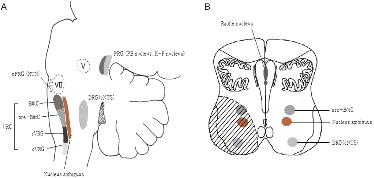 | Figure 2.Brainstem structures for respiration are represented neuroanatomically by a parasagittal brainstem section (A). The presumed structures of damage are expressed by hatch in transverse section of the lower medulla (B). PRG, pontine respiratory group; PB, Parabrachialis; K-F: Kölliker-Fuse; Pfrg, parafacial respiratory group; RTN, retrotrapezoid nucleus; DRG, dorsal respiratory group; cNTS, caudal nucleus tractussolitarius; VRC, ventral respiratoy column; BötC, Bötzinger complex; pre-BötC, pre-Bötzinger complex; rVRG, rostral ventral respiratory group; cVRG, caudal ventral respiratory group.
|
중추수면무호흡은 일반적으로 양측성 연수병변에 의해 발생하지만, 편측 연수병변에서도 나타날 수 있는 것으로 알려져 있다. 일측성 외측연수경색에서 중추수면무호흡의 발생빈도까지는 알려진 바가 없고, 국내외에서 몇몇 증례보고들만 있다[5-7]. 본 증례도 일측성 외측연수경색에서 중추수면무호흡으로 인해 호흡정지까지 발생하였던 증례이다. 일측성 연수병변에서 자율호흡부전이 일어나는 기전에 대해서 명확히 밝혀진 것은 없다. 다만, 이를 설명할 수 있는 가설로 연수에 위치한 호흡중추가 일부 사람에서는 편측에 주로 존재할 가능성8과, 편측 손상이라도 반대측에 교차한 신경섬유가 함께 손상되었을 가능성[9]이 제시된 바 있다.
본 증례에서 뇌경색 발생 이틀 후에 무호흡증상이 생겼는데 이는 진행성 열공경색(progressive lacunar infarction)에서처럼 혈전전파(thrombi propagation), 병변 주위 부종(perilesional edema) 등의 기전으로 인한 병변의 확장 때문으로 생각된다.
저자들은 일측성 외측연수경색에서 중추수면무호흡이 발생한 증례를 경험하였고, 이는 호흡정지로 인한 갑작스러운 심장정지까지 일으킬 수 있는 심각한 합병증이므로, 외측연수경색환자의 진료 시 이를 염두에 두고 환자의 호흡패턴에 대한 주의 깊은 관찰이 필요할 것으로 생각된다.
Go to :
 XML Download