Large basilar tip cerebral aneurysms are difficult to treat. Recently, endovascular treatment has mitigated much of the morbidity associated with treating these lesions. However, the morphology of aneurysms of the vertebrobasilar system can preclude endovascular treatment.
Basilar tip aneurysms are a special group of intracranial aneurysms. First, these kinds of aneurysms are rarely treated with surgery, even aneurysms with unfavorable shape are coiled, while those in the anterior circulation would be clipped. They often have a wide neck and can be found on a vascular bifurcation. Therefore, reopening of coiled basilar tip aneurysms occurs more often than those in other locations. Second, some of them have been observed to grow progressively worse, resulting in a mass effect on the brain stem [1,2].
We present the case of an 81-year-old woman who developed a rapidly growing aneurysm at the basilar tip. This patient showed sudden decreased consciousness even though she had no cerebral hemorrhage.
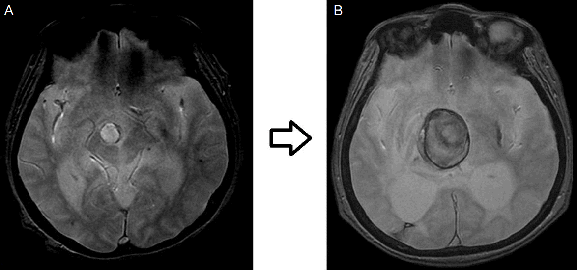
An 81-year-old woman presented to the hospital with vertigo and ataxia in April 2014. Magnetic resonance imaging (MRI) revealed acute infarction in the right pons with an aneurysm (16×14 mm) at the basilar artery tip (Fig. 1A). Considering her age, we decided to monitor her progress instead of performing an operation. Acute infarction symptoms were treated and improved. Although we decided to monitor the progress of aneurysm enlargement through computed tomographic (CT) angiography as an outpatient at regular intervals, the patient did not follow up, but only received medication at a hospital near her home.
The patient remained well until June 2015, when she again presented with sudden decline in level of consciousness. Though she had intermittent headache from several days before visited to hospital, there was no sign of increase in intracranial pressure such as nausea and she could continue daily activities without abnormalities such as gait ataxia. When the patient came to hospital, she was in drowsy mental status with no notable sign of neck stiffness or increase in intracranial pressure but left side weakness was observed. In the neurological exam, even though there was no big difference in the size between pupil of right eye (3.5 mm) and that of left (3 mm), light reflex of right eye was significantly decreased. MRI showed significant enlargement of the aneurysm (47×52 mm); midbrain compression was the presumed cause of decreased consciousness (Fig. 1B). No other specific cause was found. In laboratory test, electrocardiography and other tests, no specific cause was found for the patient’s decreased consciousness.
Although endovascular treatment was considered for the patient, uneven respiration required ventilator therapy and aggravated vital signs such as blood pressure prevented immediate performance of it. The patient was hospitalized with the intention to perform the therapy immediately after mitigation of the symptoms. After 5 days, the patient’s consciousness fell into comatose state and bilaterally showed fixed and dilated pupils with no observed light reflex, and showed decerebrate posture. Although no intracranial hemorrhage was observed in CT performed during observation of process, the aneurysm kept compressing the brainstem and 3rd ventricle which aggravated hydrocephalus and caused respiratory failure, leading to the death of the patient without being able to perform endovascular treatment.
An intracranial aneurysm is a vascular disorder which occurs in the weak point of an intracranial arterial wall to form a localized dilation. The prevalence of unruptured intracranial aneurysm (UIA) in the general population is 1.8-8.4% [3]. UIA rupture causes subarachnoid hemorrhage, which usually results in significant neurological deficits or death. Rapid aneurysm growth is associated with rupture.
UIAs occur in various locations around the Willis’ circle. Bifurcation areas, where the arterial wall is weak and hemodynamic stress is altered, are known to be vulnerable sites [4]. The most common histologic findings of UIAs are a decreased thickness of the tunica media and middle muscular layer of the artery, which leads to structural defects. These defects, combined with hemodynamic factors, lead to aneurysmal outpouching [5].
Although no clear mechanism has been known for the fast development of aneurysm, it increases pressure on the areas with permanent low wall shear stress (WSS) and on the parts of the vessel wall with decreased resist-ability. This is known to accelerate aneurysm by causing endothelial damage and we judge that basilar tip had faster development of aneurysm than other parts since it is more heavily affected by hemodynamic influence [6].
There are two possible causes for the patient’s decreased consciousness. First one is that rapid enlargement of aneurysm affected ascending reticular activating system (ARAS) by pressing midbrain, leading to worsening consciousness and second one is that obstructive hydrocephalus caused by aneurysm pressing 3rd ventricle might have decreased the patient’s consciousness. As this patient had intermittent headaches even before she came to hospital, there is high possibility that hydrocephalus had already been in slow progress. Still, as decreased consciousness appeared without such symptoms as neck stiffness or ataxia, we thought that decreased consciousness at the time she came to hospital was caused more by midbrain compression. Rapid decline of consciousness and worsening symptoms after hospitalization, however, were deemed to be caused by fast progress of hydrocephalus.
Considering that the early size was not big, patient was in old age of 81 and the location of aneurysm was at tip of basilar artery, we decided to observe the progress rather than performing endovascular treatment with risks involved. Still, since the patient did not visit our hospital afterwards, it was impossible to observe the progress and the aneurysm became bigger much faster than expected, which is supposed to be caused by the fact that the patient’s arterial wall became much weaker due to her old age and the aneurysm was located in the position which was more susceptible to hemodynamic stress.
In general, when basilar artery aneurysm is accompanied by hemorrhage, it causes decreased consciousness [7]. In this case, although there was no hemorrhage, aneurysm at the tip of basilar artery became bigger quickly and pressed brain stem and 3rd ventricle, caused hydrocephalus, and leading to decreased consciousness of the patient.
When there is an aneurysm at the tip of basilar artery in old age, endovascular treatment should be positively considered since the aneurysm may grow bigger and have effect on brain stem which may lead to negative prognosis.
REFERENCES
1. van Rooij WJ, Sluzewski M. Opinion: imaging follow-up after coiling of intracranial aneurysms. AJNR Am J Neuroradiol. 2009; 30:1646–8.
2. van Eijck M, Bechan RS, Sluzewski M, Peluso JP, Roks G, van Rooij WJ. Clinical and imaging follow-up of patients with coiled basilar tip aneurysms up to 20 years. AJNR Am J Neuroradiol. 2015; 36:2108–13.

3. Foutrakis GN, Yonas H, Sclabassi RJ. Saccular aneurysm formation in curved and bifurcating arteries. AJNR Am J Neuroradiol. 1999; 20:1309–17.
5. Li J, Shen B, Ma C, Liu L, Ren L, Fang Y, et al. 3D contrast enhancement-MR angiography for imaging of unruptured cerebral aneurysms: a hospital-based prevalence study. PloS One. 2014; 9:e114157.


 PDF
PDF Citation
 Print
Print


 XML Download