INTRODUCTION
The cause of ischemic infarction of the spinal cord usually involves the anterior spinal artery territory. The clinical presentation of an ischemic spinal cord infarction is referred to as anterior spinal artery syndrome (ASAS), which is characterized by bilateral limb weakness and impaired pain and temperature sensation with bladder dysfunction [1]. Symptom such as unilateral limb weakness is possible with spinal cord infarction, but this is a rare phenomenon. Previous study reveals that two of twenty-eight patients with acute spinal cord infarction appear to have unilateral weakness [2]. So the unilateral weakness as a result of cervical cord infarction is sometimes misdiagnosed as a pure motor stroke. We report a case of cervical spinal cord infarction presenting with pure unilateral limb weakness as the initial manifestation.
CASE REPORT
A 66-year-old woman with a history of hypertension visited ER complaining of chest discomfort. Her symptom had begun twenty minutes ago and she described her pain as feeling like a heavy weight had been placed on the right chest and radiating to the posterior neck. Soon after, she found it difficult to raise her right arm and leg. On examination the patient was alert, oriented, and cooperative. Her speech was well-articulated and fluent. Naming, repetition, and comprehension were normal. Cranial nerve functions were normal. Pinprick and temperature sensation were normal. Proprioception and vibratory sensation were also normal. There was normal resistance to passive movement of the right arm and leg. Mild weakness without fasciculation or wasting was present in the right arm and leg.
Tendon reflexes were normal and Babinski sign was absent on the right side. And there was no bladder dysfunction. The patient’s blood pressure was 161/89 mmHg, heart rate was 73 beats per minutes, and blood sugar level was 154mg/dL. Cardiac enzymes based on initial specimen were normal (CK 92 IU/L, CK-MB 0.88 ng/mL, Troponin I 0.035 ng/mL). Two hours later, CK and CK-MB levels were normal (CK 74 IU/L, CK-MB 0.94 ng/mL).Troponin I was slightly higher than upper normal limit (Troponin I 0.052 ng/mL) but it was normalized after one day.
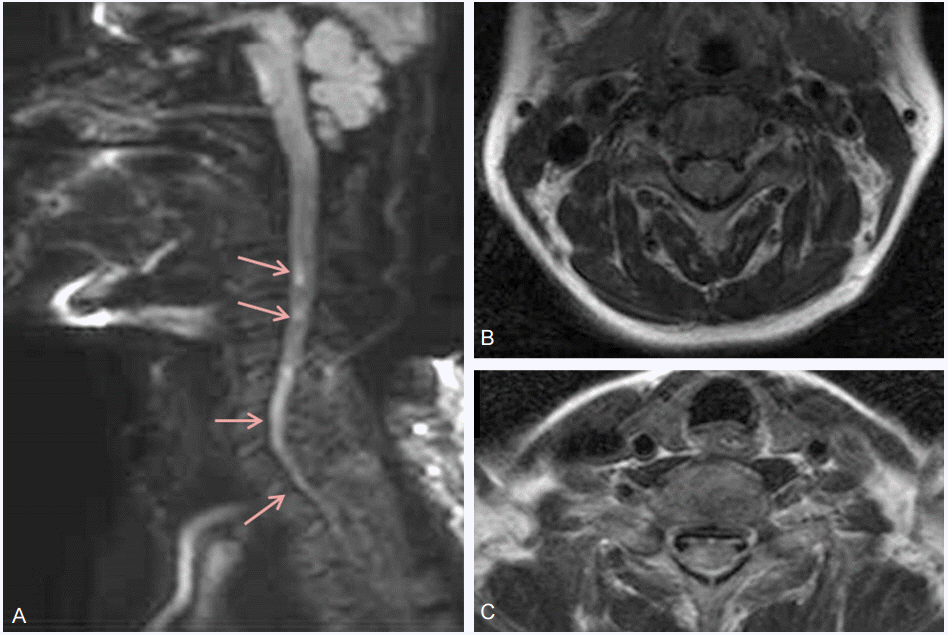
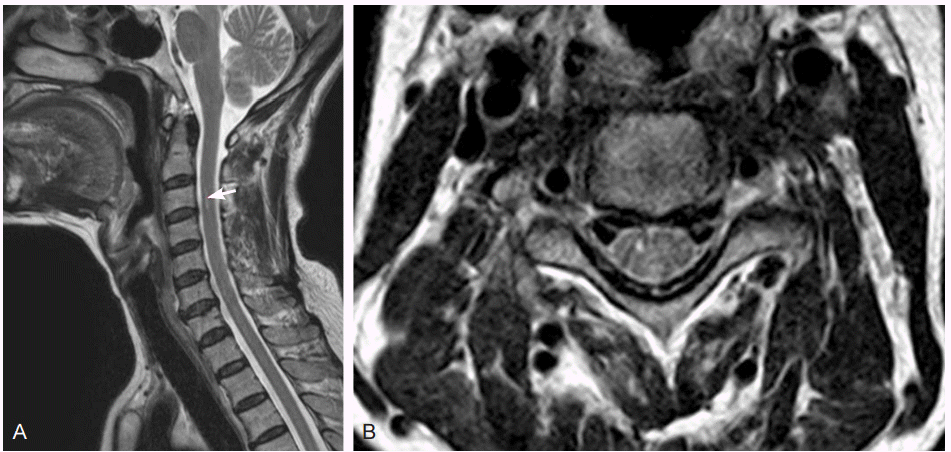
Electrocardiogram showed normal sinus rhythm. According to the protocol of acute stroke management guideline, suspicious acute stroke patients should take a brain magnetic resonance imaging (MRI). Her brain MRI did not show any acute and leg, but not on the neck or face. Proprioception and vibratory sensation were normal. The patient also felt voiding difficulty. Sagittal diffusion-weighted spinal MRI showed high signal intensities extending from segments C3 to C7. Axial T2-weighted spinal MRI showed high signal intensities localized to the right anterior horn (Fig. 1). Therefore, the patient was diagnosed with cervical spinal cord infarction. There was no abdominal aneurysm or dissection on the computed tomography scan. After diagnosis, she received conservative treatment with aspirin. Powers of her right arm and leg were gradually improved, so she was able to walk with a cane by 1 month after onset. Also she could urinate without difficulty, but she continually had sensory impairment. Follow up MRI, obtained 44 days after the onset, showed decreased extent of the lesion, extending from C3 to C4 (Fig. 2).
DISCUSSION
Spinal cord infarction is usually accompanied by sudden paraparesis or quadraparesis depending on the level of the lesions, and also often present with sensory impairment and voiding difficulty. While various symptoms and signs are possible according to the levels of involved spinal cord, unilateral weakness is a rare symptom [2]. We report a rare case of spinal cord infarction accompanied by unilateral limb weakness without sensory impairment and bladder dysfunction as the initial manifestation of the disease that could mimic a pure motor stroke. The pure motor stroke usually have symptoms like facial palsy, dysarthria, but the patients who have unilateral limb weakness without facial palsy or dysarthria are approximately 19 percents [3]. In our case, patient had acute unilateral limb weakness with chest discomfort. She could have been misdiagnosed as the pure motor stroke. Previous study revealed that two of twenty-eight patients with acute spinal cord infarction appear to have unilateral weakness, but it has never been reported in South Korea. Unilateral spinal cord involvement is explained by the incomplete linking of posterior systems and by duplication of the anterior system [4-6]. Cervical spinal cord infarction may be considered in patients with sudden unilateral weakness as an initial manifestation, even though sensory or voiding impairments were absent.


 PDF
PDF Citation
 Print
Print


 XML Download