This article has been
cited by other articles in ScienceCentral.
Abstract
Background:
Cardiac arrhythmias in critically ill patients often cause severe hemodynamic impairment or medical complications, precipitating rapid deterioration of patients’ conditions. This manuscript describes common or fatal cardiac arrhythmias occurring in patients hospitalized at intensive care units.
Results:
Differential diagnosis of wide QRS tachycardias includes ventricular tachycardias (VTs), supraventricular tachycardias with aberrant conduction and noise artifacts. Idiopathic VTs in patients with structurally normal hearts are responsive to antiarrhythmic agents and are cured by catheter ablation. Ventricular tachycardias in structural heart diseases require amiodarone for acute suppression and implantable defibrillator for longterm management. Polymorphic VTs or Torsades de Pointes are effectively suppressed by intravenous infusion of magnesium. Discontinuation of offending agents and, in some refractory cases, installation of temporary pacing is necessary. Atrial fibrillation (AF) is the most commonly observed tachyarrhythmias in intensive care settings. When combined with sepsis or congestive heart failure, AF with rapid ventricular response induces hypotension and sympathetic stimulation, which again aggravates rapid ventricular responses. This viscious cycle continues until adequate rate control measures are initiated. Intravenous amiodarone and anticoagulation are important initial management. In rare cases, electrical noises mimic ventricular tachycardias. Noises are distinguished from true VTs by identification of sharp QRS notches and stable hemodynamics during the events.
Conclusion:
Proper interpretation of monitoring or standard ECG is crucial for differential diagnosis of cardiac arrhythmias occurring in intensive care units. Management should be tailored based upon arrhythmia characteristics and individual medical conditions.
Go to :

Keywords: Tachycardia, Bradycardia, Intensive care
서 론
중환자실 환자에서 나타나는 부정맥은 기저 심질환 혹은 내과적인 전신질환이 있는 상황에서 나타나는 경우가 많고, 부정맥이 발생하면 심한 혈역학적 변화가 나타나서 임상 경과를 급격히 악화시키는 원인이 될 수 있으므로 중환자실에서 부정맥 진료는 중요한 의미를 갖는다.
Figure 1의 도표는 2012.7.1부터 2012.9.30까지 3개월간 서울아산병원 내, 외과계, 그리고 신경/심장계 중환자실 환자에서 발생한 부정맥의 빈도를 표로 정리한 것이며 중환자실 환자에서 다양한 부정맥이 관찰되는 것을 보여주고 있다. 가장 흔하게 나타나는 부정맥은 심방세동이며 심실빈맥은 치명적이기는 하지만 비교적 드물게 나타난다. 신경계 중환자실에서는 중추신경계 손상 후 나타나는 neurogenic stunned myocardium과 관련되어 심부전이 나타나거나 심장의 재분극이 연장되고(QT prolongation) 다형 심실빈맥을 포함한 다양한 부정맥을 유발하기도 한다. 본고에서는 중환자실에서 흔하게 관찰되는 Torsades de Pointes (TdP) 형태의 다형 심실빈맥과 심방세동의 치료에 대하여 주로 기술하고 말미에는 서맥을 보이는 환자에서의 중환자실에서 필요한 진료를 간략히 추가하겠다.
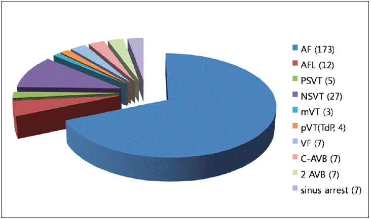 | Figure 1.Prevalence of cardiac arrhythmias in the intensive care units. Cardiac arrhythmias observed in patients admitted at intensive care units of Asan Medical Center are shown. Numbers in the parentheses denote the number of arrhythmias that occurred during the period between 2012.7.1 to 2012.9.30. Abbreviations are as follows. AF: atrial fibrillation, AFL; atrial flutter, PSVT: paroxysmal supraventricular tachycardia, NSVT: nonsustained ventricular tachycardia, mVT: monomorphic ventricular tachycardia, pVT: polymorphic ventricular tachycardia, VF: ventricular fibrillation, C-AVB: complete atrioventricular block, 2-AVB: second degree atrioventricular block. 
|
Go to :
본 론
심실성 부정맥(ventricular tachyarrhythmias) 개요
중환자실 감시 심전도(ECG monitoring)에서 넓은 폭의 QRS파를 보이는 빈맥(wide QRS tachycardia, WQRST)이 나타나면 다양한 감별진단이 필요하며 크게는 심실빈맥과 심실상 빈맥의 편위전도를 감별하는 것이 중요하다. 심전도 파형에서 방실해리현상(
Fig. 2), 흉부유도 전체에서 QRS파가 음성 혹은 양성을 보이는 precordial concordance 소견이 관찰되면 심실빈맥을 강력하게 시사하므로 쉽게 감별이 가능하나 이러한 소견이 없으면 QRS파의 형태를 보고 감별하여야 하며 경험이 필요하다. 응급실이나 중환자실에서 QRS파 폭이 넓고 규칙적인 빈맥의 감별진단 목적으로 아데노신을 주사할 수는 있으나 이는 간혹 관상동맥병 환자에서 심실세동을 일으킬 수 있고 Wolff-Parkinson-White 증후군 환자에서는 심방세동을 유발할 수 있으므로 심전도에서 이러한 소견이 보이는 환자에서는 아데노신 사용에 주의를 요하여야 한다[
1].
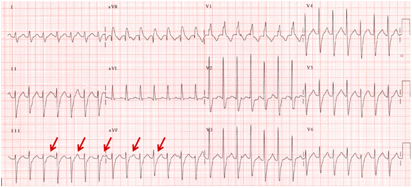 | Figure 2.Monomorphic ventricular tachycardia in a patient with structurally normal heart. A 12 year-old girl was admitted at the cardiac intensive care unit for the evaluation of recurrent palpitation and syncope. A 12-lead ECG taken during palpitation revealed a wide QRS tachycardia with right bundle branch block and left axis deviation. Small indentations (arrows) in the limb lead III suggest ventriculoatrial dissociation, a marker of ventricular tachycardia. The absence of structural heart disease and ECG morphologic characteristics (right bundle branch block and left axis) of VT confirmed the diagnosis of idiopathic fascicular tachycardia. Tachycardia was terminated by intravenous infusion of verapamil.
|
일반적으로 단형 심실빈맥이 관찰되면 기저 심질환이 없었던 환자에서는 특발성 심실빈맥의 가능성이 많은데, 특징적으로 좌각차단, 우축편위를 보이거나 우각차단, 좌축편위의 심전도 특성으로 보이면 더욱 확실하게 심실빈맥을 진단할 수 있다(
Fig. 2). 이러한 특발성 심실빈맥은 대부분 약물치료에 잘 반응하고 장기적인 예후도 양호하므로 그 임상적 경과가 양호하다. 하지만 심근경색, 심근병증(비후성, 확장성) 등의 심질환이 있는 환자에서 나타나는 wide QRS tachycardia는 그 약물 치료도 복잡하고 장기적인 치료전략이 다양하게 달라지므로(약물치료, 도자절제술, 제세동기, 심장이식 등) 부정맥 전문의와 치료방향을 논의하는 것이 좋다. 다형 심실빈맥은 전해질장애, 심근허혈 등의 원인으로도 나타날 수 있으나 그보다 더 흔하게는 약물(QT간격을 연장시키는 약물), 전해질 이상, 일시적으로 나타난 스트레스성 심근병증(stress cardiomyopathy, SCM), neurogenic stunned myocardium 등과 관련하여 나타나는 TdP가 더 임상적으로 중요하다. 이러한 TdP은 대부분 심전도상 저명한 QT간격의 연장소견이 관찰되고, 특히 빈맥의 시작 직전에 잦은 조기심실수축(premature ventricular contraction, PVC)과 giant T wave와 같이 현저하게 큰 T파가 나타나면서 빈맥이 시작되는 것이 특징적이다(
Fig. 3). 일단 빈맥이 시작되면 연속적으로 되풀이되어 나타나서(반복적으로 심실빈맥이 나타나는 것을 electrical storm이라고 함) 혈역학적 허탈(collapse)이 동반되므로 중환자실 환자 진료 시 상당히 당혹스럽게 느껴지고 적절한 치료가 제대로 이루어지지 않아서 반복적인 전기충격만 지속하다가 환자가 사망에 이를 수도 있다. 반면, 치료 가이드라인에 따라 적절한 치료를 시작하면 약물에 대한 반응이 좋고 단 시간 내에 뚜렷하게 부정맥의 호전을 볼 수 있으므로 TdP에 대한 적절한 인지와 치료에 대한 지식이 무엇보다도 중요한 부정맥이다[
2].
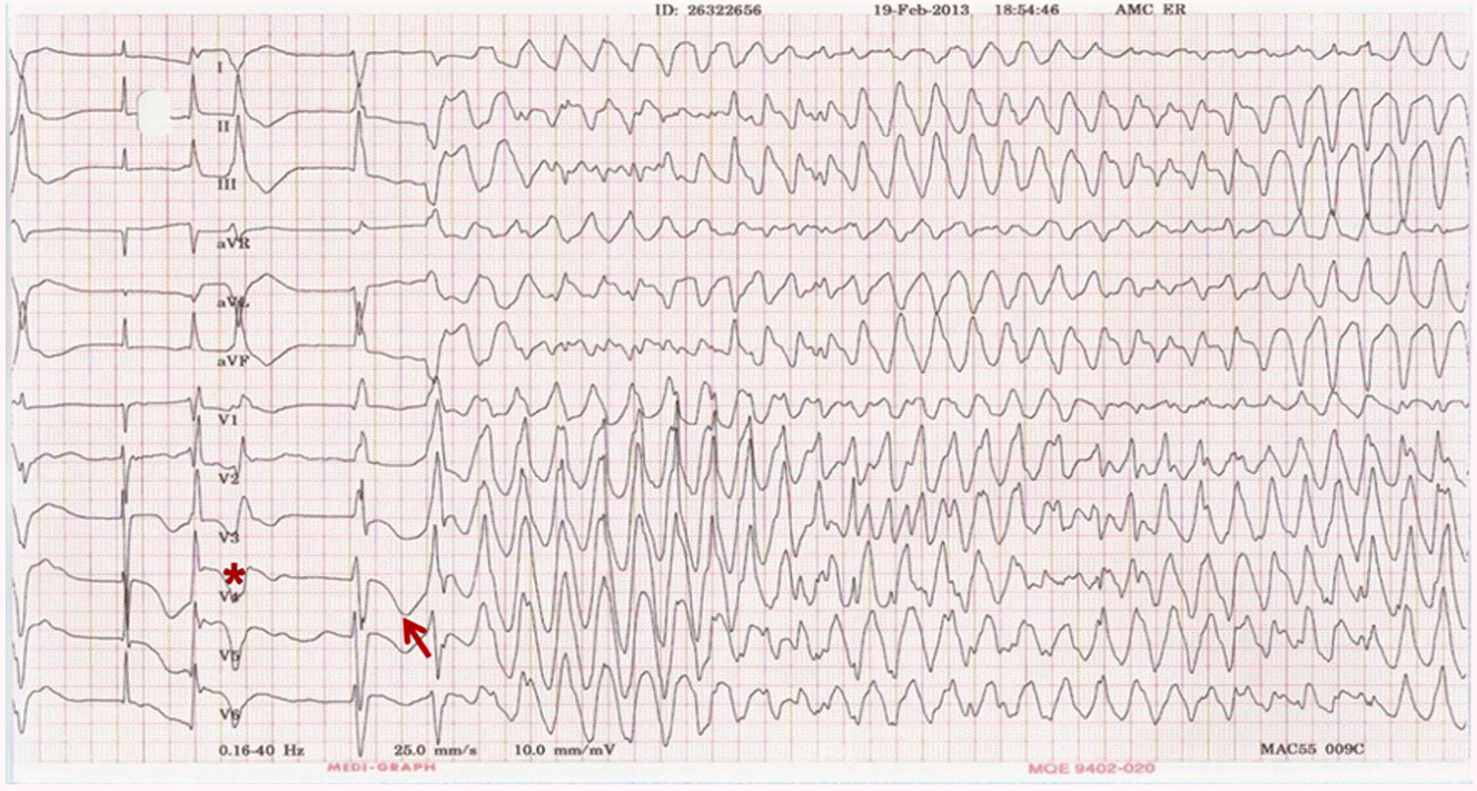 | Figure 3.Polymorphic ventricular tachycardia with QT prolongation in a patient hospitalized for subarachnoid hemorrhage. A 57 year-old female patient was referred for the management of subarachnoid hemorrhage. A marked prolongation of QT interval and polymorphic ventricular tachycardia is noted. Tachycardia was initiated after a pause following premature ventricular beat (star) in a short-long-short sequence of activation. A giant T wave precedes ventricular tachycardia (arrow). Tachycardia was completely suppressed by intravenous infusion of magnesium (2.0 gram). Quinolone antibiotics and quetiapine were discontinued because of the potential for prolongation of QT intervals.
|
단형 심실빈맥의 치료
기저 심질환이 없는 환자에서는 약물치료가 효과적이며, 특히 우각차단, 좌축편위를 보이는 특발성 좌심실 빈맥은 verapamil에 잘 들어서 일명 verapamil-sensitive ventricular tachycardia로도 불리운다. 좌각차단, 우축편위를 보이는 우심실 유출로 빈맥은 일반적으로 다양한 항부정맥제(beta-blocker, Caantagonist, propafenone, flecainide, pilsicainide, sotalol, amiodarone)에 잘 반응하므로 약물치료가 수월하다. 반면 심질환이 있는 환자에서 나타나는 단형 심실빈맥은 사용할 수 있는 약물이 아미오다론에 국한되고 일부 허혈성 심질환 환자에서는 리도카인을 써볼 수 있는 정도이므로 약물치료에서 그 선택의 폭이 넓지 않다. 일반적으로 리도카인은 그 작용 효과가 빨라서 심실 종료 효과를 수 분 내에 기대할 수 있으나 실제적으로 부정맥 종료의 효과는 아미오다론에 비하여 약한 편이다. 아미오다론은 그 효과는 리도케인에 비하여 뛰어나나 주사약을 쓰더라도 그 효과가 상당히 느리게 나타나고(적어도 수시간, 보통 수일), 간장애, 심한 서맥이나 저혈압 등의 부작용이 드물지만 심하게 나타날 수 있어 그 사용에 주의를 요한다(
Table 1). 부작용의 발생은 아미오다론을 주사제로 사용할 경우 첫 주에는 1-2일 간격으로 간기능 검사와 흉부방사선을 확인하는 것이 좋고, 혈압 저하가 나타나면 주입속도를 1/2 혹은 그 이하로 줄여서 투여한다. 드물게 서맥 혹은 심정지가 나타날 수 있으며, 임시박동조율기 삽입하고 수일 기다리면 회복되는 것이 일반적이다.
Table 1.
Loading and maintenance doses of the common antiarrhythmic drugs
|
Loading dose |
Onset of action |
Maintenance dose |
|
Diltiazem |
0.25 mg/kg IV over 2 min |
5 min |
5–15 mg/hour |
|
Verapamil |
0.075–0.15 mg/kg IV over 2 min |
5 min |
no data |
|
Esmolol |
0.5 mg/kg over 1 min |
5 min |
0.05–0.2 mg/kg/min |
|
Propranolol |
0.15 mg/kg IV |
5 min |
no data |
|
Digoxin |
0.25 mg IV each 2 h, up to 1.5 mg |
2 hour |
0.125–0.25 mg daily |
|
Lidocaine*
|
1-2 mg/Kg over 2 min (half dose 30 min later if not responsive) |
|
1-4 mg/min |
|
Amiodarone†
|
5–7 mg/kg over 30–60 min, then 1.2–1.8 g per day continuous IV or in divided oral doses until 10 g total, then 200–400 mg per day maintenance |
hours |
|

다형 심실빈맥의 치료
다형 심실빈맥은 QT간격이 정상이면서 나타나는 형태도 있으나 일반적으로는 QT간격의 현저한 연장과 함께 나타나는 TdP이 더 흔하며, 심전도를 통하여 빠르게 인지만 된다면 그 치료는 그리 어렵지 않고 치료에 대한 반응도 좋은 편이다. 원인으로는 선천성/후천성 QT연장 증후군(congenital or acquired long QT syndrome)이 있으며 후천성 QT연장 증후군은 각종 약물(astemizole, terfenadine, ketoconazole, erythromycin, pentamidine), 전해질 이상(hypokalemia, hypomagnesemia), 심근허혈, 심부전 등의 원인에 의하여 이차적으로 나타난다.
다형 심실빈맥을 보이는 환자에서 빈맥 중 축이 여러 방향으로 변화하고(twisting of points) 그 시작이 PVC후 서맥에 의하여 나타나며 빈맥의 첫 박자가 giant T파로 보이면 TdP의 가능성이 많다(
Fig. 3). 이러한 TdP은 magnesium에 의하여 억제가 잘 되므로 가장 먼저 해야 할 것은 정맥주사로 magnesium을 주입하는 것이다. 대략 MgSO4 1-2 gram을 5% 포도당용액에 섞어서 1-2분에 걸쳐서 서서히 주입하고 이후 부정맥의 발생양상을 관찰하여 PVC가 완전히 억제되지 않으면 5-10분 후 동량을 다시 주입할 수 있다. 보통 magnesium의 혈중 농도는 조직 내 농도를 반영하지 못하므로 혈액검사 상 정상 농도일지라도 magnesium을 주사하는 것이 좋다. 보통 중환자실에 입실한 환자는 경구투여가 불가능하여 만성적으로 영양 결핍 상태이고 심부전이나 기타 원인으로 이뇨제를 쓰고 있는 경우가 많아 체내 전해질이 많이 부족한 경우가 대부분이며 항생제/진정제 등 QT간격의 연장을 초래시킬 수 있는 약물을 복합적으로 사용하므로 TdP의 발생에 취약하다. 따라서 혈중레벨이 정상이어도 TdP이 발생한 당일에 약 10 g 정도의 magnesium을 주입(0.5-1.0 g/hr 혹은 하루에 걸쳐 서서히 주입)하는 것이 좋고, 이후 부정맥이 없어질 때까지 혈중레벨이 정상 혹은 약간 높을 정도로 유지하는 것이 바람직하다. 단, 신부전 환자에서는 소변을 통한 magnesium 배출이 지연되므로 magnesium에 의한 독성 (서맥, 저혈압)이 나타날 가능성을 고려하여 신중히 투여한다. 혈중 칼륨레벨을 높이면 세포막을 통한 칼륨 배출이 증가하므로 재분극을 촉진시켜 QT간격을 줄일 수 있어서 칼륨레벨을 정상범위 내에서 높게 유지하는 것도 좋다. 리도케인을 쓸 수는 있으나 magnesium보다는 그 효과가 뚜렷하지 않으므로 magnesium 주입 후 반응이 없을 경우에 고려해 볼 수 있다. 단, amiodarone은 QT간격을 더 증가시키고 부정맥을 악화시키므로 사용하지 않는 것이 좋다.
이러한 치료와 동시에 QT연장의 원인이 될 수 있는 약물은 중단하고 전해질 이상소견을 교정하도록 하면 TdP은 대부분 교정되는 것이 일반적이다. 이러한 치료에도 불구하고 빈맥이 지속되는 경우에는 임시박동조율기를 통하여 심방 혹은 심실조율을 하면 QT간격이 줄어들어 TdP의 발생을 줄일 수 있다.
Go to :
상심실성 부정맥(supraventricular tachyarrhythmias) 개요
QRS파 폭이 좁고 불규칙적인 빈맥(regular narrow QRS tachycardia)
대부분 상심실빈맥(paroxysmal supraventricular tachycardia) 혹은 심방조동(atrial flutter)이며, 경동맥동 마사지(carotid sinus massage), adenosine 혹은 verapamil 주사에 의하여 종료되거나 심실 박동수가 줄어드는 효과를 볼 수 있다. 약물 주입 후 재발하면 diltiazem, verapamil, esmolol등의 주사제를 연속주입하거나 경구약물로 전환하여 유지할 수 있으며, 장기적으로는 전극카테터절제법을 고려하는 것도 좋다[
1].
QRS파 폭이 좁고 불규칙적인 빈맥(irregular narrow QRS tachycardia)
일반적으로 심방세동(atrial fibrillation, AF)이 가장 흔한 원인이고, 호흡기 환자에서는 다원 심방빈맥(multifocal atrial tachycardia)이 그 원인이 되기도 한다. 중환자실 환자에서 심방세동이 갑자기 발생하면 이러한 환자에서는 패혈증, 심부전 등 다양한 원인으로 혈압이 낮게 유지되는 환자가 많으므로 부정맥으로 인하여 추가적인 혈압강하가 나타나면 교감신경 흥분제를 투여하여야 할 경우가 많다(
Fig. 4). 이러한 경우 심방세동의 심박수가 더 빨라지고 심박수가 더 빨라지면 혈압강하가 악화되는 악순환을 밟게 되므로, 중환자실에서 심방세동이 나타나면 환자의 경과가 급격히 나빠지는 경우가 많다. 심방세동이 갑자기 발생하였을 경우 증상이 심하고 혈역학적으로 불안정하다면 직류전류 심장율동 전환(DC cardioversion)을 시행한다. 에너지는 단상 파형(monophasic waveform shock)의 경우 200 J (혹은 300 J)에서 시작하여 반응이 없을 경우 300 J, 360 J로 증가시킨다. 이상파형(biphasic shock)의 경우 100 J에서 시작하여 150 J, 200 J로 증가시킨다.
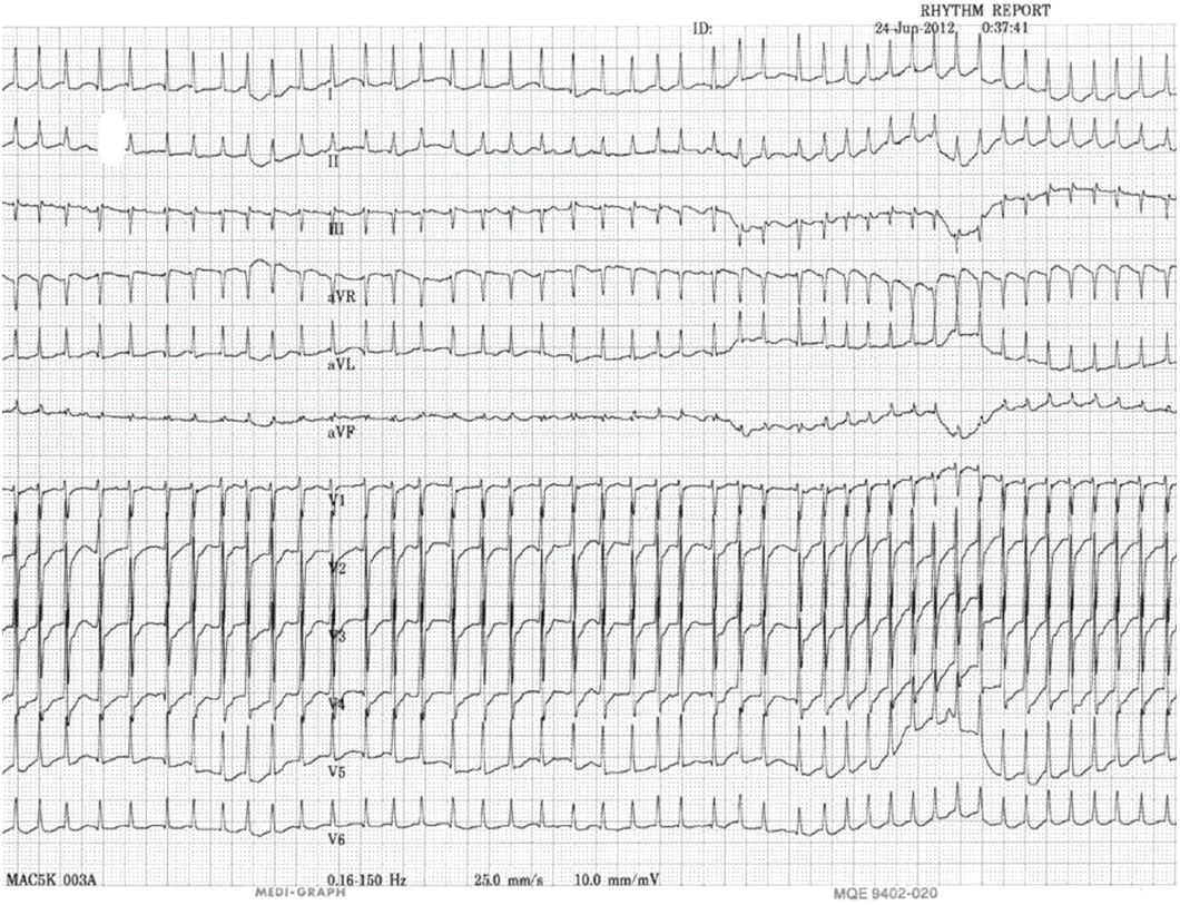 | Figure 4.A narrow QRS tachycardia in a 78 year-old male patient with biliary sepsis. Tachycardia was irregularly irregular with a heart rate of about 260 beats per minutes. Blood pressure decreased from 100 to 70mmHg due to the rapid heart rate. After infusion of intravenous diltiazem, heart rate was decreased to 160 beats per minutes, but blood pressure remained low at 70-80mmHg. Change to intravenous amiodarone (150mg over 2 hours, then continuous infusion at a rate of 0.5mg/min) further decreased heart rate to 130 beats per minutes with blood pressure increased to 90mmHg.
|
심한 증상이나 혈역학적 변화가 크지 않을 경우에는 일단 관찰하거나 심박수 조절을 시도할 수 있다. 심박수 조절은 diltiazem, esmolol, digoxin 등의 주사제를 써서 시도할 수 있으며 맥박수가 대략 분당 60-90회 정도 유지되는 것을 목표로 하나 환자의 상태를 고려하여 개별화하는 것이 필요하다. 하지만 diltiazem, esmolol 등으로 혈압 저하가 나타나는 경우가 많아서 약물을 중단하여야 할 경우가 많은데, 특히 심부전이나 패혈증이 동반되어 있을 경우 자주 나타나며 이러한 경우에는 digoxin이나 amiodarone이 효과적이다. 주사로 주입할 경우 amiodarone은 부하용량 주입(loading) 중 혈압 저하가 나타날 수 있으므로 혈압에 따라 주사 속도를 반으로 혹은 그 이하로 줄여서 주입하는 것이 필요하다. 대개 2-3일 이내에 맥박수와 혈압이 안정되므로 이 때 경구제제로 교체할 수 있다.
Go to :
전기잡음(electrical noise)의 감별진단
간혹 심전도, 특히 감시 심전도에 전기잡음이 들어갈 경우 심실빈맥과 유사하게 나타나는 경우가 있어서 주의를 요한다(
Fig. 5). 실제 심실빈맥과 달리 전기잡음에서는 잡음 중간 중간에 QRS complex의 날카로운 파가 정상 QRS파의 간격과 동일하게 유지되므로 이러한 파형이 관찰되면 잡음을 쉽게 구별할 수 있다. 혹은 동시에 기록된 혈량측정(plethysmography) 파형이 정상 심장율동일 때와 변화가 없이 지속되거나, 동시에 기록된 다른 유도의 심전도가 정상소견이면 심실빈맥처럼 보였던 심전도 파형이 잡음이라는 것을 유추할 수 있다.
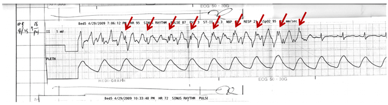 | Figure 5.Monomorphic ventricular tachycardia in a patient with structurally normal heart. A 12 year-old girl was admitted at the cardiac intensive care unit for the evaluation of recurrent palpitation and syncope. A 12-lead ECG taken during palpitation revealed a wide QRS tachycardia with right bundle branch block and left axis deviation. Small indentations (arrows) in the limb lead III suggest ventriculoatrial dissociation, a marker of ventricular tachycardia. The absence of structural heart disease and ECG morphologic characteristics (right bundle branch block and left axis) of VT confirmed the diagnosis of idiopathic fascicular tachycardia. Tachycardia was terminated by intravenous infusion of verapamil.
|
Go to :
서맥성 부정맥(bradyarrhythmias) 개요
동결절 부전(sinus node dysfunction)과 방실 전도장애(AV conduction disturbance) 구별의 중요성
동결절 부전은 실신의 위험성은 있지만 급사의 위험성은 높지 않고 대개 원인되는 약물을 줄이거나 중단함으로써 호전되는 경우가 많다. 반면에 완전 방실차단은 실신의 위험 외에도 급사의 가능성을 높이므로 인공심장조율기를 삽입하는 것이 필요하다. 따라서 같은 서맥이지만 그 예후와 치료가 상이하게 달라지므로 이 두 가지 상태의 심전도 감별은 임상적으로 대단히 중요하다(
Fig. 6).
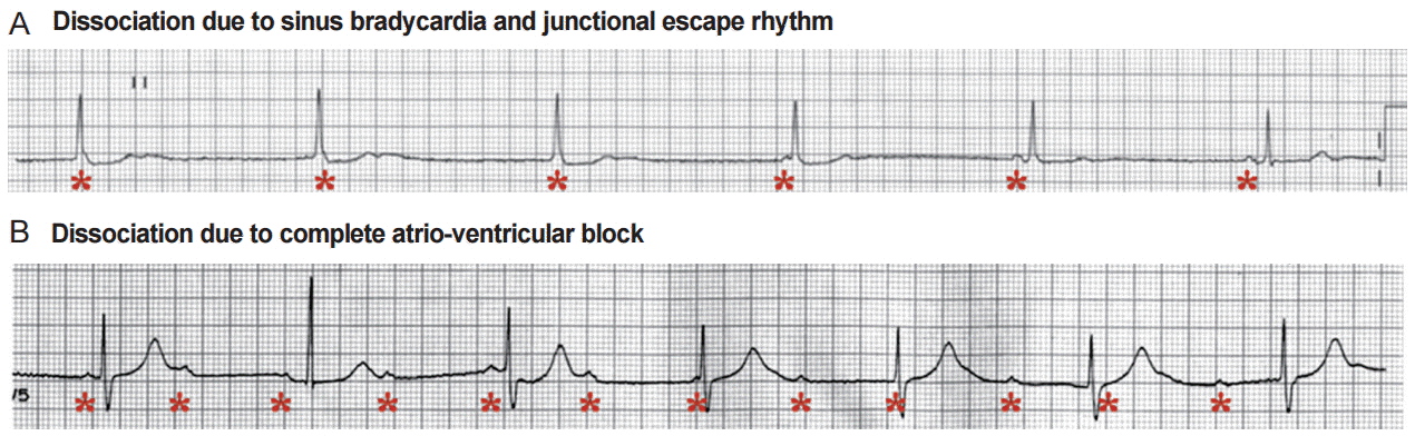 | Figure 6.Differential diagnosis of atrioventricular dissociation. The rhythm strips show two different causes of atrioventricular (AV) dissociation. In the upper tracing (A), the cause of AV dissociation is slowed sinus rate (stars) with escape junctional rhythm, while the cause of dissociation in the bottom tracing (B) is complete AV block. Syncope or sudden cardiac deaths are rare in sinus bradycardia and junctional rhythm, whereas the risk is high in patients with bradycardia due to complete AV block. Temporary and/or permanent pacemaker is necessary in the latter condition.
|
반사성 서맥(reflex bradycardia)
동방결절이나 방실결절, His-Purkinje system의 이상 없이 자율신경계의 항진/억제에 의하여 동성서맥이나 전도 장애가 나타날 수 있다. 전형적인 예는 혈관미주신경 실신이며 이와 비슷한 반사 반응(reflex vagotonia)이 복부/골반부위의 시술, 간조직검사, 복강경, 안과수술, 기관삽관, 기관지 자극, 두개내압 상승, 소변, 대변, 구토 등 다양한 원인에 의하여 초래될 수 있고, 특히 시술과 관련되어 환자의 불안이 크거나 시술부위의 통증이 심할 경우에도 이러한 반사반응이 나타날 수 있다. 대부분 일과성이므로 치료를 필요로 하지 않지만 치료가 필요한 경우에는 atropine, epinephrine을 사용할 수 있고, 장기적인 예후는 양호하다.
Go to :
서맥성 부정맥 환자의 치료
약물치료
서맥의 치료는 서맥에 의한 증상이 심하게 존재하거나 서맥으로 인한 혈역학적 변화가 초래될 때 적응이 된다. 이때 반드시 서맥의 원인(가역적 원인)들의 가능성을 검토하고 교정이 가능하면 교정하는 것이 중요하다. 대표적인 가역적 원인들로는 약물(digitalis, beta-blocker, Ca-channel blocker, class IA, IC antiarrhythmic agent), 전해질 이상, 개심술 후의 일과성 방실전도계 손상(hypothermia, inflammation), 급성 심근경색, 일과성 심근허혈, 부교감 신경 항진 등이 있다.
서맥성 부정맥 치료에서 약물치료의 역할은 대부분 심장조율 전 임시 조치로 필요한 경우가 대부분이다. Isoproterenol, atropine이 이용되며, 동기능의 호전이나 방실결절 전도 차단 시 전도 장애의 호전에는 도움이 되나, 방실결절 하부(infranodal block) 전도차단에서는 전도장애 호전 정도가 뚜렷하지 않아 큰 기대를 하기 어렵고 허혈성 심질환이 있을 경우에는 사용에 주의를 요한다. Atropine (0.5-1.0 mg)은 5분 간격으로 주사할 수 있고(최고 0.03-0.04 mg/Kg), dopamine (5-20 g/Kg/min), isoproterenol (2-10 g/mirn)을 연속주사 하면서, 빠르게 원인 질환에 대해 평가하고 경정맥 박동조율기의 필요성 등을 판단한다[
3].
경피적 박동조율기(transcutaneous pacing)
경피적 박동조율기는 빠른 시간 내에 전극(electrode pad)을 피부에 부착하고 심박조율을 실시할 수 있는 유용한 장치로서, 단기간 내에 서맥이 회복될 가능성이 많을 경우, 서맥이 예상되나 예방적으로 경정맥 박동조율기를 삽입하기 곤란할 경우, 혹은 안정적으로 경정맥 조율을 실시하기 전까지 임시로 심박조율이 필요할 경우에 특히 유용하다. 전극 pad는 흉곽의 앞, 뒤로 부착하되 전방 전극(anterior electrode, cathode)은 흉골좌측에 맥박이 최대로 촉지되는 점 혹은 심전도의 V3 위치에 일치시키고 후방전극(posterior electrode, anode)은 전방전극의 반대편으로 좌측 혹은 우측의 견갑골(scapula)-척추 사이 공간에 부착한다. 일반적으로 20-40 msec의 자극파를 0-200 mA 범위에서 투여할 수 있도록 되어있으며, 성인에서는 대략 40-70 mA의 전류로 조율이 가능하다. 종종 조율역치(capture threshold)가 높거나 조율에 따른 동통이 문제가 되나 전극과의 접촉 피부면을 알코올로 닦거나 면도 후 pad를 부착하면 조율 시의 통증을 줄일 수 있고, 의식이 있는 환자에서는 diazepam, morphine을 투여하는 것이 좋다. 조율은 조율역치보다 10% 정도 높은 전류로 유지하고 반드시 맥박과 혈압(혹은 동맥압 monitor)을 측정하여 효과적으로 조율이 되는지를 확인하여야 한다. (맥박은 조율기에 의한 근육수축과의 혼동을 피하기 위하여 우측 경동맥이나 우측 고동맥에서 촉지한다)[
4,
5].
경정맥 심박조율(transvenous temporary pacing)
경정맥 심박조율은 경피적 심박조율에 비하여 좀 더 안정적으로 심박조율을 시행할 수 있지만, 어느 정도 숙련된 술자 및 시설이 필요하고, 간혹 합병증이 발생할 수 있어 장단점이 있다.
전극은 주로 대퇴정맥, 쇄골하정맥, 내경정맥을 통하여 삽입되며, 6 Fr bipolar 혹은 quadripolar electrode가 가장 흔하게 이용된다. 박동조율기는 수일에서 수주까지도 유지할 수 있으나 오래될수록 감염의 위험이 증가하므로 가역적 원인이 아니라면 빨리 영구형 박동조율기로 교체한다. 매일 threshold와 심전도를 확인하고 전극삽입부위의 감염 여부도 관찰한다[
4,
5].
Go to :
결 론
중환자실 입원 환자에서 나타나는 부정맥은 심전도를 통한 감별진단이 그 치료에 있어 가장 중요하며 부정맥 자체의 특성뿐 아니라 환자의 다양한 전신 질환 및 심혈관 상태를 고려하여 접근하여야 하는 자세가 필요하다.
Go to :


 PDF
PDF Citation
 Print
Print


 XML Download