Journal List > Cancer Res Treat > v.53(2) > 1154759

Sun, Wei, Chen, Hu, Ji, Xu, Du, Zhao, Lu, Sang, Zhong, Yang, and Mao: Comparison of the Prognostic Value of Platelet-Related Indices in Biliary Tract Cancer Undergoing Surgical Resection

Abstract

Purpose

Platelet-related indices, including mean platelet volume (MPV) and plateletocrit (PCT), have been reported as new prognostic factors of overall survival (OS) in many cancers, but not yet in biliary tract cancer (BTC). We intended to assess these indices in predicting OS in BTC patients with the aim to build a new prognostic model for patients with BTC after surgical resection.

Materials and Methods

Survival analysis and time receiver operating characteristic analysis were applied to screen the platelet indices. Univariate and multivariate Cox analyses were used to identify independent prognostic factors and develop a new prognostic model. Harrell’s C-statistics, calibration curves, and decisive curve analysis were used to assess the model.

Results

MPV and platelet distribution width (PDW)/PCT showed the best prognostic accuracy among the platelet indices. In multivariable analysis, factors predictive of poor OS were presence of nodal involvement, Non-radical surgery, poor tumor differentiation, carbohydrate antigen 19-9 > 100 U/mL, MPV > 8.1 fl, and PDW/PCT > 190. The new model was found to be superior to the TNM staging system and our new staging system showed higher discriminative power.

Conclusion

MPV and PDW/PCT have high prognostic value in BTC patients, and the novel staging system based on these two indices showed good discrimination and accuracy compared with the American Joint Committee on Cancer 7th TNM staging system.

Introduction

Biliary tract cancer (BTC) is a group of biliary malignancies mainly comprising intrahepatic cholangiocarcinoma (ICC), extrahepatic cholangiocarcinoma (ECC), and gallbladder cancer (GBC). ICC is estimated to account for about 0.59% of all new cancer cases in 2018, while GBC containing ECC accounts for 1.2% worldwide [1]. Despite the low incidence, the mortality of BTC is relatively high, comprising up to 2% of total cancer deaths per year [2]. BTC is acknowledged to be aggressive with a dismal prognosis; according to the RARECAREnet project, based on cases between 2000 and 2007 in Europe, survival for ICC was 25% at 1 year, 8% at 3 years, and 5% at 5 years, while the 5-year survival rate of GBC was less than 5% [3]. Survival for ECC was marginally better, albeit dramatically reducing from 48% at 1 year to 23% at 3 years and 17% at 5 years [4]. Increased incidence has been observed in cholangiocarcinoma and GBC [5] worldwide. Unfortunately, the only available curative treatment for BTC is surgery.
The prognosis of BTC is highly dependent on tumor stage, as the estimated 5-year survival is 50% for American Joint Committee on Cancer (AJCC) stage I, 30% for stage II, 10% for stage III, and 0% for stage IV [6]. Both novel biomarkers and staging systems are warranted, as no prognostic biomarkers are currently used clinically, and the AJCC and Bismuth-Corlette staging system are not accurate in predicting survival [2]. Currently, nodal involvement, tumor differentiation, preoperative and postoperative serum carbohydrate antigen 19-9 (CA19-9) [7] have been confirmed as important prognostic factors in cholangiocarcinoma patients undergoing surgery. Furthermore, lymph node status and the presence of R0 curative resection are closely related to survival in GBC [8]. During the search for novel factors, inflammation-related indexes have been extensively studied as prognostic factors, but their roles in predicting overall survival (OS) for BTC are still under discussion. The neutrophil-to-lymphocyte ratio, one of the most popular inflammation-related indices, cannot maintain its role as a dependent prognostic factor of OS in BTC [9]; thus, more accurate predictors need to be identified.
It has been widely accepted that platelets contribute to tumor progression through a variety of mechanisms, including angiogenesis, assisting metastasis, and regulating immunity. Platelets can protect tumor cells from immune attack by modulating innate and adaptive immune responses [10]. The role of platelets as regulators of inflammation has been recognized, and as a result, the prognostic ability of platelet-related indices has been investigated in many cancers. Platelet count (PLT) and morphologic indices, including the mean platelet volume (MPV), plateletocrit (PCT), and platelet distribution width (PDW), have been frequently studied in a number of cancers, including pancreatic cancer [11], invasive breast cancer [12], lung cancer [13], and esophageal cell carcinoma [14]. However, the relationship between platelet-related indices and prognosis in BTC remains unknown. Therefore, we investigated the prognostic performance of platelet indices in patients with TNM stages I–III BTC after surgical resection and found two potential prognostic factors. A new prognostic model and staging system were developed based on these two factors, which may benefit patients with BTC in surgery selection and postoperative management.

Materials and Methods

1. Patients

Patients diagnosed with BTC, either ICC, ECC, or GBC, at the Peking Union Medical College Hospital (PUMCH) between December 2002 and December 2017 were included. The inclusion criteria were as follows: (1) pathologically confirmed BTC; (2) no other malignant tumors; and (3) patients who underwent resection without antitumor treatment before or during surgical intervention. The exclusion criteria were as follows: (1) patients with AJCC 7th stage IV; (2) patients with incomplete follow-up data; and (3) patients with active inflammatory diseases. A total of 527 patients were identified. Data, including patient demographics, operative outcomes, and pathologic features, were collected. Hematologic parameters, such as CA19-9, albumin (ALB), alkaline phosphatase (ALP), and γ-glutamyl transpeptidase (GGT) were collected within 5 days before surgery. OS was measured from the date of surgery to the date of death or the final follow-up. All patients were randomly split 7:3 into training (371) and validation (156) sets using createDataPartition from the R package’caret’, which is based on simple random sampling. To use this package, all patients were numbered with Arabic numerals and the seed was set as 2020.

2. Definitions of platelet-related indices

The 10 platelet-related indices that were considered were PLT (×109/L), MPV (fl), PCT (%), PDW (%), platelet-to-lymphocyte ratio (PLR), red blood cell distribution width-to-platelet ratio (%/×10–9/L), and ratios of PDW/PLT, PDW/PCT, MPV/PLT, and MPV/PLT. PLT, MPV, PCT, PDW were checked by SYSMEX XN automatic hematological analysis (SYSMEX, Kobe, Japan). When results were doubtful, microscopic examination was still the golden standard. All values of platelet indices were collected before operation.

3. Statistical analysis

The Shapiro-Wilk normality test was used to identify non-normal continuous variables, which were presented as median (interquartile range). Categorical variables were expressed as counts and percentages, and baseline characteristics were summarized as categorical variables. Acknowledged optimal cut-off values for platelet indices, total bilirubin (TBIL) and tumor diameter were absent. Therefore, these values were determined by ‘surv cutpoint’ function from ‘survminer’ R package using the maximally selected rank statistics to provide a value of cutpoint that correspond to the most significant relation with survival outcome. The remaining cut-off values were determined based on the median values. Comparisons of the two groups were performed with the Kruskal-Wallis rank test for non-normal continuous variables and the chi-square test or Fisher exact test for categorical variables, as appropriate.
Kaplan-Meier analysis was used to perform survival tests, and the log-rank test was used for statistical comparisons between curves. Areas under time-dependent receiver operating characteristic (ROC) curves (time-AUC) curves were used to predict the efficacy of platelet indices. The association between variables and OS was assessed by univariate and multivariate Cox proportional hazards regression analyses. All statistically significant variables in the univariate analysis were included in the multivariate analysis. All Cox analyses were performed using the R package ‘survival’.
A nomogram was constructed according to the multivariate Cox model. Patients with assigned prognostic scores derived from the nomogram were stratified equally into three risk groups. The predictive accuracy of the new model was evaluated by calculating Harrell’s concordance index (C-index) and plotting calibration curves in training and validation cohorts. Decisive curve analysis (DCA) was also used to assess the clinical benefit. The results were compared to those of the AJCC 7th edition TNM staging system. All statistical analyses were performed using R software ver. 3.6.2 (R Foundation for Statistical Computing, Vienna, Austria). In all analyses and tests, a two-sided p < 0.05 was considered statistically significant.

Results

1. Baseline characteristics

A total of 527 patients met the inclusion criteria. The median follow-up was 21 months (range, 10 to 41 months), and 328 patients (62.2%) were confirmed dead. Table 1 summarizes the demographic, clinical, and pathological characteristics of the patients. Among the 527 patients, 296 (56.2%) were diagnosed with ECC, followed by GBC (n=122) and ICC (n=109). A total of 235 patients (44.6%) were female, and 292 (55.4%) were male. According to the AJCC 7th TNM staging system, 9 (1.7%), 179 (34.0%), 174 (33.0%), and 165 (31.3%) patients were classified as TNM stage 0, I, II, and III, respectively. A total of 322 patients (61.7%) underwent radical surgery, defined as R0 resection, and 200 patients (38.3%) underwent non-radical surgery. Overall, 110 out of 463 patients received postoperative chemotherapy, most common chemo regimens were as follows: 42 patients (38.1%) received interventional therapy mainly consists of fluorouracil with pirarubicin or epirubicin, 15 patients (13.6%) received single-agent chemotherapy with oxaliplatin or tegafur or gemcitabine or capecitabine, 23 (20.9%) received multiagent chemotherapy with a combination of two or more agents above. The training cohort consisted of 371 patients, and the validation cohort consisted of 156 patients; these two cohorts were comparable with respect to all characteristics. Among the training cohort, 321 were included in the model construction.

2. Comparison of the prognostic efficacy of platelet indices

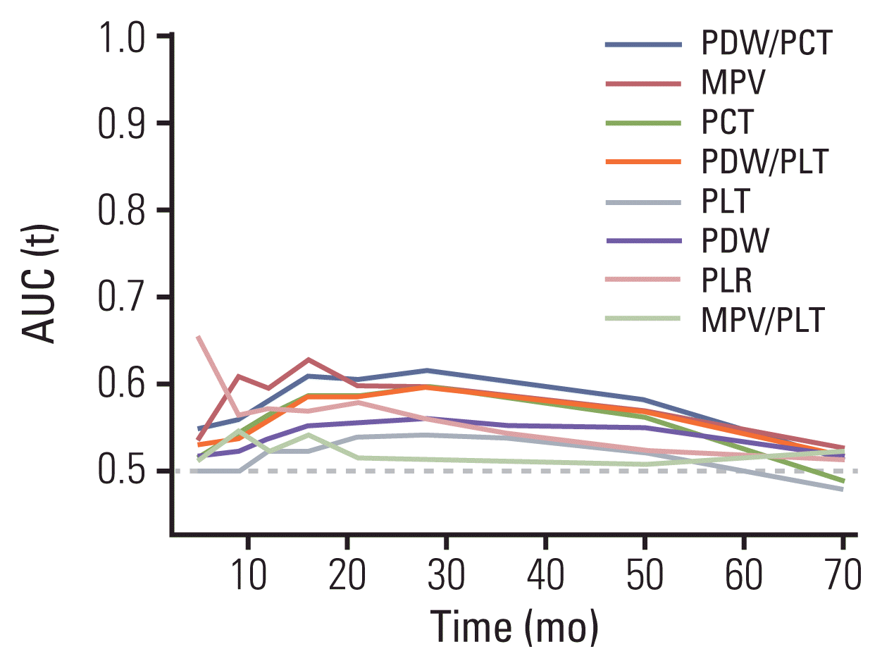
Of the 10 platelet indices, eight indices, including PLT, PDW, MPV, PCT, PLR, PDW/PLT, PDW/PCT, and MPV/PLT, were found to be significantly associated with OS in both Kaplan-Meier analysis (S1 Fig.) and univariate Cox analysis (S2 Table). Time-AUC curves were generated to further evaluate the prognostic accuracy of these eight platelet indices (Fig. 1). According to the curves, PDW/PCT had the highest predictive accuracy, followed by MPV. At any point of time, every subject has two outcomes, time-dependent ROC curve depicts the specificity and sensitivity of each marker in predicting survival outcome. Therefore, time-AUC curves indicate the predicting accuracy of different markers in survival outcomes. According to Fig. 1, from 10 to 60 months, time-AUC curves of PDW/PCT and MPV maintain to be on the top of all other curves representing higher predicting accuracy.
In univariate analysis of all the remaining baseline characteristics, factors associated with worse OS included nodal involvement, emaciating, non-radical surgery, ALP > 200 U/L, ALB ≤ 35 g/L, TBIL > 106 μmol/L, CA19-9 > 100 U/mL, and poorer tumor differentiation. An initial multivariable analysis revealed that the presence of nodal involvement, non-radical surgery, CA19-9 > 100 U/mL, and poorer tumor differentiation were independent adverse prognostic factors. After integration into those four factors, MPV > 8.1 fl (hazard ratio [HR], 1.33; p=0.045) and PDW/PCT > 190 (HR, 0.78; p=0.046) remained as independent factors (Table 2). These two platelet indices were added into the final multivariable analysis instead of the initial one in order to minimize potential confounding with respect to their association with a number of baseline characteristics as described below.

3. Relationships between PDW/PCT, MPV, and clinicopathological features

Patients with low PDW/PCT were characterized by emaciation, high levels of alanine aminotransferase, aspartate aminotransferase, GGT, ALP, and low levels of ALB, which suggested poor nutritional status and impaired liver function. In addition, these patients had high levels of CA19-9, TBIL, and poor TNM staging, representing quick tumor progression. High levels of TBIL may be derived from severe biliary tract obstruction (S3 Table). In addition to most of the characteristics mentioned above, patients with high MPV were prone to present nodal involvement and large tumor size, both of which indicate malignant behavior. It seemed that these two platelet indices were associated with a number of baseline characteristics, including patient-related factors and pathological features of the tumor. In our sub-analysis, we detected the relationship between these two indices with operation outcomes, including hospitalization day, biliary leakage, infection, and other complications. S4 Table shows that MPV and PDW/PCT were only significantly correlated to hospitalization days, suggesting that platelet indices may not be eligible for predicting operation outcomes.

4. Development and validation of a novel staging system

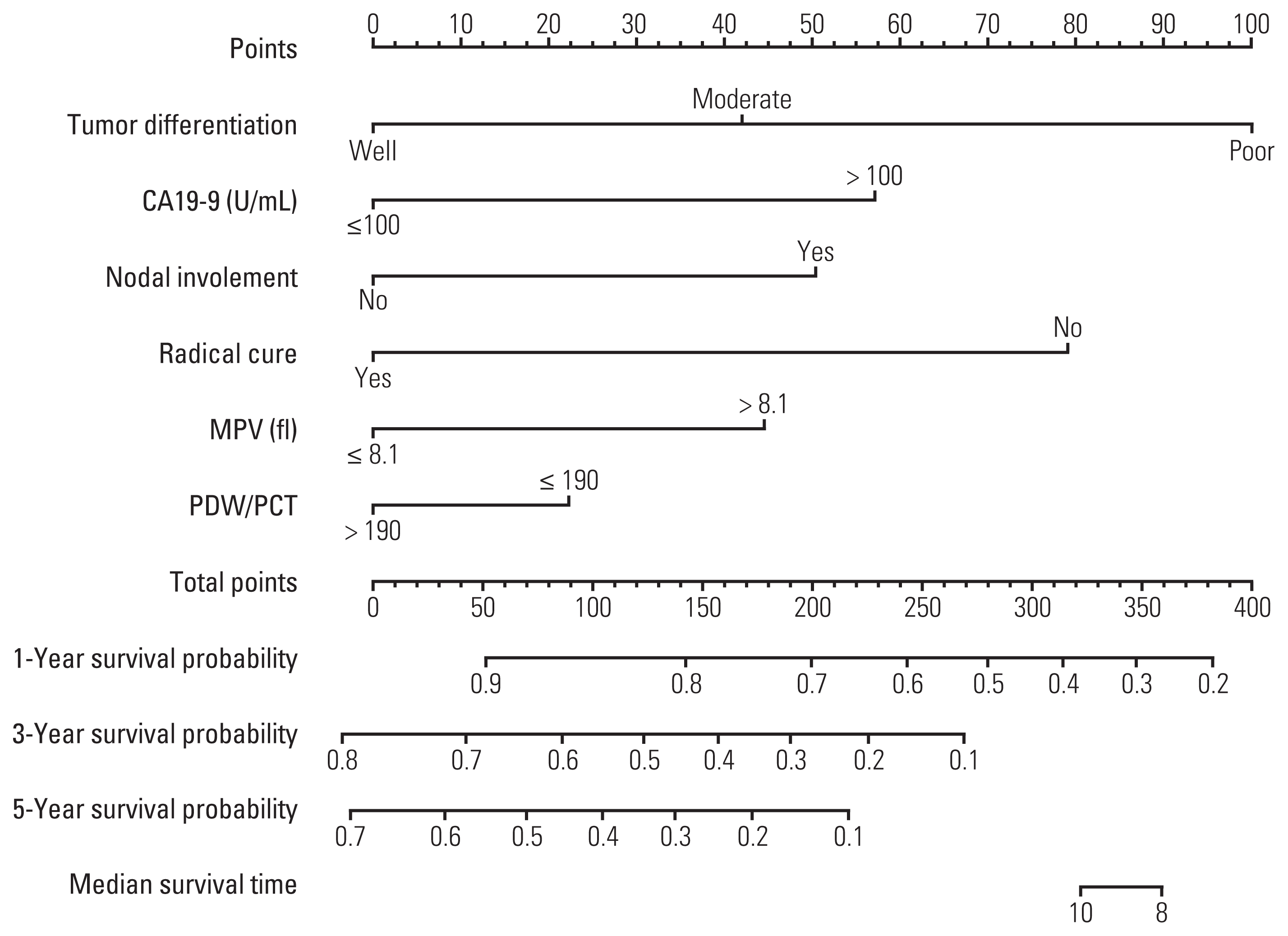
A nomogram comprising nodal involvement, radical surgery, CA19-9 level, tumor differentiation, MPV, and PDW/PCT was created based on the multivariable Cox analysis (Fig. 2) to predict OS individually. Subsequently, patients in the whole cohort were stratified into three stages almost equally, stage I (n=153, score < 107.5), stage II (n=154, score 107.5–152.0), and stage III (n=154, score > 107.5) according to their scores. The calibration curves for 3- and 5-year OS (S5 Fig.) illustrated good consistency of our new model in the training cohort and suboptimal in the validation cohort.
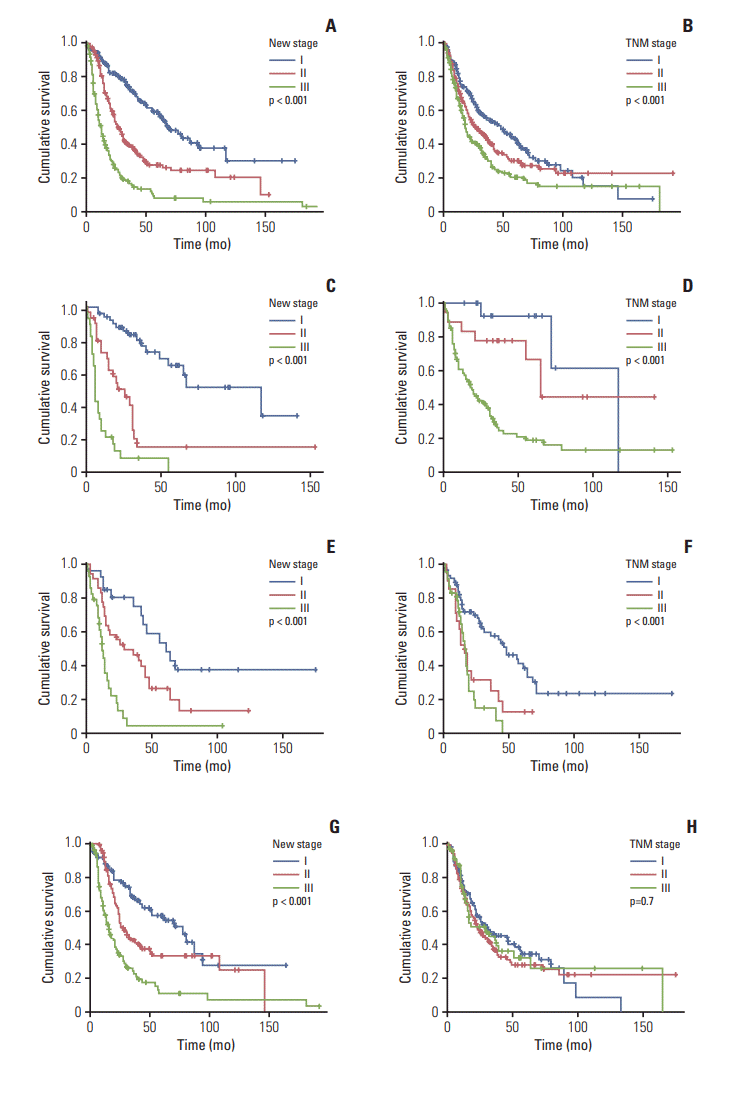
In survival analysis, patients were classified into GBC, ECC, and ICC groups according to their tumor sites to investigate the discriminative capacity of our novel staging system in each subset of BTC patients. For the convenience of comparison between our new staging system and the AJCC 7th TNM staging system, TNM stage 0 was merged into TNM stage I. In the whole cohort, the median OS for AJCC TNM stages I, II, and III was 48, 27, and 18 months, respectively (Fig. 3B), with HRs of 1.0 (reference), 1.30 (1.00–1.70), and 1.76 (1.35–2.30), respectively (p < 0.001). The median OS for the novel staging system (Fig. 3A) was 68, 27, and 12 months, respectively, with HRs of 1.0 (reference), 2.15 (1.57–2.96), and 4.81 (3.51–6.58), respectively (p < 0.001). With regards to specific types of BTC, the new staging system (Fig. 3C) had a performance similar to that of the TNM staging system for GBC patients (Fig. 3D). However, for ICC patients, the new staging system (Fig. 3E) performed better than the TNM staging system (Fig. 3F). Noticeably, for ECC patients, the differences between groups classified by the new staging system (Fig. 3G) remained significant, while the TNM staging system (Fig. 3H) failed to discriminate between patients with different OS.
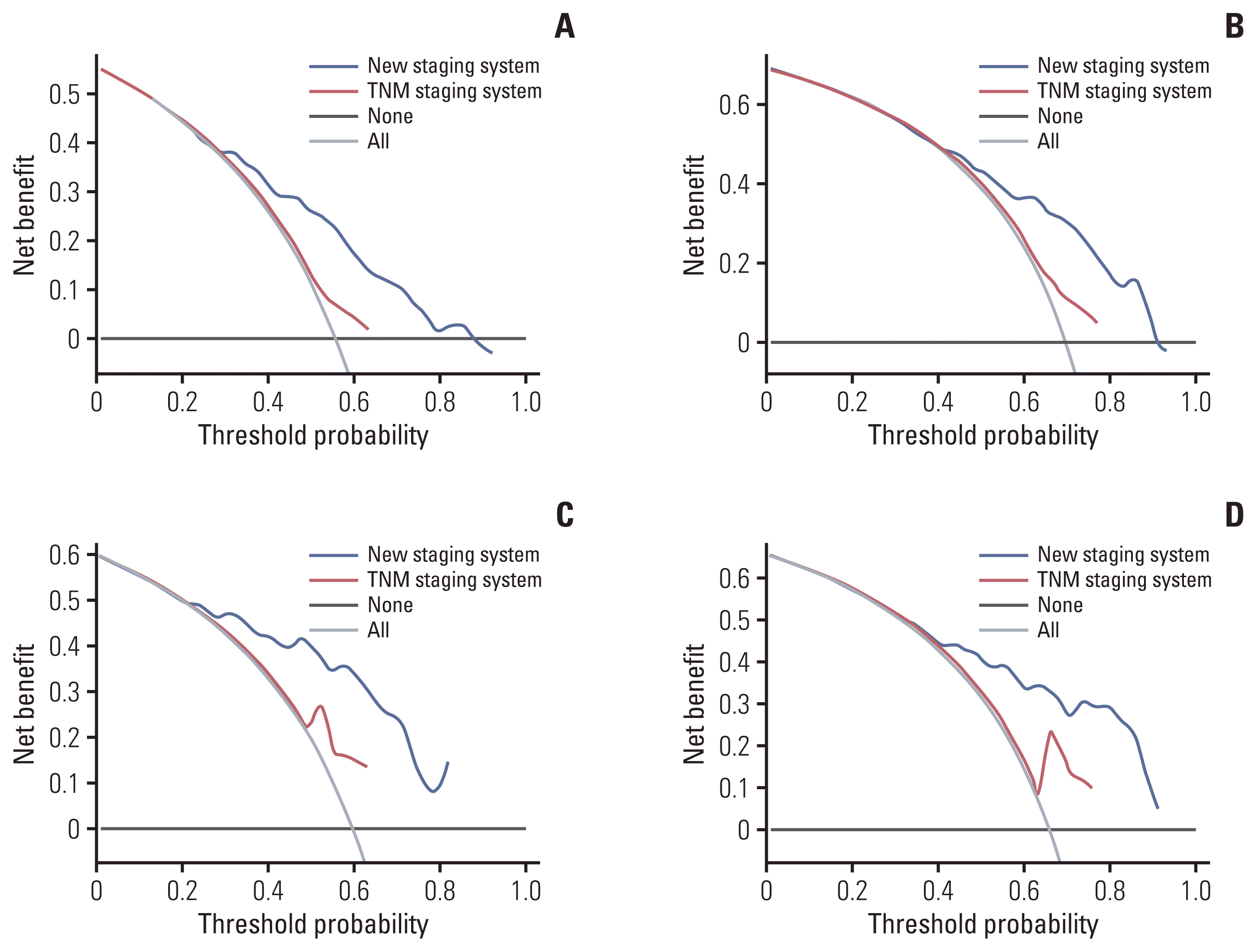
The C-index for the new staging system was 0.703 in the training cohort and 0.728 in the validation cohort using the bootstrapping method (500 repetitions). The C-index for AJCC TNM stage was 0.561 in the training cohort and 0.618 in the validation cohort using the same method. Based on the results of the survival analyses and C-index, we can conclude that the new staging system has superior discrimination capacity to the AJCC TNM staging system, regardless of the specific type of BTC nor the whole entity of BTC. In addition, DCA for 3- and 5-year OS (Fig. 4) indicated that the new prognostic model had more clinical benefits.

Discussion

BTC is a type of entity tumor with distinctive characteristics with respect to high heterogeneity, poor diagnosis, and quick progression without diverse effective treatment. Radical surgery is the only acknowledged curative treatment, as the role of adjuvant chemotherapy in treating BTC remains to be confirmed. Consequently, a broader and more effective prognostic model is required for the prognostication of patients with BTC undergoing surgical resection. Therefore, we constructed and internally validated a new prognostic model to predict OS and to stratify patients with BTC into different stages after surgery. The model was presented as a nomogram based on six independent prognostic factors, including two platelet indices, nodal involvement, CA19-9, radical surgery, tumor differentiation, PDW/PCT, and MPV. By integrating tumor features, hemostatic parameters, and operation correlated factors, our novel staging system based on the model showed good discrimination and accuracy.
Recently, an increasing number of prognostic factors for cancer have been defined, including platelet indices. The platelet indices selected in the current study were indices depicting platelet morphology including PLT, PCT, MPV, and PDW, and the ratios in between, including PDW/PCT, PDW/PLT, MPV/PLT, and MPV/PCT. PLR, an indicator of inflammation, which is drawing increasing attention, was also included. Prognostic values of most of these indices have not been investigated for BTC. Univariate Cox analysis revealed that eight out of 10 indices were associated with OS in BTC, suggesting a marked impact of platelets on tumor progression. According to time-AUC curves of these eight indices (Fig. 1), we found that MPV and PDW/PCT have the highest potential to be independent prognostic factors of BTC, which was subsequently confirmed by the final multivariable Cox analysis. MPV, representing platelet size, has been demonstrated to be an independent predictor for OS in many cancers, and a high MPV has been reported as a poor indicator of OS in pancreatic cancer [11], invasive breast cancer [12], lung cancer [13] and esophageal carcinoma [14]. Our findings add to the accumulating evidence that high MPV is an independent negative predictor of OS. PCT as determined by MPV and PLT, reflects the number and size of platelets simultaneously. In our study, high PLT and high MPV were both observed in patients with poor survival, resulting in a reasonably high PCT. Furthermore, a previous study found that increased PCT levels were indicative of poor prognosis in metastatic colorectal cancer [15]. Although the relationship between PCT and OS in cancers remains to be investigated, based on the evidence that MPV and thrombocytosis are associated with poor survival in many cancers [16], we can speculate that PCT has the potential to be a good predictor of tumor progression. The role of PDW in predicting OS is controversial. Low PDW was found to be correlated with poor OS in non-small cell lung cancer [17], colorectal cancer [18], and gastric cancer [19]. The mechanism underlying high PDW with poor tumor stage remains to be elucidated. An emerging notion is that the interplay between PDW and MPV should be considered when evaluating their prognostic performance. Overall, the correlation between PDW and tumor progression remains to be investigated, and whether the underlying mechanisms are unique for heterogeneous tumors is unknown. In our study, by combining the PDW and PCT. PDW/PCT showed better performance in terms of predicting prognosis than PDW or PCT alone, while the prognostic performance of PDW/PCT has not yet been revealed in cancers.
An increasing body of evidence has highlighted the role of platelets in various stages of tumorigenesis, including tumor growth, invasion, metastasis, and chemotherapy resistance. The interaction between platelets and tumor cells is mutual, and recent studies have demonstrated that cancer can influence platelet’ s, physiology, and activation, even in the RNA profile; thus, providing potential markers for cancer prognosis [20]. Mechanisms of cancer-stimulated thrombocytosis have been elucidated, and primary tumors can influence megakaryopoiesis and thrombopoiesis by modulating a variety of cytokines directly or indirectly. Granulocyte colony-stimulating factor and macrophage colony-stimulating factor secreted by tumor cells can stimulate megakaryopoiesis [20], while elevated interleukin-6 released from cancer cells can increase the ploidy of megakaryocytic nuclei, resulting in an elevated platelet volume [21]. The ploidy of megakaryocytes has been observed to correlate with the MPV [22]. Moreover, platelets with a high MPV have been confirmed to be a potential indicator of platelet activation [23]. Larger platelets contain higher absolute amounts of proteins and more receptors on the surface, which enables them to interact with tumor cells [22]. For example, during the process of platelet activation, granules containing ADP released by platelets [20] interact with P2Y12 receptors on platelets, triggering epithelial-mesenchymal transition, followed by invasion and metastasis [24]. Platelets with high MPV can also release more growth and prothrombotic factors, promoting angiogenesis and tumor progression [21]. Furthermore, activated platelets can crosslink, forming clots, and protecting circulative tumor cells against shear stress or immune attack [24]. In addition, increased platelet turnover is often observed in cancer patients, possibly arising from increased platelet consumption or sequestration. When platelet turnover increases, new platelets are produced with a larger size [22]. Whether larger platelets are more prothrombotic remains unclear. Evidence clarifying the role of small platelets in cancer-associated thrombosis is limited. Different mechanisms for platelets with different sizes interfering with cancer cells may exist, considering the diversity of cancer and the extensive functions of platelets. Lower PDW represents lower heterogeneity of platelets, which can be interpreted by a higher proportion of large new platelets under the stimulation of cancer cells. In contrast, platelets with a high MPV have more potential to interfere with cancer cells, assisting tumor progression in many stages. Based on these evidences and our findings, we propose that high MPV and low PDW/PCT are indicators of poor prognosis in BTC.
After integrating MPV and PDW/PCT into the multivariable Cox model, we developed a new prognostic model and a novel staging system. The OS of patients belonging to different stages were significantly different, either limited to specific types of BTC or the entire BTC cohort. Meanwhile, the AJCC 7th TNM staging system presented modest prognostic power in patients with ECC (Fig. 3H), and has been shown to have poor prognostic performance in several subsets of patients with BTC [25]. The C-index of our novel prognostic model was 0.703 in the training cohort and 0.728 in the validation cohort. Compared to the AJCC TNM stage, with a C-index of 0.561 and 0.618, our prognostic model has superior discrimination to the AJCC TNM stage. DCA also implied increased clinical benefits with the utility of our new staging system. Based on these results, we can conclude that our new prognostic model outperformed the AJCC TNM staging system in many aspects. An obvious advantage of our new prognostic model is the universal application to patients with BTC without the need to adjust the prognostic standard for any specific type. The AJCC TNM staging system is applicable for all subclasses of BTC, whereas the staging standards are not uniform with respect to different types of BTC. For example, T stage in GBC represents the infiltration depth of the tumor, while in ECC, T stage depends on the extent of invasion. In contrast, our model performed well in predicting the survival of patients with all types of BTC, which allows for greater convenience and wider application in comparison to the TNM staging system. Prognostic models have been proposed for GBC [26] and ICC [27], respectively, while relatively few models have been developed for BTC independent of a tumor site. Schweitzer et al. [28] proposed a prognostic score for patients with BTC at the time of diagnosis before any intervention. Bridgewater et al. [29] developed a model to predict OS in advanced BTC with limited predictive prognostic accuracy. A suitable method for survival prediction of patients with BTC undergoing resection has not yet been determined. Our novel staging system had extraordinary discriminative capability in predicting the OS of patients with BTC independent of the tumor site. The nomogram cannot only inform patients after surgery about their prognosis but also help to identify candidates for aggressive surgery among patients with BTC beforehand. With respect to the involvement of platelet indices, our new prognostic model can be applied to stratify patients in clinical trials intended to explore the efficacy of anti-platelet therapy, since post-diagnosis aspirin use has been reported to be associated with prolonged BTC survival [30].
One thing needs to be explained is the relatively high proportion of non-radical surgery (38.3%). This could be attributed to several reasons. Symptoms of biliary tumors were of insidious onset and most patients were found to be in advanced stage during the first medical visit. In our research, 31.3% patients were classified as TNM stage III with diverse severity of perinephric tissue invasion which increased the difficulty of surgery. Another situation was that preoperative assessment underestimate the range of tumor invasion where only radical surgery had the chance to remove all lesions. However, due to patient intolerance or family disagreement, non-radical surgery was performed instead of radical surgery. Another reason was that surgeries were performed between December 2002 and December 2017, intraoperative frozen-section was not routinely performed until recent years. From 2002 to 2012, 111 patients out of 213 (52.1%) received radical surgery while from 2013 to 2017, 168 patients out of 245 (68.6%) received radical surgery. The development of new technology elevated the probability of radical surgery.
Our study has several limitations. First, our data need to be completed, as detailed features of tumors, including invasion or local extension, are absent. However, the prognostic ability of perineural invasion and papillary tumors are inconsistent in BTC due to correlation with other factors [6], the absence of these data probably had a slight influence on the outcome. Second, our study was a single-center retrospective study with inevitable selection bias; multi-center and prospective studies are needed before generalization to a broader population. Third, the lack of external validation may decrease the credibility of the model, although we performed internal validation and obtained a high C-index value in the validation cohort.
To the best of our knowledge, our study is the first to systematically evaluate platelet indices as prognostic factors for BTC. We found that MPV and PDW/PCT are both independent prognostic factors for OS in patients with BTC. Subsequently, a new prognostic model and novel staging system were proposed for patients with BTC undergoing surgical resection, whose prognostic ability outperformed TNM staging in many aspects.

Supplementary Information

Supplementary materials are available at Cancer Research and Treatment website (https://www.e-crt.org).

Notes

Ethical Statement

This study was approved by the Medical Ethics Committee of PUMCH (S-K1110), and written informed consent was obtained from all patients. The study conformed to the ethical guidelines of the 1975 Declaration of Helsinki.

Author Contributions

Conceived and designed the analysis: Sun L, Yang H, Mao Y.

Collected the data: Chen Y, Hu W, Ji X, Xu H, Du S, Zhao H, Lu X, Sang X, Zhong S.

Contributed data or analysis tools: Sun L, Chen Y.

Performed the analysis: Wei Y.

Wrote the paper: Wei Y.

Conflicts of Interest

Conflict of interest relevant to this article was not reported.

ACKNOWLEDGMENTS

The study was supported by grants from Chinese Academy of Medical Sciences (CAMS) Innovation Fund for Medical Sciences (CIFMS) [No.2016-I2M-1-001] and Tsinghua University-Peking Union Medical College Hospital Cooperation Project [PTQH201904552].

References

1. Bray F, Ferlay J, Soerjomataram I, Siegel RL, Torre LA, Jemal A. Global cancer statistics 2018: GLOBOCAN estimates of incidence and mortality worldwide for 36 cancers in 185 countries. CA Cancer J Clin. 2018; 68:394–424.
crossref
2. Macias RI, Kornek M, Rodrigues PM, Paiva NA, Castro RE, Urban S, et al. Diagnostic and prognostic biomarkers in cholangiocarcinoma. Liver Int. 2019; 39(Suppl 1):108–22.
crossref
3. Yang P, Javle M, Pang F, Zhao W, Abdel-Wahab R, Chen X, et al. Somatic genetic aberrations in gallbladder cancer: comparison between Chinese and US patients. Hepatobiliary Surg Nutr. 2019; 8:604–14.
crossref
4. Squadroni M, Tondulli L, Gatta G, Mosconi S, Beretta G, Labianca R. Cholangiocarcinoma. Crit Rev Oncol Hematol. 2017; 116:11–31.
crossref
5. de Aretxabala X. Biliary spillage a new prognostic factor in gallbladder cancer? Hepatobiliary Surg Nutr. 2019; 8:537–8.
crossref
6. Valle JW, Lamarca A, Goyal L, Barriuso J, Zhu AX. New horizons for precision medicine in biliary tract cancers. Cancer Discov. 2017; 7:943–62.
crossref
7. Kondo N, Murakami Y, Uemura K, Sudo T, Hashimoto Y, Sasaki H, et al. Elevated perioperative serum CA 19-9 levels are independent predictors of poor survival in patients with resectable cholangiocarcinoma. J Surg Oncol. 2014; 110:422–9.
crossref
8. Chen M, Lin J, Cao J, Zhu H, Zhang B, Wu A, et al. Development and validation of a nomogram for survival benefit of lymphadenectomy in resected gallbladder cancer. Hepatobiliary Surg Nutr. 2019; 8:480–9.
crossref
9. McNamara MG, Templeton AJ, Maganti M, Walter T, Horgan AM, McKeever L, et al. Neutrophil/lymphocyte ratio as a prognostic factor in biliary tract cancer. Eur J Cancer. 2014; 50:1581–9.
crossref
10. Stoiber D, Assinger A. Platelet-leukocyte interplay in cancer development and progression. Cells. 2020; 9:855.
crossref
11. Zhang K, Gao HF, Mo M, Wu CJ, Hua YQ, Chen Z, et al. A novel scoring system based on hemostatic parameters predicts the prognosis of patients with advanced pancreatic cancer. Pancreatology. 2019; 19:346–51.
crossref
12. Gu M, Zhai Z, Huang L, Zheng W, Zhou Y, Zhu R, et al. Pre-treatment mean platelet volume associates with worse clinicopathologic features and prognosis of patients with invasive breast cancer. Breast Cancer. 2016; 23:752–60.
crossref
13. Omar M, Tanriverdi O, Cokmert S, Oktay E, Yersal O, Pilanci KN, et al. Role of increased mean platelet volume (MPV) and decreased MPV/platelet count ratio as poor prognostic factors in lung cancer. Clin Respir J. 2018; 12:922–9.
crossref
14. Song Q, Wu JZ, Wang S, Chen WH. Elevated preoperative platelet distribution width predicts poor prognosis in esophageal squamous cell carcinoma. Sci Rep. 2019; 9:15234.
crossref
15. Chang J, Lin G, Ye M, Tong D, Zhao J, Zhu D, et al. Decreased mean platelet volume predicts poor prognosis in metastatic colorectal cancer patients treated with first-line chemotherapy: results from mCRC biomarker study. BMC Cancer. 2019; 19:15.
crossref
16. Haemmerle M, Stone RL, Menter DG, Afshar-Kharghan V, Sood AK. The platelet lifeline to cancer: challenges and opportunities. Cancer Cell. 2018; 33:965–83.
crossref
17. Cui MM, Li N, Liu X, Yun ZY, Niu Y, Zhang Y, et al. Platelet distribution width correlates with prognosis of non-small cell lung cancer. Sci Rep. 2017; 7:3456.
crossref
18. Li Y, Wu H, Xing C, Hu X, Zhang F, Peng Y, et al. Prognostic evaluation of colorectal cancer using three new comprehensive indexes related to infection, anemia and coagulation derived from peripheral blood. J Cancer. 2020; 11:3834–45.
crossref
19. Zhang X, Cui MM, Fu S, Li LL, Liu YS, Liu ZP, et al. Platelet distribution width correlates with prognosis of gastric cancer. Oncotarget. 2017; 8:20213–9.
crossref
20. Plantureux L, Mege D, Crescence L, Dignat-George F, Dubois C, Panicot-Dubois L. Impacts of cancer on platelet production, activation and education and mechanisms of cancer-associated thrombosis. Cancers (Basel). 2018; 10:441.
crossref
21. Korniluk A, Koper-Lenkiewicz OM, Kaminska J, Kemona H, Dymicka-Piekarska V. Mean platelet volume (MPV): new perspectives for an old marker in the course and prognosis of inflammatory conditions. Mediators Inflamm. 2019; 2019:9213074.
crossref
22. Handtke S, Thiele T. Large and small platelets: (when) do they differ? J Thromb Haemost. 2020; 18:1256–67.
23. Jagroop IA, Tsiara S, Mikhailidis DP. Mean platelet volume as an indicator of platelet activation: methodological issues. Platelets. 2003; 14:335–6.
crossref
24. Leblanc R, Peyruchaud O. Metastasis: new functional implications of platelets and megakaryocytes. Blood. 2016; 128:24–31.
crossref
25. Groot Koerkamp B, Wiggers JK, Gonen M, Doussot A, Allen PJ, Besselink MGH, et al. Survival after resection of perihilar cholangiocarcinoma-development and external validation of a prognostic nomogram. Ann Oncol. 2015; 26:1930–5.
crossref
26. Yadav S, Tella SH, Kommalapati A, Mara K, Prasai K, Mady MH, et al. A novel clinically based staging system for gallbladder cancer. J Natl Compr Canc Netw. 2020; 18:151–9.
27. Raoof M, Dumitra S, Ituarte PH, Melstrom L, Warner SG, Fong Y, et al. Development and validation of a prognostic score for intrahepatic cholangiocarcinoma. JAMA Surg. 2017; 152:e170117.
crossref
28. Schweitzer N, Fischer M, Kirstein MM, Berhane S, Kottas M, Sinn M, et al. Risk estimation for biliary tract cancer: Development and validation of a prognostic score. Liver Int. 2017; 37:1852–60.
crossref
29. Bridgewater J, Lopes A, Wasan H, Malka D, Jensen L, Okusaka T, et al. Prognostic factors for progression-free and overall survival in advanced biliary tract cancer. Ann Oncol. 2016; 27:134–40.
crossref
30. Jackson SS, Pfeiffer RM, Liu Z, Anderson LA, Tsai HT, Gadalla SM, et al. Association between aspirin use and biliary tract cancer survival. JAMA Oncol. 2019; 5:1802–4.
crossref

Fig. 1
Time-AUC curves of the platelet indices for overall survival prediction. AUC, area under curve; MPV, mean platelet volume; PCT, plateletocrit; PDW, platelet distribution width; PLR, platelet-to-lymphocyte ratio; PLT, platelet count; Time-AUC, areas under time-dependent ROC curves.
crt-2020-833f1.gif
Fig. 2
Nomogram for predicting the 1-, 3-, 5-year, and median survival in patients with biliary tract cancer lines are drawn upward to determine the points for each variable. Total points represent the sum of points corresponding to each variable. Another line is drawn downward to determine the survival probability for total points. CA19-9, carbohydrate antigen 19-9; MPV, mean platelet volume; PCT, plateletocrit; PDW, platelet distribution width.
crt-2020-833f2.gif
Fig. 3
Kaplan-Meier curves for OS in different subsets of patients with BTC. BTC patients (A), GBC patients (C), ICC patients (E), and ECC patients (G) were stratified by the novel staging system. BTC patients (B), GBC patients (D), ICC patients (F), and ECC patients (H) were stratified according to the AJCC TNM staging system. AJCC, American Joint Committee on Cancer; BTC, biliary tract cancer; GBC, gallbladder cancer; ECC, extrahepatic cholangiocarcinoma; ICC, intrahepatic cholangiocarcinoma; OS, overall survival.
crt-2020-833f3.gif
Fig. 4
Decisive curve analysis of the nomogram. (A) 3-Year survival of the training cohort. (B) 5-Year survival of the training cohort. (C) 3-Year survival of the validation cohort. (D) 5-Year survival of the validating cohort. The horizontal black line denotes no survival, while the gray curve represents all survival.
crt-2020-833f4.gif
Table 1
Baseline characteristics
Characteristic Total Validation datasets Training datasets p-value
No. 527 156 371
Type
 GBC 122 (23.1) 37 (23.7) 85 (22.9) 0.953
 ECC 296 (56.2) 86 (55.1) 210 (56.6)
 ICC 109 (20.7) 33 (21.2) 76 (20.5)
Age (yr)
 ≤ 60 233 (44.3) 64 (41.3) 169 (45.6) 0.423
 > 60 293 (55.7) 91 (58.7) 202 (54.4)
Sex
 Female 235 (44.6) 76 (48.7) 159 (42.9) 0.254
 Male 292 (55.4) 80 (51.3) 212 (57.1)
Blood type
 AB 35 (6.7) 10 (6.5) 25 (6.8) 0.752
 A 114 (21.7) 38 (24.5) 76 (20.5)
 B 214 (40.8) 59 (38.1) 155 (41.9)
 O 162 (30.9) 48 (31.0) 114 (30.8)
Fever
 No 451 (85.6) 131 (84.0) 320 (86.3) 0.586
 Yes 76 (14.4) 25 (16.0) 51 (13.7)
Emaciation
 No 259 (49.6) 71 (46.1) 188 (51.1) 0.346
 Yes 263 (50.4) 83 (53.9) 180 (48.9)
Debilitation
 No 467 (89.0) 135 (86.5) 332 (90.0) 0.320
 Yes 58 (11.0) 21 (13.5) 37 (10.0)
Drink
 No 402 (76.6) 120 (76.9) 282 (76.4) 0.991
 Yes 123 (23.4) 36 (23.1) 87 (23.6)
Fat liver
 No 453 (91.9) 135 (91.2) 318 (92.2) 0.859
 Yes 40 (8.1) 13 (8.8) 27 (7.8)
ALT (U/L)
 ≤ 100 298 (56.7) 91 (58.3) 207 (55.9) 0.683
 > 100 228 (43.3) 65 (41.7) 163 (44.1)
AST (U/L)
 ≤ 100 351 (69.8) 103 (69.6) 248 (69.9) > 0.99
 > 100 152 (30.2) 45 (30.4) 107 (30.1)
GGT (U/L)
 ≤ 200 232 (46.0) 70 (47.3) 162 (45.5) 0.788
 > 200 272 (54.0) 78 (52.7) 194 (54.5)
ALP (U/L)
 ≤ 200 230 (45.6) 68 (45.9) 162 (45.5) > 0.99
 > 200 274 (54.4) 80 (54.1) 194 (54.5)
ALB (g/L)
 ≤ 35 112 (21.3) 34 (21.8) 78 (21.1) 0.959
 > 35 413 (78.7) 122 (78.2) 291 (78.9)
TBIL (μmol/L)a)
 ≤ 106 323 (61.3) 99 (63.5) 224 (60.4) 0.572
 > 106 204 (38.7) 57 (36.5) 147 (39.6)
CA 19-9 (U/mL)
 ≤ 100 256 (51.0) 70 (48.3) 186 (52.1) 0.497
 > 100 246 (49.0) 75 (51.7) 171 (47.9)
Tumor differentiationb)
 Poor 51 (10.1) 20 (13.2) 31 (8.8) 0.266
 Moderate 262 (52.0) 73 (48.3) 189 (53.5)
 Well 191 (37.9) 58 (38.4) 133 (37.7)
Radical cure
 No 200 (38.3) 64 (41.6) 136 (37.0) 0.375
 Yes 322 (61.7) 90 (58.4) 232 (63.0)
T category
 Tis 9 (1.7) 2 (1.3) 7 (1.9) 0.362
 1 130 (24.7) 36 (23.1) 94 (25.3)
 2 153 (29.0) 55 (35.3) 98 (26.4)
 3 196 (37.2) 52 (33.3) 144 (38.8)
 4 39 (7.4) 11 (7.1) 28 (7.5)
Nodal involvement
 No 364 (69.3) 113 (73.4) 251 (67.7) 0.234
 Yes 161 (30.7) 41 (26.6) 120 (32.3)
TNM stage
 0 9 (1.7) 2 (1.3) 7 (1.9) 0.737
 I 179 (34.0) 57 (36.5) 122 (32.9)
 II 174 (33.0) 47 (30.1) 127 (34.2)
 III 165 (31.3) 50 (32.1) 115 (31.0)
Tumor size (cm)a)
 ≤ 2.6 303 (60.2) 87 (58.8) 216 (60.8) 0.741
 > 2.6 200 (39.8) 61 (41.2) 139 (39.2)
Postoperative chemotherapy
 No 353 (76.2) 103 (73.6) 250 (77.4) 0.441
 Yes 110 (23.8) 37 (26.4) 73 (22.6)

Values are presented as number (%). ALB, albumin; ALP, alkaline phosphatase; ALT, alanine aminotransferase; AST, aspartate aminotransferase; CA 19-9, carbohydrate antigen 19-9; ECC, extrahepatic cholangiocarcinoma; GBC, gallbladder cancer; GGT, γ-glutamyl transpeptidase; ICC, intrahepatic cholangiocarcinoma; TBIL, total bilirubin.

a) Cut-off values of TBIL (106 μmol/L) and tumor sice (2.6 cm) were determined by ‘maxstat’ R package,

b) Tumor differentiation was judged by pathologists. Poor includes undifferentiated or poorly differentiated, which means disordered arrangement of cancer cells, high cellular atypia and high proportion of cancer cells in division phase. Well means almost normal arrangement of cancer cells, low cellular atypia and low proportion of cancer cells in division phase. Moderate means somewhere in between.

Table 2
Univariate and multivariate analyses for OS in BTC patients
Univariate analysis Multivariate analysis


HR (95% CI) p-value HR (95% CI) p-value
Type

 GBC Reference

 ECC 0.97 (0.74–1.27) 0.799

 ICC 1.12 (0.81–1.54) 0.496

Age (yr)

 ≤ 60 Reference

 > 60 1.21 (0.97–1.51) 0.087

Sex

 Female Reference

 Male 0.97 (0.78–1.21) 0.785

Blood typea)

 AB Reference

 A 0.67 (0.42–1.07) 0.091

 B 0.85 (0.56–1.31) 0.467

 O 0.85 (0.55–1.32) 0.467

Fever

 No Reference

 Yes 1.02 (0.75–1.39) 0.902

Emaciation

 No Reference

 Yes 1.35 (1.08–1.68) 0.007b)

Debilitation

 No Reference

 Yes 1.10 (0.78–1.55) 0.582

Drink

 No Reference

 Yes 0.91 (0.70–1.18) 0.491

Fat liver

 No Reference

 Yes 1.19 (0.80–1.77) 0.393

ALT (U/L)

 ≤ 100 Reference

 > 100 1.17 (0.94–1.45) 0.167

AST (U/L)

 ≤ 100 Reference

 > 100 1.17 (0.92–1.48) 0.198

GGT (U/L)

 ≤ 200 Reference

 > 200 1.23 (0.98–1.53) 0.073

ALP (U/L)

 ≤ 200 Reference

 > 200 1.40 (1.12–1.75) 0.003b)

ALB (g/L)

 ≤ 35 Reference

 > 35 0.64 (0.50–0.81) < 0.001b)

TBIL (μmol/L)

 ≤ 106 Reference

 > 106 1.61 (1.30–2.01) < 0.001b)

CA19-9 (U/mL)

 ≤ 100 Reference

 > 100 2.01 (1.60–2.53) < 0.001b) 1.60 (1.25–2.05) < 0.001b)

Tumor differentiation

 Poor Reference

 Moderate 0.68 (0.47–0.98) 0.040b) 0.72 (0.59–0.87) < 0.001b)

 Well 0.44 (0.30–0.65) < 0.001b)

Radical cure

 No Reference

 Yes 0.40 (0.32–0.50) < 0.001b) 0.47 (0.37–0.60) < 0.001b)

T category

 Tis Reference

 1 5.38 (0.75–38.71) 0.095

 2 6.43 (0.90–46.14) 0.064

 3 9.03 (1.26–64.60) 0.028a)

 4 6.07 (0.82–45.13) 0.078

Nodal involvement

 No Reference

 Yes 1.99 (1.58–2.50) < 0.001b) 1.68 (1.31–2.15) < 0.001b)

Tumor size (cm)

 ≤ 2.6 Reference

 > 2.6 0.90 (0.72–1.13) 0.364

Postoperative chemotherapy

 No Reference

 Yes 0.93 (0.71–1.22) 0.624

MPV (fl)

 ≤ 8.1 Reference

 > 8.1 1.52 (1.19–1.95) < 0.001b) 1.33 (1.01–1.75) 0.045b)

PDW/PCT

 ≤ 190 Reference

 > 190 0.65 (0.52–0.82) < 0.001b) 0.78 (0.61–1.00) 0.046b)

ALB, albumin; ALP, alkaline phosphatase; ALT, alanine aminotransferase; AST, aspartate aminotransferase; BTC, biliary tract cancer; CA19-9, carbohydrate antigen 19-9; CI, confidence interval; ECC, extrahepatic cholangiocarcinoma; GBC, gallbladder cancer; GGT, γ-glutamyl transpeptidase; HR, hazard ratio; ICC, intrahepatic cholangiocarcinoma; MPV, mean platelet volume; OS, overall survival; PCT, plateletocrit; PDW, platelet distribution width; TBIL, total bilirubin.

a) All patients were Rh positive,

b) Statistically significant (p < 0.05).

TOOLS
Similar articles