Journal List > Cancer Res Treat > v.53(1) > 1154689

Jung, Kim, Kim, Park, Ko, Han, Kim, Lee, and Lee: Sequential Treatment with an Immune Checkpoint Inhibitor Followed by a Small-Molecule Targeted Agent Increases Drug-Induced Pneumonitis

Abstract

Purpose

Immune checkpoint inhibitors (ICI) and targeted small-molecule drugs are mainstay elements of lung cancer chemotherapy. However, they are associated with development of pneumonitis, a rare, but potentially life-threatening event. We analyzed lung cancer patients treated with ICI to evaluate the effect of sequential therapeutic administration on the incidence of pneumonitis.

Materials and Methods

In this retrospective study, 242 patients were included. Serial radiologic findings taken during and immediately after ICI treatment were reviewed. Factors that increased pneumonitis and the relationship between peri-ICI chemotherapy and the development of pneumonitis were evaluated.

Results

Pneumonitis developed in 23 patients (9.5%); severe pneumonitis (grade ≥ 3) occurred in 13 of 23 patients (56%); pneumonitis-related death occurred in six. High-dose thoracic radiation (≥ 6,000 cGy) revealed a tendency toward high risk of pneumonitis (odds ratio, 2.642; 95% confidence interval, 0.932 to 7.490; p=0.068). Among 149 patients followed for ≥ 8 weeks after the final ICI dose, more patients who received targeted agents within 8-weeks post-ICI experienced pneumonitis (3/16, 18.8%) compared with patients who received cytotoxic agents (4/54, 7.4%) or no chemotherapy (4/79, 5.1%) (p=0.162). Targeted therapy was associated with earlier-onset pneumonitis than treatment with cytotoxic agents (35 vs. 62 days post-ICI, p=0.007); the resulting pneumonitis was more severe (grade ≥ 3, 100% vs. 0%, p=0.031).

Conclusion

Sequential administration of small-molecule targeted agents immediately after ICI may increase the risk of severe pneumonitis. The sequence of chemotherapy regimens that include ICI and targeted agents should be carefully planned to reduce the risk of pneumonitis in lung cancer patients.

Introduction

Since their initial development, immune checkpoint inhibitors (ICI) have changed the paradigm of treatment for a diverse range of malignancies [1]. They have not only improved clinical outcomes, but have solidified immune modulation as a fundamental strategy for cancer treatment [2]. In the treatment of advanced lung cancer, for which systemic therapy is required, platinum-based chemotherapy has traditionally been the mainstay of care [3]. However, the efficacy of platinum-based chemotherapy is limited by the development of dose-limiting toxicities [4,5]. Although small-molecule targeted agents against epidermal growth factor receptor (EGFR) and anaplastic lymphoma kinase (ALK) have demonstrated improved efficacy and tolerability than conventional chemotherapy, their use is available only to patients with specific genetic alterations [6-8]. On the other hand, treatment with ICI leads to substantial improvements in efficacy and tolerability for all types of lung cancer [9,10].
Although uncommon, drug-induced pneumonitis is an important, potentially life-threatening immune-related adverse event caused by ICI [11]. In patients with non–small cell lung cancer (NSCLC) who receive ICI, drug-induced pneumonitis occurs in 1.1%-3.6% of patients, and the mortality rate among infected patients is as high as 8%-9.4% [12,13]. Risk factors that predispose patients to drug-induced pneumonitis include cigarette smoking, prior thoracic radiotherapy, and prior pulmonary comorbidities. However, the most recent studies on this issue were performed retrospectively with relatively small sample sizes or in patients with mixed types of cancer [14,15].
Small-molecule agents that target EGFR are another potential cause of pneumonitis in patients with lung cancer. The incidence of drug-induced pneumonitis associated with gefitinib and erlotinib is 1.2%-1.6%, with a relatively high mortality of 22.8% [13]. In addition, a post-marketing survey of crizotinib found that it is associated with pneumonitis in 5.77% of ALK-positive NSCLC patients, of which 3.45% were pneumonitis of at least grade 3 [16]. Oshima et al. [17] also reported that the incidence of pneumonitis after treatment with EGFR-targeted agents was increased in patients receiving nivolumab; however, the dataset used for that study did not consider the duration in between treatments. These targeted agents have become a crucial element of treatment for patients with specific markers such as EGFR mutation or ALK rearrangement. However, the emergence of resistance to targeted therapies can complicate treatment. In such cases, ICI can provide an important backup therapeutic option [18]. However, there is still much to learn about the clinical implications of the interactions between ICI and targeted small-molecule agents. Therefore, this study aimed to evaluate the relationship between the use of chemotherapy drugs with ICI treatment and the resulting impacts on the incidence and severity of drug-induced pneumonitis.

Materials and Methods

1. Study population

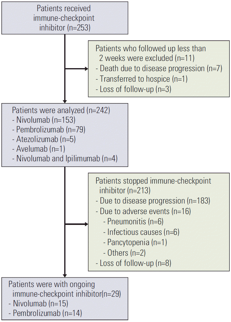
This single-center retrospective study initially enrolled 253 patients with histologically diagnosed lung cancer who received ICI between November 2015 and October 2018 at the National Cancer Center, Korea. The inclusion criteria were age of ≥ 18 years at diagnosis, received at least 1 cycle of ICI that included programmed death-1 (PD-1) inhibitors, programmed death ligand-1 (PD-L1) inhibitors, and cytotoxic T-lymphocyte-associated protein-4 inhibitors, and available baseline clinical data and images after diagnosis. The exclusion criterion was a follow-up duration of less than 2 weeks after initiation of ICI. By this criterion, 11 patients were excluded (Fig. 1). Monotherapy with nivolumab (3 mg/kg) was administered intravenously once every 2 weeks, and pembrolizumab (200 mg) was given once every 3 weeks. Atezolizumab (1,200 mg) was administered every 3 weeks, and avelumab (10 mg/kg) was given every 2 weeks. A combined regimen of nivolumab (3 mg/kg once every 2 weeks) and ipilimumab (1 mg/kg every 6 weeks) was used according to clinical judgment. The regimen was decided by each physician depending on each patient’s medical condition, stage, PD-L1 expression, and insurance coverage. Evaluations of response to therapy were performed with computed tomography (CT) scans every 2-3 cycles, as appropriate. The medical records of the 242 patients included in this analysis were comprehensively reviewed, and CT scans of each patient were independently reviewed by a thoracic radiologist (H.Y.K.). The definitive diagnosis of drug-induced pneumonitis was established based on a consensus between the radiologist and medical oncologist. For accurate decision, independent analysis was performed by the radiologist and medical oncologist. The diagnosis was established only if both of them assumed drug-induced pneumonitis. Especially, for patients who received radiotherapy in lung field previously, the clinical information such as radiation field and interval time between radiotherapy and the onset of pneumonitis was assessed with collaboration of radiation oncologist to avoid false interpretation. Radiologic patterns of pneumonitis were classified as pure ground-glass opacity (GGO), pure consolidation, combined GGO and consolidation, and others. The Common Terminology Criteria for Adverse Events ver. 5.0 was used to classify the grade of pneumonitis.

2. Statistical analysis

To identify risk factors associated with drug-induced pneumonitis, continuous variables such as age and the dose of radiation received prior to the initiation of ICI were dichotomized (> 65 vs. ≤ 65 for age; ≥ 6,000 vs. < 6,000 cGy for radiation dose). Considering the previously known pharmacokinetics of ICI, the threshold for sequential chemotherapy was defined as treatment within 8 weeks after the last dose of ICI [19]. Immediate chemotherapy prior to ICI was defined as treatment within 8 weeks before the first administration of ICI. Estimates of odds ratios (ORs) and 95% confidence intervals (CIs) were obtained with logistic regression. Variables with a p-value of less than 0.1 in univariate analysis were included in a multivariable analysis. All statistical analyses were performed using R software (ver. 3.5.1, R Foundation for Statistical Computing, Vienna, Austria), and differences were considered significant at a 2-sided p-value of < 0.05.

Results

1. Patient characteristics

After exclusion, 242 patients were included in an analysis of the incidence and risk factors of drug-induced pneumonitis after treatment with ICI. The baseline characteristics of patients are described in Table 1. Most patients had stage IV NSCLC based on 8th edition of TNM staging. Among 32 patients with stage III NSCLC who were initially indicated for locoregional curative modalities, 18 received concurrent chemoradiotherapy as initial treatment, while others could not due to diverse reasons such as poor lung function, extensive lung field, and cancer progression during neoadjuvant chemotherapy (S1 Table). Prior to the administration of ICI, radiation therapy in the lung field was performed in 78 patients. Among them 22 received 6,000 cGy, while 36 received less than 6,000 cGy—most of them were performed with palliative purpose with median dose of 3,000 cGy except one patient who stopped concurrent chemoradiotherapy due to newly developed brain metastasis. The median number of previous chemotherapy treatments was 1 (range, 0 to 5), and 14 patients (5.8%) received ICI as the first line therapy. More than 60% of patients (n=153) received monotherapy with nivolumab, and pembrolizumab was used in 79 patients (32.6%). Atezolizumab and avelumab were prescribed for five (2.1%) and one (0.4%) patients, respectively. Four patients (1.7%) received a combined regimen with nivolumab and ipilimumab. Baseline characteristics of patients and clinical outcomes according to the ICI regimen were described in S2 and S3 Tables. The median number of cycles of ICI administered was 3 (range, 1 to 20) with a median ICI duration of 44 days (range, 1 to 855). Twenty-nine patients were still being treated with ICI at the time of analysis, however the other 213 patients discontinued ICI after disease progression (n=183), adverse events (n=16), or loss to follow-up (n=8) (Fig. 1).

2. Incidence and risk factors for drug-induced pneumonitis

During a median follow-up of 13.3 months after ICI started, drug-induced pneumonitis occurred in 23 cases (9.5%). The median time from the first administration of ICI to onset of pneumonitis was 85 days (range, 11 to 321 days), and it developed within 2 months in 43.5% of patients. The median interval between the last dose of ICI and the onset of pneumonitis was 27 days (range, 9 to 105 days) with the exception of one patient who experienced drug-induced pneumonitis 263 days after the last dose of ICI. Among the 153 patients who received nivolumab, pneumonitis was diagnosed in 15 (9.8%). The incidence of pneumonitis was similar after treatment with pembrolizumab (8.9%, 7/79). One case of pneumonitis was observed in a patient treated with atezolizumab (20.0%, 1/5). The radiologic appearance of pneumonitis is described in S4 Table, in which chest CT scan of drug-induced pneumonitis revealed pure GGO patterns in more than 60%.
Among 23 cases of pneumonitis, 13 (56.5%) were grade ≥ 3 (Table 2). All patients with grade-1 pneumonitis and two patients with grade-2 pneumonitis were treated in the outpatient setting with oral steroids. However, hospitalization and administration of intravenous steroids were required for 16 patients (69.6%). The median duration of hospitalization was 15.5 days (range, 2 to 45 days). One patient who presented grade 4 pneumonitis was recovered while six patients (26.1%) died despite of high-dose systemic intravenous steroid therapy.
In univariate logistic regression, a radiation dose of 6,000 cGy or higher prior to ICI was significantly associated with the occurrence of pneumonitis (OR, 3.455; 95% CI, 1.346 to 8.867; p=0.010), but a lower dose of radiation was not (Table 3). After adjustment for any cigarette smoking (vs. never smokers) and stage IV (vs. stage III), which showed p value less than 0.1 in univariable analysis, a radiation dose of ≥ 6,000 cGy prior to ICI revealed a tendency toward high risk of pneumonitis although it was not statistically significant (OR, 2.642; 95% CI, 0.932 to 7.490; p=0.068). The factors related to thoracic radiation such as the dose schedule, targeted volume, and techniques were additionally analyzed in S5 Table, in which there was no significant factor to predict the development of pneumonitis. Immediate chemotherapy prior to ICI was included in the analysis to determine how the sequence of chemotherapy influences the development of drug-induced pneumonitis. However, no drug-induced pneumonitis occurred in patients who received a targeted agent within 8 weeks before ICI. In 123 patients who were treated with cytotoxic agents prior to ICI within 8 weeks, pneumonitis developed in 10 (8.1%). The factors related to the previous cytotoxic agents such as regimen, intensity, and interval time to the administration of ICI were assessed in S6 Table, in which there was no significant factor to influence the occurrence of pneumonitis. There was no statistical difference in incidence of drug-induced pneumonitis between patients treated with PD-1 inhibitor and PD-L1 inhibitor.

3. Drug-induced pneumonitis was associated with post-ICI sequential chemotherapy

The effects of post-ICI sequential chemotherapy on the risk of drug-induced pneumonitis were analyzed in 149 patients who were observed for at least 8 weeks after the last dose of ICI. Among 79 patients who did not receive chemotherapy during that period, pneumonitis occurred in four cases (5.1%). In the 54 patients who received a cytotoxic agent within 8 weeks of their last ICI dose, pneumonitis developed in four cases (7.4%), of which two were treated with docetaxel and one patient each with pemetrexed monotherapy or pemetrexed with carboplatin. Of note, three of 16 patients (18.8%) who received a targeted agent within 8 weeks of their last ICI dose were diagnosed with pneumonitis, although this relationship was not statistically significant (p=0.162). The relationship between drug-induced pneumonitis and sequential chemotherapy are shown in Table 4. Patients with treated with a sequential targeted agent tended to experience a significantly more rapid onset of pneumonitis after the last administration of ICI compared to those treated with a sequential cytotoxic agent (35.0 days vs. 62.0 days, p=0.007). Further, all cases of drug-induced pneumonitis that occurred after sequential treatment with ICI and targeted agents were grade ≥ 3, in stark contrast with the finding of no grade ≥ 3 pneumonitis in patients treated with sequential ICI and cytotoxic agents (p=0.031). Of those grade ≥ 3 patients, one patient died 2 days after the onset of pneumonitis even despite hospitalization and intensive treatment with intravenous high dose steroids (Table 5). The CT images of those patients presented GGO patterns (Fig. 2). Among the targeted agents, gefitinib showed the strongest relationship with drug-induced pneumonitis (2/6, 33.3%), followed by osimertinib (1/6, 16.7%). There were no cases of drug-induced pneumonitis in the few patients treated with erlotinib (n=2), crizotinib (n=1), or glesatinib (n=1). To assess the effect of risk factors in this population, we performed separate logistic regression. Although there was no statistical significance, patients who were treated with targeted agent sequentially after ICI presented tendency toward increased risk of drug-induced pneumonitis (OR, 5.296; 95% CI, 0.981 to 28.582; p=0.053) (S7 Table).

Discussion

This retrospective study assessed risk factors associated with drug-induced pneumonitis related to the use of ICI in a real-world setting. It also examined the impact of sequential chemotherapy on the occurrence of pneumonitis. The overall incidence of any grade of drug-induced pneumonitis was 9.5% in our cohort, somewhat higher than previous results [12,13]. Strikingly, 18.8% of patients who were treated with a targeted agent after ICI therapy experienced pneumonitis, markedly higher than the known risk of pneumonitis after treatment with targeted agents alone [13,16]. These findings suggest a possible synergistic effect between ICI and targeted agents in the development of drug-induced pneumonitis. Interestingly, this relationship existed through 8 weeks, significantly beyond the half-life of ICI.
Our results are in agreement with the findings of several previous studies [17]. Schoenfeld et al. [20] reported that severe immune-related adverse events were observed in 24% of patients who received ICI followed by osimertinib within 3 months. Although the TATTON trial (combination treatment with osimertinib and durvalumab) initially demonstrated encouraging clinical activity, enrollment for the phase III study was terminated due to high incidence of interstitial lung disease [21]. Lin et al. [22] assessed the increased hepatotoxicity associated with sequential treatment ICI and crizotinib in patients with NSCLC, and found that patients who received sequential ICI and crizotinib had a significantly higher incidence of elevated liver enzymes compared to patients who were treated with crizotinib alone. In that study, the median interval between the last administration of ICI and crizotinib was 30 days. And drug-induced pneumonitis occurred within 8 weeks in seven of 11 patients. The half-life of nivolumab is approximately 3 weeks, but pharmacodynamic studies of ICI have revealed sustained activity for more than 2 months after infusion [19]. Pembrolizumab has a half-life of around 4 weeks, but response times vary greatly, with most responses occurring by 12 weeks [23]. Thus, it is likely that the effects of ICI persist even 8 weeks after the final dose, enabling overlapping toxicity after treatment with targeted agents within this period. Interestingly, no immune-related adverse events were observed when the targeted agent was administered prior to ICI in previous reports or in our cohort [20]. The known half-life of targeted agents is around 15-60 hours [24]. This likely explains the decreased incidence of drug-induced pneumonitis when ICI is administered after small-molecule targeted agents. Remarkably, every case of drug-induced pneumonitis that occurred after the sequential use of ICI and targeted agents was grade ≥ 3. This suggests that the synergistic effects of ICI and targeted agents contribute to both increased incidence and severity of pneumonitis.
Our study found that the dose of thoracic radiation is a strong predictor of drug-induced pneumonitis. This is in contrast with previous studies that have studied any history of thoracic radiation as a predictive factor for drug-induced pneumonitis [14,25,26]. The patients who received ICI for stage III lung cancer experienced a 18.8% rate of drug-induced pneumonitis, in contrast with an 8.1% incidence among patients with stage IV cancer. This finding indirectly suggests the predictive role of thoracic radiation in the development of drug-induced pneumonitis in lung cancer patients; the radiation dose for patients with stage III disease was higher than stage IV (median, 6,000 vs. 5,000 cGy; p=0.011). Indeed, the dose of thoracic radiotherapy is a known risk factor for pneumonitis [27]. Although we simply categorized on the basis of radiation dose and did not assess the dose-volume relationship, as in previous studies, we chose a practical cutoff value (6,000 cGy), which is a recommended dose of definitive thoracic radiotherapy [28]. Further study is warranted to investigate the possible relationships between ICI and radiotherapy dose in larger, prospective studies.
Our study had several limitations. First, this study was performed retrospectively. Thus, we had a considerable number of patients that were lost to follow-up, which might have affected the results (such as incidence of pneumonitis). However, we plan to perform a prospective observational study of the use of ICI on the basis of this work. Approximately 95% of the patients in this study were treated with PD-1 inhibitors, and there was no significant difference in drug-induced pneumonitis between patients treated with PD-1 inhibitors and PD-L1 inhibitors. However, this comparison requires further verification, as prior reports have indicated a higher risk of pneumonitis after treatment with PD-1 inhibitor relative to treatment with a PD-L1 inhibitor [15,29]. Although our overall sample size was sufficient, the number of patients who received sequential therapy with targeted agents was relatively small. Therefore, despite the observation of a higher average incidence of drug-induced pneumonitis in this group relative to patients treated sequentially with cytotoxic agents, the difference was not statistically significant. Additionally, patients in our cohort were treated with multiple different targeted agents, which makes analysis of the risks associated with specific therapeutics difficult. Future studies should investigate the impacts of each separate targeted agent when administered after ICI therapy. Although we analyzed the impact of sequential chemotherapy at 8 weeks, a timepoint chosen on the basis of previously known pharmacodynamics of ICI, we cannot currently determine what interval is required to reduce the likelihood of synergistic toxicity. Recently, Shinno et al. [30] conducted liquid chromatography-mass spectrometry analyses to check the concentration of serum nivolumab in patients who had immunologic adverse events after sequential treatment with nivolumab and osimertinib. Three patients with adverse events were identified, of whom two developed interstitial lung disease. The durations between the final nivolumab administration and osimertinib were 46 and 22 days, but the time intervals between the last nivolumab and the onset of pneumonitis were 96 and 56 days, respectively. The blood analyses proved that nivolumab remained for months at sufficient level to cause pneumonitis. This result shows that it would be difficult to define the exact point to determine the risk of drug induced pneumonitis after the last dose of ICI. However, delaying treatment for more than 8 weeks after discontinuation of ICI is not often feasible in real practice. Therefore, we expect that knowledge of the risks posed by treatment within 8 weeks of discontinuation of ICI will be valuable for the treatment of patients with advanced lung cancer.
In conclusion, treatment with small-molecule targeted agents within 8 weeks of ICI therapy could increase the risk of drug-induced pneumonitis in patients with advanced lung cancer. Therefore, the sequence of treatment with ICI and targeted agents should be carefully planned to minimize the risk of pneumonitis in patients with lung cancer.

Electronic Supplementary Material

Supplementary materials are available at Cancer Research and Treatment website (https://www.e-crt.org).
S1 Table.
Initial treatment of patients with stage III lung cancer
crt-2020-543-suppl1.pdf
S2 Table.
Baseline characteristics of patients according to the regimen of immune-checkpoint inhibitor
crt-2020-543-suppl2.pdf
S3 Table.
Clinical outcomes of patients according to the regimen of immune-checkpoint inhibitor
crt-2020-543-suppl3.pdf
S4 Table.
Radiologic patterns of drug-induced pneumonitis related to ICI
crt-2020-543-suppl4.pdf
S5 Table.
Univariable logistic regression of risk factors related to thoracic radiation theapy for drug-induced pneumonitis in patients who received radiation therapy prior to the administration of ICI
crt-2020-543-suppl5.pdf
S6 Table.
Univariable logistic regression of risk factors related to the use of cytotoxic agents for drug-induced pneumonitis in patients who received systemic chemotherapy within 8 weeks prior to the administration of ICI
crt-2020-543-suppl6.pdf
S7 Table.
Univariable and multivariable logistic regression of risk factors for drug-induced pneumonitis in patients who observed for at least 8 weeks after the last dose of ICI
crt-2020-543-suppl7.pdf

Notes

Ethical Statement

This study was approved by the institutional review board (IRB) and conducted in accordance with the Declaration of Helsinki (NCC 2020-0005). Written informed consent was exempted from IRB because no intervention was involved due to the nature of the retrospective study.

Author Contributions

Conceived and designed the analysis: Jung J, Kim DG, Han JY, Kim HT, Lee JS, Lee Y.

Collected the data: Jung J, Kim DG, Ko AR.

Contributed data or analysis tools: Jung J, Kim HY, Kim DG, Park SY, Lee Y.

Performed the analysis: Jung J, Lee Y.

Wrote the paper: Jung J, Lee Y.

Conflicts of Interest

Conflicts of interest relevant to this article was not reported.

ACKNOWLEDGMENTS

This research was supported by National Cancer Center research grant (1710230-3).

References

1. Lonberg N, Korman AJ. Masterful antibodies: checkpoint blockade. Cancer Immunol Res. 2017; 5:275–81.
crossref
2. Tolcher AW, Sznol M, Hu-Lieskovan S, Papadopoulos KP, Patnaik A, Rasco DW, et al. Phase Ib study of utomilumab (PF-05082566), a 4-1BB/CD137 agonist, in combination with pembrolizumab (MK-3475) in patients with advanced solid tumors. Clin Cancer Res. 2017; 23:5349–57.
crossref
3. Azzoli CG, Baker S Jr, Temin S, Pao W, Aliff T, Brahmer J, et al. American Society of Clinical Oncology Clinical Practice Guideline update on chemotherapy for stage IV non-small-cell lung cancer. J Clin Oncol. 2009; 27:6251–66.
crossref
4. Ardizzoni A, Boni L, Tiseo M, Fossella FV, Schiller JH, Paesmans M, et al. Cisplatin- versus carboplatin-based chemotherapy in first-line treatment of advanced non-small-cell lung cancer: an individual patient data meta-analysis. J Natl Cancer Inst. 2007; 99:847–57.
crossref
5. Azzoli CG, Kris MG, Pfister DG. Cisplatin versus carboplatin for patients with metastatic non-small-cell lung cancer--an old rivalry renewed. J Natl Cancer Inst. 2007; 99:828–9.
crossref
6. Shi Y, Au JS, Thongprasert S, Srinivasan S, Tsai CM, Khoa MT, et al. A prospective, molecular epidemiology study of EGFR mutations in Asian patients with advanced non-small-cell lung cancer of adenocarcinoma histology (PIONEER). J Thorac Oncol. 2014; 9:154–62.
crossref
7. Soria JC, Ohe Y, Vansteenkiste J, Reungwetwattana T, Chewaskulyong B, Lee KH, et al. Osimertinib in untreated EGFR-mutated advanced non-small-cell lung cancer. N Engl J Med. 2018; 378:113–25.
8. Mok TS, Wu YL, Thongprasert S, Yang CH, Chu DT, Saijo N, et al. Gefitinib or carboplatin-paclitaxel in pulmonary adenocarcinoma. N Engl J Med. 2009; 361:947–57.
crossref
9. Reck M, Rodriguez-Abreu D, Robinson AG, Hui R, Csoszi T, Fulop A, et al. Pembrolizumab versus chemotherapy for PD-L1-positive non-small-cell lung cancer. N Engl J Med. 2016; 375:1823–33.
crossref
10. Borghaei H, Paz-Ares L, Horn L, Spigel DR, Steins M, Ready NE, et al. Nivolumab versus docetaxel in advanced nonsquamous non-small-cell lung cancer. N Engl J Med. 2015; 373:1627–39.
crossref
11. Topalian SL, Hodi FS, Brahmer JR, Gettinger SN, Smith DC, McDermott DF, et al. Safety, activity, and immune correlates of anti-PD-1 antibody in cancer. N Engl J Med. 2012; 366:2443–54.
crossref
12. Ma K, Lu Y, Jiang S, Tang J, Li X, Zhang Y. The relative risk and incidence of immune checkpoint inhibitors related pneumonitis in patients with advanced cancer: a meta-analysis. Front Pharmacol. 2018; 9:1430.
crossref
13. Skeoch S, Weatherley N, Swift AJ, Oldroyd A, Johns C, Hayton C, et al. Drug-induced interstitial lung disease: a systematic review. J Clin Med. 2018; 7:356.
crossref
14. Cui P, Liu Z, Wang G, Ma J, Qian Y, Zhang F, et al. Risk factors for pneumonitis in patients treated with anti-programmed death-1 therapy: a case-control study. Cancer Med. 2018; 7:4115–20.
crossref
15. Nishino M, Giobbie-Hurder A, Hatabu H, Ramaiya NH, Hodi FS. Incidence of programmed cell death 1 inhibitor-related pneumonitis in patients with advanced cancer: a systematic review and meta-analysis. JAMA Oncol. 2016; 2:1607–16.
16. Gemma A, Kusumoto M, Kurihara Y, Masuda N, Banno S, Endo Y, et al. Interstitial lung disease onset and its risk factors in Japanese patients with ALK-positive NSCLC after treatment with crizotinib. J Thorac Oncol. 2019; 14:672–82.
crossref
17. Oshima Y, Tanimoto T, Yuji K, Tojo A. EGFR-TKI-associated interstitial pneumonitis in nivolumab-treated patients with non-small cell lung cancer. JAMA Oncol. 2018; 4:1112–5.
crossref
18. Lim ZF, Ma PC. Emerging insights of tumor heterogeneity and drug resistance mechanisms in lung cancer targeted therapy. J Hematol Oncol. 2019; 12:134.
crossref
19. Brahmer JR, Drake CG, Wollner I, Powderly JD, Picus J, Sharfman WH, et al. Phase I study of single-agent anti-programmed death-1 (MDX-1106) in refractory solid tumors: safety, clinical activity, pharmacodynamics, and immunologic correlates. J Clin Oncol. 2010; 28:3167–75.
crossref
20. Schoenfeld AJ, Arbour KC, Rizvi H, Iqbal AN, Gadgeel SM, Girshman J, et al. Severe immune-related adverse events are common with sequential PD-(L)1 blockade and osimertinib. Ann Oncol. 2019; 30:839–44.
crossref
21. Moya-Horno I, Viteri S, Karachaliou N, Rosell R. Combination of immunotherapy with targeted therapies in advanced non-small cell lung cancer (NSCLC). Ther Adv Med Oncol. 2018; 10:1758834017745012.
crossref
22. Lin JJ, Chin E, Yeap BY, Ferris LA, Kamesan V, Lennes IT, et al. Increased hepatotoxicity associated with sequential immune checkpoint inhibitor and crizotinib therapy in patients with non-small cell lung cancer. J Thorac Oncol. 2019; 14:135–40.
crossref
23. Longoria TC, Tewari KS. Evaluation of the pharmacokinetics and metabolism of pembrolizumab in the treatment of melanoma. Expert Opin Drug Metab Toxicol. 2016; 12:1247–53.
crossref
24. Mizoguchi K, Nakamura Y, Sano K, Sato S, Ikegami Y, Motoshima K, et al. Pharmacokinetic parameters of gefitinib predict efficacy and toxicity in patients with advanced non-small cell lung cancer harboring EGFR mutations. Cancer Chemother Pharmacol. 2016; 78:377–82.
crossref
25. Palma DA, Senan S, Tsujino K, Barriger RB, Rengan R, Moreno M, et al. Predicting radiation pneumonitis after chemoradiation therapy for lung cancer: an international individual patient data meta-analysis. Int J Radiat Oncol Biol Phys. 2013; 85:444–50.
crossref
26. Nakahama K, Tamiya A, Isa SI, Taniguchi Y, Shiroyama T, Suzuki H, et al. Association between imaging findings of airway obstruction adjacent to lung tumors and the onset of interstitial lung disease after nivolumab. In Vivo. 2018; 32:887–91.
crossref
27. Katsui K, Ogata T, Watanabe K, Katayama N, Soh J, Kuroda M, et al. Dose-volume parameters predict radiation pneumonitis after induction chemoradiotherapy followed by surgery for non-small cell lung cancer: a retrospective analysis. BMC Cancer. 2019; 19:1144.
crossref
28. Rodrigues G, Choy H, Bradley J, Rosenzweig KE, Bogart J, Curran WJ Jr, et al. Definitive radiation therapy in locally advanced non-small cell lung cancer: executive summary of an American Society for Radiation Oncology (ASTRO) evidence-based clinical practice guideline. Pract Radiat Oncol. 2015; 5:141–8.
crossref
29. Fukihara J, Sakamoto K, Koyama J, Ito T, Iwano S, Morise M, et al. Prognostic impact and risk factors of immune-related pneumonitis in patients with non-small-cell lung cancer who received programmed death 1 inhibitors. Clin Lung Cancer. 2019; 20:442–50.
crossref
30. Shinno Y, Goto Y, Ohuchi M, Hamada A, Nokihara H, Fujiwara Y, et al. The long half-life of programmed cell death protein 1 inhibitors may increase the frequency of immune-related adverse events after subsequent EGFR tyrosine kinase inhibitor therapy. JTO Clin Res Rep. 2020; 1:100008.
crossref

Fig. 1.
Flow diagram of the study population.
crt-2020-543f1.tif
Fig. 2.
Axial computed tomography (CT) images of drug-induced pneumonitis with sequential use of immune checkpoint inhibitor and small molecular targeted agent. (A) CT image of 76-year-old man who received gefitinib after nivolumab shows diffuse ground-glass opacities (GGOs) in both lungs. (B) CT image of 70-year-old man who received gefitinib after nivolumab shows patchy GGOs in left lung and right lower lobe. (C) CT image of 55-year-old woman who received osimertinib after pembrolizumab shows multifocal patchy GGOs and reticular opacities in both lungs.
crt-2020-543f2.tif
Table 1.
Baseline characteristics of patients treated with ICI
Characteristic Value
No. 242
Age (yr) 63.1±10.0
 ≤ 65 142 (58.7)
 > 65 100 (41.3)
Sex
 Male 174 (71.9)
 Female 68 (28.1)
Smoking
 Ever-smoker 173 (71.5)
 Never-smoker 69 (28.5)
Stage
 IIIA 16 (6.6)
 IIIB 15 (6.2)
 IIIC 1 (0.4)
 IV 210 (86.8)
ECOG PS
 0 52 (21.5)
 1 79 (32.6)
 2 86 (35.5)
 ≥ 3 25 (10.3)
Histology
 Non–small cell lung cancer
  Adenocarcinoma 153 (63.2)
  Squamous cell carcinoma 60 (24.8)
  Other NSCLC 24 (9.9)
 Small cell carcinoma 5 (2.1)
Previous lung resection
 Lobectomy 26 (10.7)
 Wedge resection 2 (0.8)
 Pneumonectomy 2 (0.8)
 Segmentectomy 1 (0.4)
 Not performed 211 (87.2)
Pulmonary comorbidities
 None 239 (98.8)
 Asthma/COPD 2 (0.8)
 IPF 1 (0.4)
Previous thoracic radiation
 Performed 78 (32.2)
 Not performed 164 (67.8)
Previous number of chemotherapy
 None 14 (5.8)
 1 99 (40.9)
 ≥ 2 129 (53.3)
Immediate chemotherapy prior to ICIa)
 Cytotoxic agent 123 (50.8)
 Targeted agent 15 (6.2)
 None 104 (47.1)
ICI regimen
 Anti-PD-1 antibody (single)
  Nivolumab 153 (63.2)
  Pembrolizumab 79 (32.6)
 Anti-PD-L1 antibody (single)
  Atezolizumab 5 (2.1)
  Avelumab 1 (0.4)
 Combined (nivolumab+ipilimumab) 4 (1.7)

Values are presented as mean±SD or number (%). COPD, chronic obstructive pulmonary disease; ECOG PS, Eastern Cooperative Oncology Group performance status; ICI, immune-checkpoint inhibitor; IPF, interstitial pulmonary fibrosis; NSCLC, non–small cell lung cancer; PD-1, programmed death-1; PD-L1, programmed death ligand-1; SD, standard deviation.

a) Included chemotherapy performed within 8 weeks before immune-checkpoint inhibitor started.

Table 2.
Management and outcomes of drug-induced pneumonitis related to ICI according to CTCAE grades
CTCAE grade Management
Outcomes
No. (%) Observation Oral steroid IV steroid No. (%) Resolved Stable Worsened
1 5 (21.7) 5 (100) 0 0 5 (21.7) 4 (80.0) 1 (20.0) 0
2 5 (21.7) 0 2 (40) 3 (60) 5 (21.7) 4 (80.0) 1 (20.0) 0
3 6 (26.1) 0 0 6 (100) 6 (26.1) 6 (66.7) 0 0
4 1 (4.4) 0 0 1 (100) 1 (4.4) 1 (100) 0 0
5 6 (26.1) 0 0 6 (100) 6 (26.1) 0 0 6 (100)a)
Total 23 5 (21.7) 2 (8.7) 16 (69.6) 23 15 (65.2) 2 (8.7) 6 (26.1)

Values are presented as number (%). CTCAE, Common Toxicity Criteria for Adverse Events; ICI, immune checkpoint inhibitor; IV, intravenous.

a) Death due to pneumonitis.

Table 3.
Univariable and multivariable logistic regression of risk factors for drug-induced pneumonitis related to ICI
Risk factor Pneumonitis, n (%) Univariate analysis
Multivariable analysis
OR 95% CI p-value OR 95% CI p-value
Age (yr)
 ≤ 65 16/142 (11.3) - - - - - -
 > 65 7/100 (7.0) 0.593 0.234-1.499 0.269 - - -
Sex
 Female 4/68 (5.9) - - - - - -
 Male 19/174 (10.9) 1.961 0.642-5.992 0.237 - - -
Smoking
 Never smoker 3/69 (4.3) - - - - - -
 Ever smoker 20/173 (11.6) 2.876 0.826-10.011 0.097 2.178 0.604-7.859 0.234
Stage
 III 6/32 (18.8) - - - - - -
 IV 17/210 (8.1) 0.382 0.138-1.055 0.063 0.682 0.217-2.148 0.513
ECOG PS
 0-1 12/131 (9.2) - - - - - -
 > 1 11/111 (9.9) 1.091 0.461-2.578 0.843 - - -
Radiation dose prior to ICI (cGy)a)
 Not done 12/164 (7.3) - - - - - -
 < 6,000 2/36 (5.6) 0.745 0.159-3.484 0.708 0.784 0.167-3.692 0.758
 ≥ 6,000 9/42 (21.4) 3.455 1.346-8.867 0.010 2.642 0.932-7.490 0.068
Immediate chemotherapy prior to ICIb)
 None 13/104 (12.5) - - - - - -
 Targeted agent 0/15 (0) - - - - - -
 Cytotoxic agent 10/123 (8.1) 0.619 0.260-1.478 0.280 - - -
ICI regimen
 PD-1 inhibitor 22/232 (9.5) - - - - - -
 PD-L1 inhibitor 1/6 (16.7) 1.909 0.213-17.084 0.563 - - -

CI, confidence interval; ECOG PS, Eastern Cooperative Oncology Group performance status; ICI, immune-checkpoint inhibitor; OR, odds ratio; PD-1, programmed death-1; PD-L1, programmed death ligand-1.

a) Included all of radiotherapies which covered thoracic area before ICI started,

b) Defined as chemotherapy performed within 8 weeks before ICI started.

Table 4.
Comparison of clinical courses of drug-induced pneumonitis according to sequential treatment after ICI (n=149)
Sequential treatment within 8 weeks No. of patients No. of prior CTx before ICI, lines Time on ICI (day) Interval between the last ICI and sequential CTx (day) No. of pneumonitis, any grade (%) Interval between the last ICI and the onset of pneumonitis (day) Interval between sequential CTx to the onset of pneumonitis (day) Proportion of grade ≥ 3 pneumonitis, n (%) Hospitalization required, n (%) Duration of admission (day)
No further CTx 79 1 (0-7) 78.0 (1-697) 4 (5.1) 81.5 (43-263) 2 (50) 2 (50) 12.5 (11-14)
Cytotoxic agent 54 2 (0-5) 44.5 (1-452) 23.0 (7-55) 4 (7.4) 62.0 (61-67) 41.0 (29-44) 0 1 (25) 20
Targeted agent 16a) 2 (1-6) 29.0 (1-176) 32.5 (16-56) 3b) (18.8) 35.0 (32-94) 12 (7-54) 3 (100) 3 (100) 17 (2-33)

CTx, chemotherapy; ICI, immune-checkpoint inhibitor.

a) Received gefitinib (6), erlotinib (2), osimertinib (6), crizotinib (1), glesatinib (1),

b) Received gefitinib (2), osimertinib (1).

Table 5.
Description of pneumonitis in patients who received sequential targeted agent following ICI
No. Age (yr)/Sex ICI regimen No. of prior CTx before ICI, lines Time on ICI (day) Targeted agent Interval between the last ICI and targeted agent (day) Time on targeted agent (day) CTCAE grade Radiologic patterns Initial treatment Duration of admission (day) Result of treatment
1 76/M Nivolumab 1 15 Gefitinib 28 7 3 Diffuse GGO IV mPd 1 mg/kg/day 33 Resolved
2 70/M Nivolumab 2 29 Gefitinib 20 12 3 Patchy GGO IV mPd 1 mg/kg/day 2 Death
3 55/F Pembrolizumab 2 78 Osimertinib 40 54 3 Multifocal patchy GGO IV mPd 40 mg/day 17 Resolved

CTCAE, common toxicity criteria for adverse events; CTx, chemotherapy; GGO, ground-glass opacity; ICI, immune-checkpoint inhibitor; IV, intravenously; mPd, methylprednisolone.

TOOLS
Similar articles