

 PDF
PDF Citation
 Print
Print


INTRODUCTION
Nonconvulsive status epilepticus (NCSE) is a disorder with varying prognoses depending on the underlying etiology. NCSE is increasingly recognized as a cause of altered mental status or coma and accounts for 25% of all cases of status epilepticus. NCSE is difficult to diagnose without continuous electroencephalogram (EEG) monitoring. NCSE due to paraneoplastic autoimmune encephalitis associated with SOX1 antibodies has been rarely reported.
Go to : 
CASE REPORT
A 75-year-old woman visited our emergency department with acute altered mental status preceded by a headache, nausea, and vomiting for 3 days. Except for a drowsy mental status, there were no focal neurological signs. Her initial vital signs were blood pressure 220/120 mm Hg, body temperature 36.1°C, heart rate 51 per minute, respiratory rate 20 per minute, and O2 saturation 95%. No abnormalities were noted on brain computed tomography. A cerebrospinal fluid (CSF) analysis revealed elevated opening pressure (210 mm Hg) and mononuclear pleocytosis (60 cells/mm³) with elevated proteins (183 mg/dL) and decreased glucose (30 mg/dL) with a low CSF-to-serum glucose ratio (Table 1). The EEG showed an evolving pattern of rhythmic delta activity (>2.5 Hz) with response to intravenous benzodiazepine, a finding compatible with NCSE (Fig. 1). Antiepileptic treatment for NCSE was started with valproic acid 2,000 mg as a loading dose and 1,000 mg twice daily for maintenance. Burst suppression was achieved on EEG using midazolam (0.2 mg/kg/hr) continuous infusion treatment. Brain magnetic resonance imaging showed diffuse meningeal enhancement (Fig. 2). Through further evaluation, a lesion of suspected primary lung cancer was noted on chest computed tomography (Fig. 3), but no masses were noted in the abdomen or pelvis. Bronchoscopy-guided biopsy failed because it was difficult to approach to the lesion’s site. Instead, bronchial washing cytology was done, but no malignant cells were detected. In a tumor marker screening test, elevated levels of carbohydrate antigen 19-9 (CA19-9), CA125, and CA 15-3 were detected (Table 2). CSF cytopathology revealed metastatic adenocarcinoma, which was confirmed as being of lung origin (Fig. 4). Autoimmune synaptic encephalitis antibodies (N-methyl-D-aspartate receptor [NMDAR], α-amino-3-hydroxy-5-methyl-4-isoxazolepropionic acid receptor [AMPA1/2], leucine-rich glioma inactivated 1 [LGI1], contactin-associated protein-like 2 [CASPR2], gamma-aminobutyric acid [GABA]-B) and paraneoplastic syndrome antibodies (anti-ANNA-1, antineuronal nuclear antibody-type 1 [anti-Hu], anti-PCA-1, Purkinje cell cytoplasmic anti-body type 1 [anti-Yo], ANNA-2, antineuronal nuclear antibody-type2 [anti-Ri], anti-paraneoplastic antigen MA2 [anti-PNMA2; Ma2/Ta], collapsing response-mediator protein-5 [CV2/CRMP5], antiamphiphysin, antirecoverin, antititin [MGT-30]) were negative; only SOX1 antibodies were detected (Table 2). Despite the use of midazolam continuous infusion treatment and burst suppression being achieved on EEG, the patient was unresponsive to the first intrathecal chemotherapy (methotrexate 15 mg/day, cytarabine 30 mg/day, cortisol 50 mg/day) and expired suddenly 2 days later due to uncontrolled increased intracranial pressure.
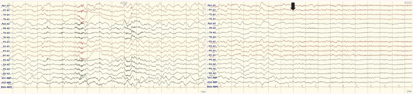 | Figure 1.Initial waking electroencephalography showed semirhythmic delta activity which showed responsiveness (arrow) after 2 mg intravenous lorazepam injection.
|
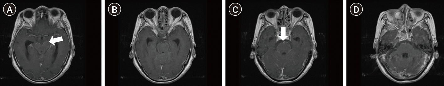 | Figure 2.(A-D) Brain magnetic resonance imaging shows meningeal enhancement around brainstem with focal nodular enhancement (arrows).
|
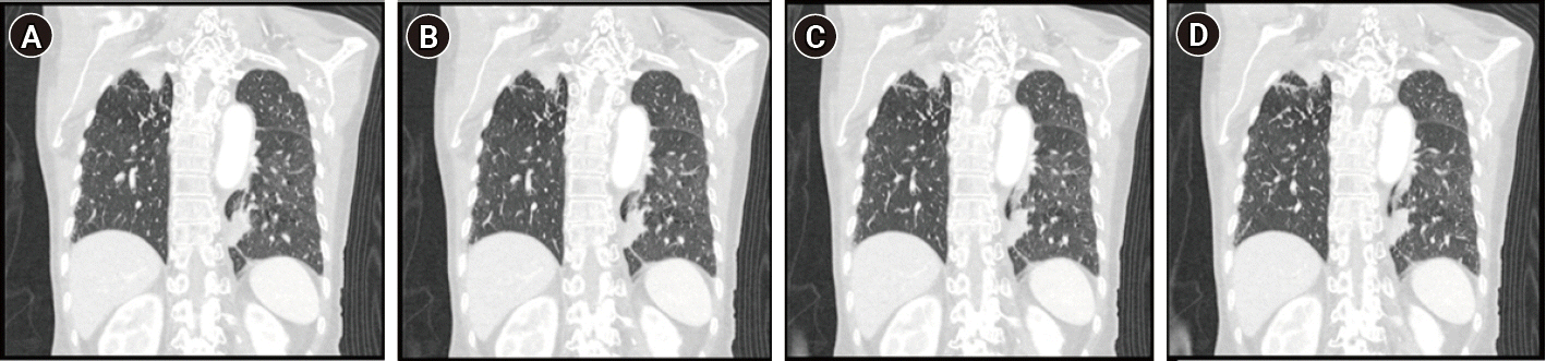 | Figure 3.(A-D) Chest computed tomography showing focal mass like conolidation in left lower lobe posterior basal segment medial aspect.
|
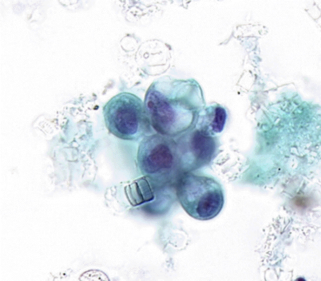 | Figure 4.Cytologic examination of the cerebrospinal fluid shows a few clusters of cells with enlarged eccentric nucleoli, prominent nucleoi and bubbly cytoplasm, suggesting metastatic adenocarcinoma (Papanicolaou stain, ×100).
|
Table 1.
Cerebrospinal fluid analysis findings
![]()
Table 2.
Paraneoplastic syndrome antibodies, autoimmune synaptic encephalitis antibodies, and serum tumor markers
Hu, ANNA-1, antineuronal nuclear antibody-type 1; Yo, PCA-1, purkinje cell cytoplasmic antibody type 1; Ri, ANNA-2, ,antineuronal nuclear antibody-type 2; Ma2, PNMA2, paraneoplastic antigen MA2; CV2/CRMP5, collapsing response-mediator protein-5; SOX1, AGNA, anti-glial nuclear antibody; NMDAR, N-methyl-D-aspartate receptor; AMPA, α-amino-3-hydroxy-5-methyl-4-isoxazolepropionic acid receptor; LGI1, leucine-rich glioma inactivated 1; CASPR2, contactin-associated protein-like 2; GABA-B, gamma-aminobutyric acid; AFP, alphafetoprotein; CEA, carcinoembryonic antigen; CA, carbohydrate antigen.
![]()
This study was approved by the Institutional Review Board of Dong-A University Hospital (IRB No: DAIHIRB-20-089). Informed consent was waived by the board.
Go to :
DISCUSSION
Leptomeningeal metastasis (LM) from cancer was first described in 1870. The identification of malignant cells on CSF cytology has been the diagnostic gold standard, although its sensitivity is limited. An estimated 10% to 30% of patients with solid tumors develop neuraxis metastases, of which 4% to 15% represent LM [1]. Breast tumors, lung tumors, and malignant melanomas are the principal tumors responsible for LM. Adenocarcinoma is the most frequently encountered histological type [2].
The median overall survival of LM patients is 2.4 months (95% confidence interval, 1.9 to 3.1) [3]. The diagnosis and treatment of NCSE usually depend on etiology, EEG findings, and the patient’s clinical status. It is not always possible to identify to what extent the electrographic activity contributes to clinical impairment or ongoing neuronal injury.
The underlying causes of NCSE vary and differ according to the patient population being studied. Approximately one-half to two-thirds of affected patients will have a prior history of seizures or epilepsy. NCSE can be the presenting symptom of infectious or autoimmune encephalitis [4-6]. The NCSE in our case could have been caused by paraneoplastic autoimmune encephalitis associated with SOX1 antibodies rather than LM, which may have been difficult to induce NCSE considering its extent and severity.
SOX1 antibodies are rarely detected but have been reported to occur in paraneoplastic neuropathy, Lambert-Eaton syndrome, and unclear forms of neuropathy and ataxia. SOX1 antibodies are often associated with antibodies against voltage-gated calcium channel, Hu, CV2/CRMP5, or amphiphysin. The most common cancer form associated with SOX1 is small cell lung cancer, but other lung cancer forms have also been reported [7,8]. In our case, no autoantibodies other than SOX1 were detected.
In conclusion, this case showed NCSE due to paraneoplastic autoimmune encephalitis associated with SOX1 antibodies rather than LM, although primary lung cancer was not confirmed pathologically. Although the NCSE was controlled by a continuous midazolam infusion, the patient did not respond to the first intrathecal chemotherapy and expired 2 days later.
Go to :
 XML Download