

 PDF
PDF Citation
 Print
Print


INTRODUCTION
Corticosteroids (CSs) are a class of steroid hormones that are produced and secreted by the adrenal glands in response to pituitary adrenocroticotropic hormone, and regulated by hypothalamic croticotropin releasing hormone. These hormones are responsible for regulating major endocrine system functions, including managing stress and controlling homeostasis. The main CSs produced by the adrenal cortex are cortisol (glucocorticoids) and aldosterone (mineralocorticoids). Aldosterone influences sodium and water balance, while cortisol exerts its effect by preventing the release of inflammation mediators [1]. In the late 1940s, high blood cortisol levels were discovered in cushingoid patients and its anti-inflammatory effects were first demonstrated in patients with rheumatoid arthritis [2]. Since then, numerous investigators have established the ability of physiologic levels of these hormones to alter inflammation and immune functions [3-6]. Currently, CSs find use in the treatment of various neurological diseases, inflammation, pain, autoimmune disorders, and cancer. The purpose of this article is to briefly review the various chemical structural differences between natural and synthetic steroids, discuss their pharmacokinetic and pharmacodynamic profiles, and describe their use in neurocritical care clinical practice.
Go to : 
PHARMACOKINETICS
Cortisol is present within the plasma in three different forms; free cortisol, protein bound, and cortisol metabolite. About 5% of circulating cortisol is unbound and, roughly, 80% of the circulating cortisol is bound to cortisol binding globulin (CBG) or albumin. During inflammation, the binding affinity of cortisol declines, rendering a higher free cortisol concentration at the site of interest to alleviate the active inflammatory process. Most synthetic CSs are also found bound to CBG, however, these glucocorticoids analogues bind less efficiently compared to native cortisol. Cortisol metabolites are biologically inactive and bind weakly to circulating plasma proteins [7-9].
In the early 1950s, chemical modifications of natural steroids revealed a number of structural features essential for their biological activities. Cortisol includes a cyclopen-tenoperhydrophenanthrene nucleus, made up of three 6-carbon rings and a single 5-carbon pentane ring. Cortisol has 21 carbon atoms with a 2-carbon side chain attached to position 17 and methyl groups at C-10 and C-13 [10]. Chemical alterations at various positions of the steroid molecule lead to synthetic analogous of cortisol with increased glucocorticoid and/or mineralocorticoid activities [11,12]. For example, cortisone is derived from cortisol by substituting the hydroxyl group at C-11 with a carbonyl group, whereas prednisolone results from the addition of a double bond between the C-1 and C-2 positions of cortisol; methylation of prednisolone at C-6 produces methylprednisolone (Fig. 1) [10]. Glucocorticoids that are fluorinated at the 9-alpha position include dexamethasone, fludrocortisone and betamethasone [10]. Both cortisone and prednisolone must be converted to the active metabolites cortisol and prednisone, respectively, prior exerting their action [13]. A slight change in the molecular structure introduces a variety of synthetic glucocorticoids with diverse potency, half-lives, and mineralocorticoid activities. Some are far more potent than cortisol, and their different pharmacodynamic and pharmacokinetic activities gave them unique characteristic. Table 1 shows common orally available corticosteroids and compares their potency and mineralocorticoids activity. Notably, fludrocortisone acetate is classified as a mineralocorticoid as it has minimal glucocorticoid activity. It is included in the table to provide perspective on mineralocorticoid potency [1,14-18].
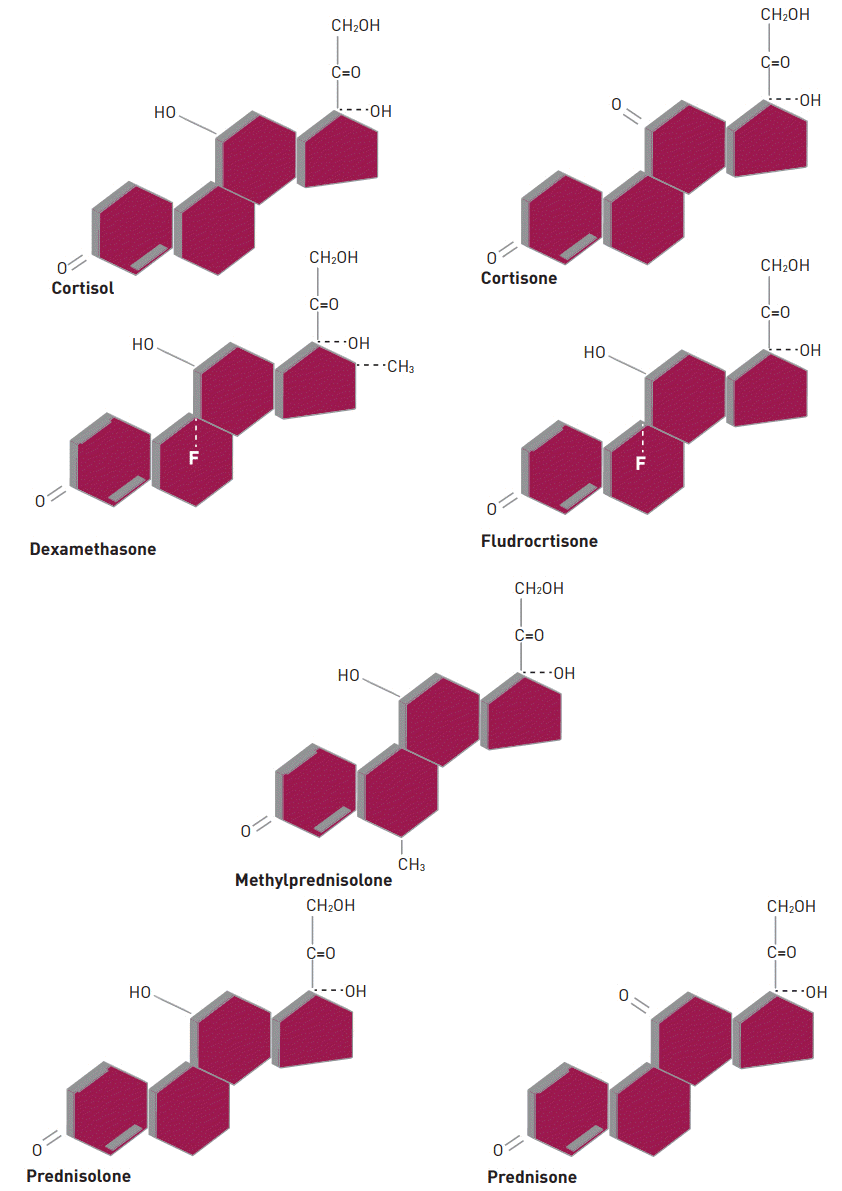
Table 1.
Corticosteroid comparison chart
| Equivalent glucocorticoid dose (mg) |
Potency relative to hydrocortisone |
Half-life duration of action (hours) | ||
|---|---|---|---|---|
| Anti-inflammatory | Mineralocorticoid | |||
| Glucocorticoids | ||||
| Short acting | ||||
| Hydrocortisone | 20 | 1 | 1 | 8-12 |
| Cortisone acetate* | 25 | 0.8 | 0.8 | 8-12 |
| Intermediate acting | ||||
| Prednisone | 5 | 4 | 0.8 | 12-36 |
| Prednisolone | 5 | 4 | 0.8 | 12-36 |
| Methylprednisolone* | 4 | 5 | 0.5 | 12-36 |
| Long acting | ||||
| Dexamethasone* | 0.75 | 30 | 0 | 36-54 |
| Mineralocorticoid | ||||
| Fludrocortisone | 0 | 15 | 150 | 24-36 |
![]()
Go to :
PHARMACODYNAMIC OF CORTICOSTEROIDS
Corticosteroids exert their anti-inflammatory and immunosuppressive effects by interrupting multiple steps in the up-regulation of the immune system. Their suppressive activity is predominantly restricted to cell-mediated immunity [19]. It is believed that CS inhibit antigen presentation, cytokine production and lymphocytes proliferation by binding to glucocorticoid receptors found throughout the body [19]. In response to CS administration, lymphocytopenia is induced as a result of redistribution of circulating lymphocytes into other lymphoid compartments. A single dose of CS produces lymphocytopenia within 4 hours of administration which normalizes through redistribution into the circulation away from periphery, rather than destruction, within 24 to 48 hours. Similarly, monocytes and eosinophils, which normally accumulate at the inflammatory site, decrease upon administration of CSs. In contrast, CSs induce neutrophilia via the release of neutrophils from the bone marrow into the circulation, reduction in the migration of neutrophils out of the circulation, and demargination of neutrophils from the vascular lining [19,20].
The anti-inflammatory process mediated by CSs is multi-modal and begins with synthesis of lipocortin-1, which then suppresses phospholipase A2, thereby blocking eicosanoid production, and further inhibiting various leukocyte inflammatory events. The end result of this process is inhibition of prostaglandin synthesis and cyclooxygenase (COX-1 and COX-2), thus potentiating the anti-inflammatory effect [19,20].
Go to :
USE OF CORTICOSTEROIDS IN CLINICAL PRACTICE
Because of their myriad effects on the immune system, the clinical utility of CSs is vast, but in clinical practice they are generally used for their anti-inflammatory and immunosuppressive effects. Side effects of these drugs are greater with increased duration of use and higher, supraphysiological doses, thus their use should be limited to specific conditions with careful assessment of the risk versus benefit [5,6]. The dose and duration of therapy varies based on the indication. For example, in acute conditions such as multiple sclerosis relapse, a higher dose but shorter course of therapy may be warranted versus chronic conditions such as rheumatoid arthritis where lower maintenance doses are advocated [21,22]. The application of CSs in neurologic diseases, though not limited to neuroimmunologic disorders, range from traumatic head and spinal cord injuries to central nervous system infections. The proposed benefit of CSs therapy in brain and spinal cord conditions, include neuroprotection from free radicals, reduced intracranial pressure via decrease in cerebrospinal fluid pressure, and maintenance of normal microvasculature dependability [22]. Despite these advantages and inspiring results in laboratory studies, clinical trials have been unsuccessful in showing a significant benefit of CSs administration on neurologic outcomes or mortality in patients with head injuries. The largest scale investigation published to date, the Corticosteroid Randomization after Significant Head Injury (CRASH) study, involved over 10,000 patients and was intended to determine the effects of short-term CS infusion on death and disability following significant head injury. The study revealed that the risk of death from all causes, within two weeks of severe head trauma, was higher in the CS group than the placebo group [23]. Thus, the current guidelines from the Brain Trauma Foundation do not indorse the use of CSs for improving outcome or reducing intracranial pressure in patients with severe head injury [24].
Methylprednisolone (MP) sodium succinate had been used in spinal trauma patients with the aim of mitigating inflammation, lipid peroxidation, and excitotoxicity associated with the acute injury [25]. At high doses, it functions as a free radical-scavenger and neuroprotectant, secondary to glucocorticoid receptor-mediated inhibition of phospholipase A2. When given in high doses, CSs impair cytokine generation and enter cell membranes. This alters the physicochemical properties as well as the activities of membrane associated proteins. These effects are deduced from administration of CSs in high doses to effectively treat acute exacerbations [21,26].
Multiple prospective and retrospective studies had attempted to evaluate the benefit of MP in patients who suffered acute spine injury. A study that is well known to many and which may have misled many clinicians in managing spine injury patients, was the National Acute Spinal Cord Injury (NASCI I, II, and III) multicentered double blinded randomized studies, that compared MP to placebo/control in acute spinal cord injury patients [27-29]. In the first study (NASCI 1), 330 patients were followed for 6 months. Each arm received either 100 mg bolus, then 25 mg every 6 hours for 10 days or 1,000 mg bolus, then 250 mg every 6 hours for 10 days. No significant differences in neurologic outcome between the two groups were seen. However, a statistically significant increase in wound infections was reported in the group who received a high dose [27]. Authors argued that the reason for lack of differences in the primary outcome was due to the low dose of MP used, which failed to reveal a potential beneficial effect. In NASCI II trail, 487 patients were included and were followed for 1 year. Subjects were randomized into 3 groups, the MP group received MP as a 30 mg/kg bolus, then 5.4 mg/kg/hour for 23 hours, naloxone and placebo were administered for the remaining two arms of the study. The outcome of this study was criticized due to inappropriate reporting of results and statistical analysis. Overall, the neurologic outcome was a negative result, but a post-hoc analysis reported positive results but failed to show any clinical significance. Wound infection and pulmonary embolus were twice as frequent in the treatment group vs. the naloxone and placebo groups [28]. The third NASCI trial, once again failed to show the benefit of MP in acute spinal cord injury patients. Similar to the NASCIS 2 study, there was no difference between MP and placebo in the outcome of spinal cord injury. The post-hoc analysis identified a statistically significant improvement, but as in the NASCIS 2 results, the clinical significance of this improvement was also questioned. Incidence of severe pneumonia and severe sepsis was higher in those treated longer duration with MP. There was also a six-fold higher mortality reported in those patients receiving longer treatment [29]. Of note, the results from these studies have not impacted the current guidelines that advocate the use of MP in spinal cord injury, nor did US Food and Drug Administration grant MP an indication for acute spinal cord injury.
Mortality and morbidity rates are high among patients suffering from bacterial meningitis, specifically from pneumococcal infections [30]. Adjunctive dexamethasone use in bacterial meningitis has been proposed to be beneficial in reducing mortality rates and hearing loss; but the results from a meta-analysis study do not support the advantage of combining dexamethasone with antimicrobial therapy. Dexamethasone administered at 10 mg IV every 6 hours 15 to 20 minutes before initiating antimicrobial therapy for up to 4 days was not associated with a significant reduction in death, severe neurological sequelae, including bilateral severe deafness or hearing loss, when all patients were included in the analysis. Thus, the benefit of adjunctive dexamethasone for bacterial meningitis remains unproven [31].
Parenteral administration of high CSs doses may be warranted in neurologic emergencies. High dose CSs are standard of care for those presenting with critically ill condition, such as relapse from neurologic disease crises [21,26]. Intravenous MP is the frequently utilized agent to treat major neurologic disease [32,33]. In emergency cases, the usual treatment consists of 500-2,000 mg of MP or dexamethasone 10-200 mg IV daily for 3-5 days [32]. If adequate response is achieved, the steroids can be tapered rapidly or continued for over 1-3 weeks [34]. Some patients may require a longer steroid maintenance based on the severity of the disease presentation or in patients with large tumor size remnants, the rate of steroid tapering depends on dosage, and duration of therapy [34]. Because of the many inherent side effects associated with CSs with longer term use, clinicians should carefully evaluate the risk versus benefit of their use and whether pulse steroid would be a better option [34,35]. Due to the mineralocorticoid activity of MP, this agent may not be the preferred agent in patients presenting with hypertension, congestive heart failure, or those in whom volume overload is a concern. In these cases dexamethasone, lacks mineralocorticoid activity, can serve as an alternative therapy in place of MP as a first line agent [34].
Go to :
CONCLUSIONS
CSs find a myriad of uses in neurological disorders due to their anti-inflammatory and immunosuppressive properties. Although, animal studies have suggested potential benefit of CSs as neuroprotective agents, clinical trials have yet to show convincing beneficial outcomes in this setting. Because of the negative outcomes seen in these large randomized studies, and the vast unwanted side effects associated with CSs, a careful risk benefit analysis should be exercised in the treatment of certain types of neurologic disorders.
Go to :
 XML Download