INTRODUCTION
Bacterial meningitis is a life-threatening critical illness which needs urgent antibacterial treatment. Without therapy, the mortality approaches up to 100 %, and even under the empirical therapy, the mortality and morbidity is still high enough making physicians to have concern about treating meningitis. Rapid deterioration of consciousness is the characteristic sign of fulminant bacterial meningitis which predicts worsening of clinical outcome [1]. Streptococcus pneumoniae is one of the most common pathogen causing bacterial meningitis in adult [2]. In this case report, we would like to represent one case of patient with fulminant bacterial meningitis caused by Streptococcus pneumonia(e) who was successfully treated by aggressive management with adjuvant antibiotics and surgical intracranial pressure (ICP) control.
Go to : 
CASE REPORT
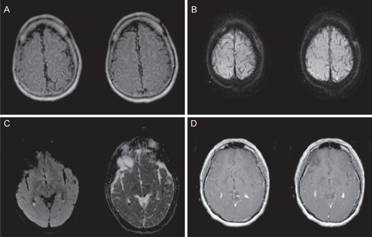
A 35-year-old male patient visited a local emergency room complaining 1 day of headache accompanied with chilling sensation and nausea. The initial body temperature was 40℃. The patient had past history of chronic recurrent otorrhea from his left ear during last 15 years. After few hours of admission, the patient became confused and irritable, and was transferred to our hospital. Upon transfer, his body temperature was 39.0o C despite using anti-pyretic. Laboratory examination of peripheral blood revealed white blood cells (WBC) count of 21,400/mm3, hemoglobin level of 15.0 g/dL, platelet count of 123,000/mm3, and C-reactive protein level of 3.53 mg/dL. Electrocardiogram was grossly normal with heart rate of 126 beats/minute and blood pressure of 140/80 mmHg. Initial non-contrast brain computed tomography (CT) did not show any specific finding. On physical exam, reflexes of all the cranial nerves were intact, but the characteristic neck stiffness was notable. Based on clinical features of decreased mentality accompanied with high fever and stiffen neck, infectious disease in central nerve system (CNS) was suspected and lumbar puncture was performed immediately for cerebrospinal fluid (CSF) study. The opening pressure was over 400 mm H2O and the CSF findings were definitely diagnostic to bacterial meningitis by showing WBC count of 3,777/mm3 (polymorphonucleocyte 96 %), protein level over 500 mg/dL and glucose level of 0 mg/dL. After the diagnosis, empirical IV antibiotics (ceftriaxone 4 g, vancomycin 4 g, ampicillin 12 g per day) were urgently administrated. Mannitol (600 mg per day) and dexamethasone (10 mg four times per day) were also administered. Gram stain of CSF did not present conclusive information for underlying pathogen, but the enzyme-linked immunosorbent assay showed positive result for Streptococcus pneumoniae. Brain magnetic resonance imaging (MRI) performed at admission day showed meningitis with pus collection in bilateral lateral ventricle (Fig. 1A, B) and coalescent otomastoiditis at left ear.
 | Figure 1.Brain magnetic resonance imaging. Brain magnetic resonance imaging performed at admission day showed high signal intensity in the sulcus on fluid-attenuated inversion recovery image (A). There are small amounts of ventricular pus collection on diffusion weighted image and apparent diffusion coefficient image (B). At seventh day of admission, follow-up brain MRI confirmed spread of diffusion high signal intensity spots in bilateral hemispheric sulci on diffusion weighted image (C) and diffuse meningeal enhancement on T1 axial enhancement image (D). MRI, magnetic resonance imaging. |
In spite of urgent medical managements, the patient’s mental status got worsened to semicoma followed by persistent fever at the next day. With impression of fulminant pneumococcal meningitis, we decided to add intrathecal administration of vancomycin (10 mg two times per day) and oral rifampicin (1,200 mg per day) as adjuvant antibiotics. Follow-up CSF study was simultaneously performed with intrathecal administration of vancomycin. The result was that opening pressure > 400 mm H2O, WBC count of 502/mm3 (polymorphonucleocyte 87 %), protein level over 59 mg/dL and glucose level of 105 mg/dL.
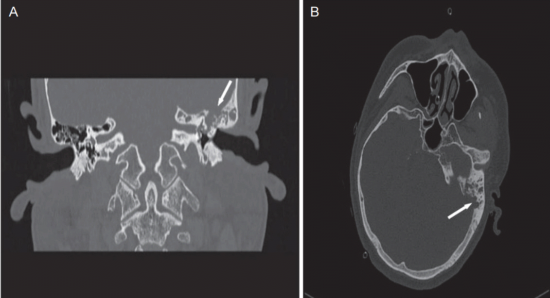
Because increased intracranial pressure (ICP) was sustained over 400 mm H2O, we also performed extra ventricular drainage operation to control and monitor the ICP. Refractory chronic otitis media at his left ear was strongly suspected as an infection source and consultation to otologist was done for further evaluation. Otoendoscopic findings showed mucosal transformation and epithelial ingrowth, with features of cholesteatoma and pocket retraction. As initial management, ventilation tube was inserted and antibiotic ear drop (tarivid) was applied to the left ear. On the third day of hospital admission, the patient gradually began to show improved consciousness. The serum vancomycin level showed 17.9 mg/L and the therapeutic level was maintained throughout the treatment. On the fifth day, patient had alert mentality without fever, nausea or headache but complained transient visual hallucinations. On the seventh day, the patient was transferred out of intensive care unit to general ward with stable clinical finding. A follow-up brain MRI demonstrated diffuse meningeal enhancement (Fig. 1C, D). Meanwhile at the same day, CSF culture incubated at admission date confirmed the presence of Streptococcus pneumoniae. The Minimum Inhibitory Concentration for this strain was as follows: 4 mg/ L for penicillin, 1 mg/L for ceftriaxone and 0.5 mg/L for vancomycin. The patient had good overall condition with clear consciousness, but he intermittently complained of mild headache and fever. Considering the residual symptom derived from left ear, we performed follow-up consultation to an otologist and temporal bone CT (Fig. 2A, B). To remove the underlying infection source, simple mastoidectomy and tympanoplasty using temporalis fascia were done at forty third day of admission. Antibiotic therapy was discontinued after the surgery and there was no recurrent headache or fever. On forty seventh day, patient was discharged from the clinic without neurological sequelae.
 | Figure 2.Temporal bone computed tomography. At forty third day of admission, temporal bone computed tomography was done. In coronal image (A), findings showed that coalescent mastoiditis (arrow) with erosion of tegmen, left. In axial image (B), findings showed that soft tissue opacification with irregular cortical dehiscence in the medial and superior aspect of mastoid. |
Go to : 
DISCUSSION
Bacterial meningitis is a medical emergency having potentials of high morbidity and mortality. Patients who are suspected to have bacterial meningitis should not be delayed more than an hour in treating with empirical therapy [3]. The initial treatment should be based on the underlying pathogen, patient’s age, risk factors and clinical features [4]. According to de Gans et al. concomitant use of dexamethasone has been shown to reduce mortality and improve neurologic outcomes in adults suffering meningitis caused by Streptococcus pneumoniae [5]. In bacterial meningitis, dexamethasone is considered to be effective to attenuation of overt inflammatory responses and reducing several harmful consequences such as cerebral edema, increased intracranial pressure, cerebral vasculitis and neuronal injury [6]. The patient in this case presented catastrophic manifestations despite of urgent medical treatment which includes IV ceftriaxone, vancomycin and dexamethasone. Decreased consciousness is one of the well-known poor prognostic factors of bacterial meningitis [7]. Increased ICP and abnormal CSF findings (high WBC count, high protein and low glucose) are associated with increased mortality after bacterial meningitis [8]. The pathogen Streptococcus pneumoniae itself and male gender are also markers for poor prognosis[7]. Because, the patient presented all of the poor prognostic factors, we tried more aggressive managements at early stage.
We used intrathecal administration of vancomycin for treating bacterial meningitis with fulminant clinical presentation. Intrathecal or intraventricular antibiotic therapy might be ideal delivery route in cases of severe CNS infection for prompt therapeutic effect [9]. A number of literatures report beneficial effects of direct administration of vancomycin into CSF since the route bypasses blood-brain barrier and achieves greater drug concentrations [10]. One randomised prospective trial showed that intraventricular vancomycin therapy is a safe and efficacious treatment modality in external ventricular drain associated ventriculitis [11]. In recent systemic review, the intraventricular vancomycin administration appears to be safe and effective in controlling CNS infection without having serious adverse effects [10]. However, there is no conclusive data from large trials. Guidelines for administration of vancomycin into the CSF are currently not available and the superiority compared to IV vancomycin has not been proven to date [12]. The effect and safety of intrathecal or intraventricular vancomycin for severe CNS infection should be further evaluated. The optimal dosage of vancomycin administrated into CSF has not been established but previous studies usually reported selected dosages of 5-20 mg/day.
We added rifampicin as an adjunctive antibiotic for Streptococcus pneumoniae. The clinical efficacy of rifampicin in patients with bacterial meningitis is not established but its use as an adjuvant to vancomycin and ceftriaxone may be beneficial for suspected pneumococcal meningitis since it is known to have synergistic effects which should result in more bactericidal effects [4]. To control excessive ICP, surgical shunt operation was performed. Recent observational data suggest that ICP-targeted therapy (mainly CSF drainage using external ventricular catheters and osmotherapy) may improve the overall outcome in patients with severe bacterial meningitis especially for those with impaired mental status [13].
Our report on the case of fulminant bacterial meningitis successfully treated with early administration of intrathecal vancomycin differs from previous reports which administered intraventricular antibiotics in the late stage, after failure of conventional management [11,14,15]. As early administration of proper antibiotics are important for successful management of bacterial meningitis, direct delivery of antibiotics for severe patients with poor prognostic factors in the early stage may be beneficial. Also in contrast to the previous reports, we administered multimodal management including IV steroids and oral rifampicin with shunt operation in the early stage.
As this is a single case report, the good outcome observed in the patient cannot be generalized as an outcome of aggressive management method used in the study, which is the main limitation of this study. However, we suggested that additional antimicrobial and surgical managements at early stage might be one of the treatment options in managing fulminant bacterial meningitis.
Go to : 
CONCLUSION
Despite the significant advances in treatment, bacterial meningitis remains to be one of the most important infectious diseases of the CNS with high mortality and morbidity. Multimodal approach including early direct administration of antibiotics into CSF and surgical ICP control may be one treatment option for fulminant bacterial meningitis.
Go to : 


 PDF
PDF Citation
 Print
Print


 XML Download