

 PDF
PDF Citation
 Print
Print


서 론
심방세동은 심장부정맥 중에 가장 흔하며, 뇌경색 및 전신색전증(systemic thromboembolism)과 같은 유해 심혈관계질환의 위험성을 높인다고 알려져 있다. 비판막성 심방세동 환자에서, 다른 혈관 위험인자가 없는 소위 고립성 심방세동일 경우(lone atrial fibrillation) 뇌경색 위험이 높지 않지만, CHA2DS2-VASc 위험인자(심부전, 고혈압, 고령, 당뇨, 뇌졸중/일과성 허혈증후군, 말초혈관질환, 관상동맥병, 대동맥궁 혈전, 여성)가 더해질 경우 정상인에 비해 최소 5배 이상의 높은 뇌경색 위험성을 갖게 된다[1]. 그러나 CHA2DS2-VASc 위험인자들은 뇌경색을 발생시키는 직접적인 원인인자들이 아니라 여러 관찰 연구를 토대로 뇌경색을 예측하기 위해 고안된 연관인자들이다.
어떻게 CHA2DS2-VASc 위험인자들이 더해질수록 뇌경색 위험성이 높아지는지는 혈전형성 기전 측면에서 Virchow’s triad로 설명되고 있다. 심방세동에서의 Virchow’s triad는 심방조직 손상(atrial tissue damage), 응고항진상태(hypercoagulable state), 혈류의 정체(blood stasis)로 구성된다[2,3]. 이 중에서 응고항진상태는 D-이합체, 트롬빈-항트롬빈복합체, 프로트롬빈, 인터루킨, C반응성 단백(C-reactive protein, CRP)의 증가로 나타나는데, 특히 D-이합체는 심방세동에서 유의하게 증가된다고 알려져 있으며[4], 또한 유해 심혈관계질환의 발생과 독립적으로 연관되어 있다고 알려져 있다[5].
심방세동에서 혈전형성의 가장 중요한 기전 중 하나인 혈류의 정체는 좌심방의 수축기능 상실(loss of left atrial systole)과 좌심방 확장(left atrial enlargement) 때문에 기인한다[2,3]. 좌심방 확장 역시 뇌경색 발생의 독립적인 예측인자일 뿐만 아니라[6-10], 심한 뇌경색을 일으켜 뇌경색의 중증도와도 관련 있음이 알려졌다[11].
이 연구의 목적은 다음과 같다. 첫째, CHA2DS2-VASc 위험인자들의 점수와 좌심방 확장의 연관성을 밝힘으로써, 좌심방 확장이 CHA2DS2-VASc 점수 증가와 혈전발생 위험의 중요한 중간 연결고리의 역할을 할 수 있음을 알아보고자 하였다. 또한 만약 연관성이 있다면 CHA2DS2-VASc 위험인자들 중에 어떤 인자들이 좌심방 확장과 가장 연관성이 있는지 알아보고자 하였다. 둘째는, CHA2DS2-VASc 위험인자들의 점수와 D-이합체의 연관성을 평가함으로써, CHA2DS2-VASc 점수의 증가가 혈전형성의 한 기전인 응고항진상태의 심화와 관련 있음을 알고자 하였다.
Go to : 
대상 및 방법
대상
이 연구과제는 서울성모병원의 윤리위원회의 승인을 받아 진행되었다. 2010년 9월부터 2013년 10월까지 비판막성 심방세동에 의해 발생한 급성 뇌경색으로 연속적으로 입원한 환자 중에 항응고제를 투여하지 않은 98명의 환자를 대상으로 하였다.
환자들은 다음의 조건들을 모두 만족하면 포함되었다: (1) 발생 2일 이내의 뇌경색이고, 이는 임상적 증상과 일치하는 뇌병변이 확산자기공명영상에서 확인되어야 함, (2) 이전에 심방세동 과거력이 있거나, 응급실, 뇌졸중 집중 치료실, 홀터모니터검사에서 심방세동이 보이는 경우, (3) 항응고제는 D-이합체 수치를 저하시키기 때문에[12], 최근 3개월 동안 항응고제를 복용하지 않았으며 입원 시 활성화부분트롬보플라스틴시간(activated partial thromboplastin time, aPTT)과 프로트롬빈시간 국제표준화비율(prothrombin time international normalized ratio, PT INR)이 정상 소견을 보이는 환자, (4) 심장초음파에서 판막질환이 없는 경우.
다음의 조건들이 한 가지라도 있는 환자들은 제외되었다: (1) 뇌경색 병변과 관련 있는 뇌혈관의 근위부에 artery-to-artery embolism을 시사하는 국소적인 동맥경화 협착이 있는 경우, (2) 감염성, 염증성, 혈관염성, 탈수초성, 또는 결합조직병(connective tissue disease)을 시사할 만한 진단검사 또는 임상 양상이 있는 경우, (3) 3개월 이내에 뇌졸중 기왕력이 있는 경우, (4) 가역적인 심방세동일 때(갑상선호르몬 이상 또는 전신마취 수술 후 2주 이내).
포함된 환자들의 나이, 성별, 흡연, 고혈압, 당뇨, 심부전, 관상동맥병, 말초혈관 질환 및 이전에 뇌졸중 기왕력의 임상 정보가 조사되었으며, 모든 환자들은 자세한 신체검사 및 신경학적 검사, D-이합체를 포함한 혈액 검사, 흉부 X선, 12리드 심전도, 심전도 지속 모니터, 홀터모니터 검사, 경흉부심장초음파(필요한 환자는 경식도심장초음파), 뇌자기공명영상, 뇌자기공명 혈관조영술(MR Angiography) 또는 전산화단층촬영 혈관조영술(CT angiography)를 시행하였다. 이 연구에서 사용한 각 환자의 CHA2DS2-VASc 점수는 급성 뇌경색 발생 전의 위험인자의 과거력(pre-stroke CHA2DS2-VASc score)으로 분석하였다.
좌심방 용적지수(left atrial volume index)의 평가
심장전문의(HJP)가 초음파를 이용하여 좌심방 용적지수를 측정하였다. 좌심방 용적은 American Society of Echocardiography standard guidelines를 준수하여 biplane area-length method를 이용하여 측정하였으며[13], 좌심방 용적지수는 이렇게 구한 좌심방 용적을 각 환자의 체표면적(body surface area)으로 나누어 얻었다.
자료 처리
통계적 분석은 Statistical Package for the Social Sciences (SPSS) software package version 15.0 (SPSS Inc., Chicago, IL, USA)을 이용하였다. CHA2DS2-VASc 점수와 좌심방 용적지수 및 D-이합체의 연관성은 상관관계분석(correlation coefficient analysis)을 이용하여 평가하였으며, CHA2DS2-VASc 위험인자들 중에 어떤 인자들이 좌심방 용적지수와 가장 독립적으로 연관성이 있는지 알아보고자 다중선형회귀분석(multiple linear regression analysis)을 이용하였다.
Go to :
결 과
전체 환자의 특성
항응고제를 투여하지 않은 비판막성 심방세동에 의한 급성 뇌경색 환자 총 98명이 대상이 되었다. 총 환자 중에 64명(65.3%)은 남자였으며, 평균 나이는 72.2±9.1 (범위, 46-92)세였다. 고혈압(n=63, 64.3%)이 가장 흔한 위험인자였으며, 그 다음으로 담배(n=37, 37.8%), 당뇨(n=25, 25.5%), 고지혈증(n=20, 20.4%), 허혈성 심장 질환(n=19, 19.4%), 허혈성 뇌졸중(n=12, 12.2%), 그리고 말초혈관질환(n=2, 2%) 순이었다.
평균 체표면적은 23.8 ± 3.5 kg/m2 (16–36)였다. 총 45명이 입원 전에 비판막성 심방세동으로 진단 받은 적이 있었으며, 이 중에 25명은 항혈소판제제를 복용하고 있었다. 평균 좌심방 용적지수는 57.1± 25.4 mL/m2 였다. 전체 환자의 특성은 Table 1에 요약되어 있다.
Table 1.
Demographic and clinical characteristics of anticoagulation-naïve patients with acute ischemic stroke due to nonvalvular AF
| Characteristic | Total (N=98) |
|---|---|
| Age (yr.) | 72.2±9.1 |
| Male, No. (%) | 64 (65.3) |
| Initial NIHSS score | 8.4±7.5 |
| Location of stroke in left hemisphere, No. (%) | 48 (49.0) |
| Posterior circulation infarction*, No. (%) | 16 (16.3) |
| Hypertension, No. (%) | 63 (64.3) |
| Diabetes mellitus, No. (%) | 25 (25.5) |
| Smoking, No. (%) | 37 (37.8) |
| Previous stroke, No. (%) | 12 (12.2) |
| Ischemic heart disease, No. (%) | 19 (19.4) |
| Peripheral vascular disease, No. (%) | 2 (2.0) |
| Body mass index | 23.8±3.5 |
| History of taking antiplatelet agents, No. (%) | 50 (51.0) |
| Statin use, No. (%) | 20 (20.4) |
| Diagnosed as AF previously, No. (%) | 45 (45.9%) |
| Pre-stroke CHA2DS2-VASc scores | |
| 0 | 8 (8.2%) |
| 1 | 17 (17.3%) |
| 2 | 12 (12.2%) |
| 3 | 26 (26.5%) |
| 4 | 18 (18.4%) |
| 5 | 9 (9.2%) |
| 6 | 4 (4.1%) |
| 7 | 2 (2%) |
| 8 | 2 (2%) |
| PT INR | 1.1±0.1 |
| D-dimer (µg/mL) – mean (SD) | 2.6±5.0 |
| Left atrium volume index (mL/m2) | 57.1±25.4 |
| Median (Interquartile range) | 52.3 (39.3-67.1) |
| Left ventricular ejection fraction (%) | 58.0±10.6 |
Values are presented as mean ± SD unless otherwise indicated.
Analyses were performed with the independent sample t-test, Fisher’s exact test, or χ2 test.
AF, atrial fibrillation; NIHSS, National Institudes of Health Stroke Scale; PT INR, prothrombin time international normalized ratio; T, total participants; M, male; F, female.
![]()
CHA2DS2-VASc 점수와 좌심방용적지수 및 D-이합체의 연관성
상관 계수 분석을 하였을 때 CHA2DS2-VASc 점수는 유의하게 좌심방 용적지수와 비례적인 관련이 있었다(r=0.325, P=0.002) (Fig. 1). 다중선형회귀분석에서는 CHA2DS2-VASc 위험인자 중에 고혈압(t=2.5, P=0.014), 심부전 (t=2.0, P=0.045), 말초혈관 질환(t=3.9, P<0.001)이 좌심방 용적지수와 독립적인 연관성을 보였다(Table 2). 혈액의 D-이합체 수치 또한 상관 계수 분석에서 CHA2DS2-VASc 점수와 비례적인 연관성이 있었다(r=0.309, P=0.006) (Fig. 2).
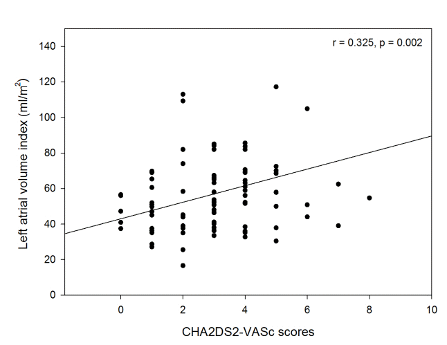 | Figure 1.Scatterplot and the result of correlation coefficient analysis between CHA2DS2-VASc scores and left atrial volume index.
|
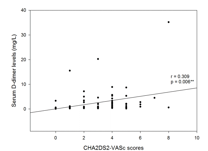 | Figure 2.Scatterplot and the result of correlation coefficient analysis between CHA2DS2-VASc scores and serum D-dimer levels.
|
Table 2.
Associations of left atrial volume index with CHA2DS2-VASc risk factors as determined by multiple linear regression analysis in patients with acute ischemic stroke due to non-valvular AF
| Variables | β ± SE | t value | P-value |
|---|---|---|---|
| Congestive heart failure | 28.615±14.034 | 2.039 | 0.045* |
| Hypertension | 12.482±4.964 | 2.515 | 0.014* |
| Age | -0.442±0.287 | -1.539 | 0.127 |
| Diabetes mellitus | 3.780±5.435 | 0.695 | 0.489 |
| Prior Stroke or TIA or thromboembolism | 12.443±7.514 | 1.656 | 0.102 |
| Ischemic heart disease | -4.091±6.140 | -0.666 | 0.507 |
| Peripheral vascular disease | 66.705±17.310 | 3.853 | 0.000† |
| Female sex | 7.319±5.004 | -1.463 | 0.147 |
![]()
Go to :
고 찰
비판막성 심방세동에 의한 급성 뇌경색 환자를 대상으로 한 이 연구에서, CHA2DS2-VASc 점수는 유의하게 좌심방 확장과 비례적인 연관성을 보였으며, CHA2DS2-VASc 위험인자 중에서도 특히 고혈압, 심부전, 말초혈관질환이 좌심방 확장과 유의한 연관성을 보였다. 또한 CHA2DS2-VASc 점수는 D-이합체의 증가와도 비례적인 연관성을 보였다.
이 연구의 결과는 CHA2DS2-VASc 점수의 증가와 좌심방 직경의 증가가 유의한 연관성을 보인 한 연구 내용과 일치한다[14]. 그러나 그 연구는 좌심방 확장을 평가하기 위해 용적보다는 직경을 측정하였는데, 이는 용적에 비해 부정확하다고 알려져 있으며[15], 그래서 The Guidelines and Standards Committee of the American Society of Echocardiography에서는 직경보다는 용적을 평가하는 것을 추천하고 있다[13]. 그러므로 좌심방 확장의 측정 측면에서 이 연구는 타 연구에 비해 강점을 가지고 있다.
이 연구의 결과에서 보인 CHA2DS2-VASc와 좌심방 확장의 유의한 연관성의 가장 큰 의미는 심방세동에서 CHA2DS2-VASc 증가가 뇌경색 위험성의 증가로 연결되는 중요한 연결고리 역할을 좌심방 확장이 할 수 있음을 나타낸다. 이 연구와 다른 연구의 결과를 종합해보면, CHA2DS2-VASc 위험인자들이 더해질수록 좌심방이 확장되고, 이렇게 커지고 정상적인 수축 기능 또한 상실된 좌심방에서는 난류가 심해지고 정체된 혈류의 용적과 표면적이 증가하면서 혈전 생성을 촉진시키는 것으로 가설을 세울 수 있다[11].
CHA2DS2-VASc 위험인자 중에서도 특히 고혈압, 심부전, 말초혈관질환이 좌심방 확장에 가장 영향력이있었다. 고혈압 및 심부전이 진행하면 좌심방이 비대해지는 것은 어떻게 보면 당연하지만, 거꾸로 생각해 본다면 CHA2DS2-VASc 위험인자들 중에 고혈압, 심부전, 말초혈관질환이 혈전 생성의 중요한 기전 중에 하나인 좌심방 확장에 있어서 가장 중요한 인자임을 시사한다고 볼 수있다.
또한 CHA2DS2-VASc 점수의 증가와 D-이합체의 증가가 유의한 상관 관계를 보였는데, 이는 아마도 좌심방이 비대해지면서 혈전 생성이 잘 만들어지는 환경을 D-이합체가 대변한 것일 수도 있고, 또는 CHA2DS2-VASc 위험인자들이 더해지면서 응고항진상태가 진행하는 것을 독립적으로 나타내는 것일 수도 있다. 이를 확인하기 위해서는 더 많은 연구가 필요하다고 생각된다.
이 연구에서의 CHA2DS2-VASc 점수와 좌심방 용적지수 및 D-이합체의 유의한 상관 관계는, 좌심방 용적지수 및 D-이합체가 CHA2DS2-VASc처럼 향후 뇌경색의 발생을 예측하는데 한 가지 예측인자로 적용할 수 있음을 시사한다. 이미 D-이합체를 CHA2DS2-VASc에 넣어서 뇌경색의 발생을 예측했을 때 더 좋은 결과를 보인 연구 결과가 있었으며[5], 본 연구는 이의 결과를 더 뒷받침한다.
이 연구는 여러 가지 한계점을 가지고 있다. 첫째는, 항응고제 효과를 배제하기 위해 항응고제를 복용하지 않는 환자를 모았는데, 예상보다 많은 환자를 모으지 못한 이유가 되었다. 둘째는 심초음파를 시행한 환자들만 포함시킴으로써, 심초음파조차 시행할 수 없을 정도의 심한 뇌졸중 환자는 배제되어 표본선택편의(selection bias)의 여지가 있다.
결론적으로 항응고제를 투여하지 않았었던 비판막성 심방세동에 의한 급성 뇌경색 환자를 대상으로 한 이 연구에서, CHA2DS2-VASc 점수의 증가는 좌심방 확장 및 D-이합체 증가와 유의한 연관성이 있었고, 좌심방 확장 및 D-이합체는 혈전형성 및 뇌경색의 발생에 있어서 향후 중요한 잠재적인 예측인자로 적용될 수 있을 것으로 생각된다.
Go to :
 XML Download