INTRODUCTION
A Swan-Ganz catheter is a useful monitoring device for the measurement of pulmonary artery pressure, pulmonary capillary wedge pressure, and cardiac output. However, its insertion can bring about many complications (3.0–4.4%), including dysrhythmia, pulmonary artery rupture, myocardial rupture, thrombosis, and infection [1].
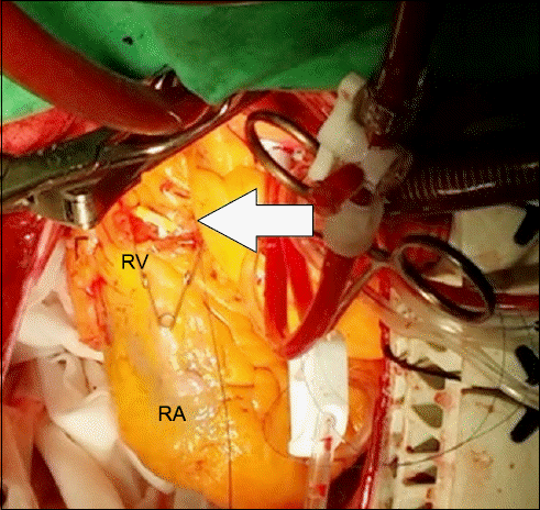
Myocardial perforation is a rare complication of pulmonary artery catheterization [2]. In heart surgery, the surgeon usually detects perforations during the handling of the heart. Therefore, the exact time and cause of the perforation are difficult to determine. We report a case of perforation of the anterior wall of the right ventricle (RV) while the Swan-Ganz catheter was in the indwelling state during a coronary artery bypass graft (CABG) surgery.
Go to : 
CASE REPORT
A 71-year-old man of 157 cm and 50 kg was admitted for persistent stabbing pain in the substernal area, which had appeared 20 days prior. The coronary angiography revealed a 70% stenosis of the left anterior descending artery and 90% stenosis of the posterolateral branch of the right coronary artery. His past medical history was unremarkable, except for an L4-5 discectomy operation for a herniated nucleus propulsus. The ejection fraction was 61%. The electrocardiograph and laboratory findings were not indicative of myocardial infarction. The patient was scheduled for elective CABG sugery for the treatment of a two-vessel coronary artery disease and unstable angina pectoris.
The patient’s vital signs upon arrival in the operating room were as follows: blood pressure (BP) 124/78 mmHg, heart rate (HR) 73 beats/min, oxygen saturation (SpO2) 99%. The anesthesia was induced with propofol (target-controlled infusion: 4.0 ug/ml) and remifentanil (target-controlled infusion: 3.0 ng/ml). Tracheal intubation was performed after the administration of rocuronium (50 mg). An arterial catheter was inserted through the right radial artery for continuous BP monitoring. The patient’s head was rotated leftwards by 20–30 degrees in a head-down position for insertion of the 9-Fr advanced venous access (Edward Lifesciences LCC, USA) via the right internal jugular vein (IJV). A 7.5 Fr Swan-Ganz catheter (Edward Lifesciences LCC, USA) was inserted through the advanced venous access. The Swan-Ganz catheter was advanced under monitoring of the pressure waveforms. No resistance or difficulty was felt during the catheterization. A sudden increase in the diastolic pressure (9–10 mmHg) confirmed that the Swan-Ganz catheter had been placed in the pulmonary artery (PA). The Swan-Ganz catheter was fixed at 42 cm after confirmation of the wedge position with 1 ml of air in the balloon. There were no significant hemodynamic changes during or immediately after the catheterization. No transesophageal echocardiography (TEE) was inserted. When the chest was opened, no bloody effusion, sign of perforation, or tip of the catheter was discovered. Prior to the cardiopulmonary bypass (CPB), the catheter was withdrawn by 5 cm in a deflated state. A cardioplegic solution (packed red blood cells + potassium + sodium bicarbonate + mannitol + 50% DW + normal saline) was used, at an infusion temperature of 6–8°C. Upon initiation of the CPB, ice was placed beside the heart to decrease the temperature, and the patient’s temperature was maintained between 31.4–31.9°C during the CPB. A stabilizer and apical positioning were used with Medtronic: Octopus 4, a tissue stabilizer with Starfish and an Urchin Heart Positioner. First, anastomosis of the left internal mammary artery and the left anterior descending coronary artery was performed. During the manipulation for anastomosis of the greater saphenous vein and the posterolateral branch of the right coronary artery, the tip of the Swan-Ganz catheter was found protruding from the anterior wall of the right ventricle (Fig. 1). The vital signs were stable and the mean arterial blood pressure was maintained between 60-80 mmHg. The catheter was completely withdrawn and the perforation was closed with 4/0 prolene and bioglue. The CABG procedure was completed without further events. The weaning off the CPB was supported with a continuous infusion of dopamine (5 ug/kg/min) and nitroglycerin (0.1 ug/kg/min). No leakage from the perforation was observed during the weaning process. The recovery was uneventful and the patient was discharged 15 days after the surgery, upon confirmation of good patency of the graft vessels through a coronary angiography.
Go to : 
DISCUSSION
The detailed risk factors associated with Swan-Ganz catheter placement include being older than 60 years, being of the female sex, pulmonary hypertension, coagulation disorders and anticoagulation therapy, hypothermia, and manipulation during surgery [3].
The serious complications related to Swan-Ganz catheters can be divided into three categories: complications associated with the insertion and placement, complications associated with indwelling catheters, and complications occurring during or after removal of the catheter [3]. In our case, the complication may have been caused by the indwelling catheter. The perforation by the catheter was discovered during the anastomosis of the vessels. Therefore, even though the catheter was withdrawn by 5 cm during the CPB, the perforation seems to have occurred during the cardiac manipulation, as the heart was displaced during the anastomosis of the greater saphenous vein and the posterolateral branch of the right coronary artery.
Swan-Ganz catheters are widely used in cardiac surgery. Although the average lengths of insertion into various chambers have been cited by several authors, the bases for the measurements are not well known. The measurement from the right IJV approach to the chambers is different between the Western population and the Indian population. The lengths measured from the right IJV approach to the PA and the wedge position are 40–45 cm and 50–60 cm respectively in the Western population [4], while those measurements are 36.0 cm and 42.8 cm respectively in the Indian population. The patient in our case measured 157 cm and weighed 50 kg. His profile was similar to that of the Indian population, which has been shown by previous studies to average 165.9 cm in height and 58.3 kg in weight. The insertion length of the Swan-Ganz in our case was 42 cm, based on the length of the wedge position in the Indian population (42.8 cm). Therefore, the insertion position of the Swan-Ganz catheter was believed to be adequate [5]. To this date, there is no absolute consensus about the withdrawal length of the Swan-Ganz catheter. Sufficient withdrawal of the catheter to the right atrium might have prevented the perforation of the right ventricle. Further research may help us determine the adequate withdrawal length and position of the Swan-Ganz catheter during CPB. Confirming the position of the Swan-Ganz catheter with a TEE after withdrawal may be an appropriate method to prevent catheter-related heart injuries.
Other known predisposing factors for ventricular perforation during catheterization include a small chamber size, a stiff catheter, outflow tract obstruction, and myocardial infarction [4,6,7]. The preoperative echocardiography of our patient showed a normal left ventricle cavity size, wall thickness, and function, with minimal pulmonary regurgitation. The heart was covered in ice and a cold cardioplegic solution was used at the beginning of the CPB, which may have caused the catheter to become stiff. Moreover, excessive manipulation may have caused the ventricular perforation by the stiff catheter. The stiffening of the catheter from hypothermia was reported as a risk factor of pulmonary artery perforation [8]. Therefore, it must be considered in the event of excessive manipulation during CPB. Gentle cardiac manipulation during surgery may be requested for prevention.
In the present case, the perforation of the right ventricle was successfully closed through primary repair, but the damage to the wall could have been bigger, and hemopericardium or dysarrhythmia may have occurred had the repair been unsuccessful. Ample withdrawal of the catheter to the right atrium under TEE guidance may prevent this complication. Prevention, caution, and a high index of suspicion could prevent the rare but potentially fatal complications arising from Swan-Ganz cathterization [9].
Go to : 


 PDF
PDF Citation
 Print
Print


 XML Download