A 76-year-old female patient was transferred to the intensive care unit (ICU) after surgery for periprosthetic fracture of femur neck. She had previous history of hypertension, diabetes mellitus, coronary artery occlusive disease, chronic kidney disease and asthma. And there was no significant adverse event during the previous anesthesia for the surgery of femur neck fracture a week before. Preoperative chest x-ray showed focal bronchiectasis at left lower lung field and there was no definite abnormal lung consolidation or collapse. Anesthesia was induced with propofol 2 mg/kg, sevoflurane, and remifentanil 0.2 μg/kg, and rocuronium 0.6 mg/kg was used to facilitate endotracheal intubation. After induction of anesthesia, vital signs were blood pressure 140/53 mmHg, heart rate 84 beats/min and pulse oximetry 99% with FiO
2 50%. Anesthesia was maintained with 1.0 minimum alveolar concentration of sevoflurane and remifentanil 0.05-2 μg/kg/min. Airway pressure was about 19-22 cmH
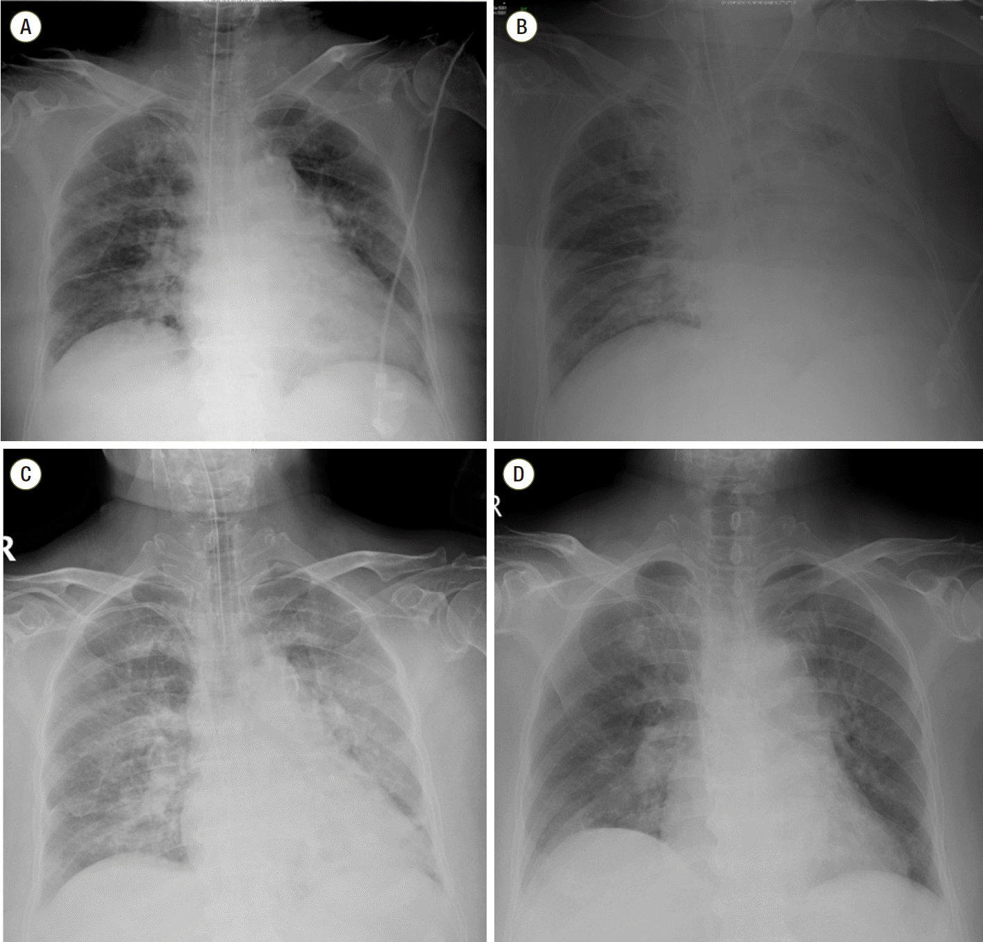
2O during the anesthetic time. After completion of the surgery and before extubation, intravenous bolus of fentanyl 50 mcg was administered, and then airway pressure was abruptly elevated to 36 mmHg and oxygen saturation fell below 95%. There was no wheezing or stridor at this point. Immediate chest x-ray showed left pleural effusion which could not explain desaturation event (
Fig. 1). Elevated airway pressure was maintained for 10 minutes and arterial blood gas analysis performed at that time did not show significant hypoxemia or hypercarbia (pH, 7.380; PaCO
2 38.8 mmHg; PaO
2 173.6 mmHg; and oxygen saturation 99.6%; FiO
2 of 50%). And there was no delayed rise of the end-tidal CO
2 (EtCO
2) on capnograph which can suggest airway obstruction. EtCO
2 was 36-38 mmHg. At this point, it was decided to transfer the patient not to the ward but to the ICU in intubated status and 25 mg of atracurium (Atra
®, Hana, Seoul, Korea) and 3 mg of midazolam were administered. On admission to ICU, FiO
2 of 50%, positive end expiratory pressure (PEEP) of 5 cmH
2O was applied and peak inspiratory pressure was 32 mmHg. And hypoxemia was noticed again by arterial blood gas analysis (ABGA) as PaO
2 55.2 mmHg; oxygen saturation 86.5%. Thus, FiO
2 was increased to 60% and PEEP was increased to 8 cmH
2O. The patient developed hypertension to 200/96 mmHg at this time, perdipine infusion was started. After ICU arrival, intravenous patient-controlled analgesia (PCA) consisting of fentanyl 20 μg/kg and 0.9% saline in a total volume of 100 mL (basal rate 2 mL/hr, bolus dose 0.5 mL, lockout time 15 minutes) was connected to the patient. One hour after ICU admission, oxygen saturation fell to 95%, tidal volume was not checked and airway pressure was elevated to 40 cmH
2O on the ventilator. After adjustment of ventilator setting as FiO
2 70% and PEEP 5 cmH
2O, ABGA showed optimal oxygenation; pH 7.393; pCO
2 35.6; pO
2 321.8; and oxygen saturation 99.9%. Auscultation revealed no evidence of wheezing or stridor at this point. Fiberoptic bronchoscopy (FOB) was performed immediately and there was no endobronchial lesion. And there was generally whitish secretion in the bronchus. There was no eosinophilia on complete blood count with differential white blood cell analysis. Eosinophil percentage was 1% and eosinophil count was 65/μL. Three hours after ICU arrival, tidal volume was not checked again and oxygen saturation fell to 60%. FiO
2 was increased to 100% and manual bag-valve-mask ventilation was initiated. Intravenous PCA was disconnected. At that time, minimal chest wall movement was observed and high airway pressure was needed to deliver tidal volume. Opioid-induced chest wall rigidity was suspected at this time, and vecuronium bromide (Norcuron
®, MSD, Seoul, Korea) 8 mg was injected, immediately. After relaxation was achieved, peak airway pressure was decreased to 19 mmHg and ventilation was feasible smoothly. Thus, cisatracurium (Nimbex
®, GSK, Seoul, Korea) infusion was started. Transthoracic echocardiography was performed for evaluation of heart failure and there were no specific findings. The next day, the patient was extubated successfully after spontaneous breathing trial.
 | Fig. 1.Perioperative chest radiograph. (A) Preoperative chest radiograph showed focal bronchiectasis at left lower lung field and there was no definite abnormal lung consolidation or collapse. (B) Postoperative chest radiograph showed left pleural effusion (C) Three hours after ICU arrival, the amount of pleural effusion was decreased. (D) On POD#2, there was no definite abnormal lung consolidation or collapse. ICU: intensive care uint. 
|


 PDF
PDF ePub
ePub Citation
 Print
Print


 XML Download