Abstract
Acute cerebral infarctions are rare in children, however they can occur as a complication of a Mycoplasma pneumoniae (MP) infection due to direct invasion, vasculitis, or a hypercoagulable state. We report on the case of a 5-year-old boy who had an extensive stroke in multiple cerebrovascular territories 10 days after the diagnosis of MP infection. Based on the suspicion that the cerebral infarction was associated with a macrolide-resistant MP infection, the patient was treated with levofloxacin, methyl-prednisolone, intravenous immunoglobulin, and enoxaparin. Despite this medical management, cerebral vascular narrowing progressed and a decompressive craniectomy became necessary for the patient’s survival. According to laboratory tests, brain magnetic resonance imaging, and clinical manifestations, the cerebral infarction in this case appeared to be due to the combined effects of hypercoagulability and cytokine-induced vascular inflammation.
Mycoplasma pneumoniae (MP) is a common respiratory pathogen in children. MP infections usually have a mild clinical course, and pneumonia is the most prominent clinical manifestation. Several types of neurologic disorders have been reported to be extrapulmonary manifestations in 0.1% of mycoplasma infections, including meningo-encephalitis, transverse myelitis, seizures, acute disseminated encephalomyelopathy, Guillain-Barre syndrome, cerebral ataxia, and stroke [1]. To date, only 12 cases of MP-associated strokes in children have been reported in the literature [2-12]. In most of these cases, the patients partially or fully recovered after treatment with antibiotics, intravenous immunoglobulin, steroids, or aspirin. In two of the cases, the children died despite medical treatment.
Herein, we report the case of an extensive and progressive acute cerebral infarction that occurred after MP infection; the patient not only received medical managements but also underwent a decompressive craniectomy.
A previously healthy 5-year-old boy presented with a productive cough and a fever for 3 days; he was found to have pneumonia and admitted to the local hospital. On the basis of the positive result of the mycoplasma-specific IgM antibody, he was treated with intravenous clarithromycin for 3 days, starting on the day of admission. His symptoms and chest radiographic findings progressively worsened despite an additional 5 days of antibiotic treatment with cefotaxime and vancomycin. During the 4 days before his transfer to our hospital, he was treated with methyl-prednisolone (2 mg/kg/d), and his fever disappeared and his chest radiography findings improved slightly. On the day of his transfer, he became drowsy, and exhibited dysarthria, limited eye movement, and irritability. He experienced a generalized tonic seizure that lasted for 3 hours despite receiving treatment with anticonvulsants including intravenous diazepam, fosphenytoin, and phenobarbital. Because of newly developed fever and neurologic symptoms, a cerebrospinal fluid (CSF) analysis and brain magnetic resonance imaging (MRI) were conducted. An acute cerebral infarction was suspected, and the patient was transferred to our hospital while receiving treatment for the management of increased intracranial pressure (IICP), including dexamethasone, mannitol, and continuous midazolam infusion. His medical and family histories were unremarkable.
When the patient arrived at our hospital, he had a temperature of 37.8℃, respiratory rate of 27 breaths/min, pulse rate of 114 beats/min, and blood pressure of 108/64 mmHg. He was receiving 4 L/min of oxygen through a nasal cannula and his oxygen saturation was 97%. His breath sounds were coarse but he had no signs of respiratory distress. He was stuporous; however, his pupils were reactive to light and isocoric. His deep tendon reflexes were normal, and he had no neck rigidity. His motor and sensory functions were not evaluated because he was sedated.
The laboratory examinations revealed a hemoglobin level of 10.6 g/dl, leukocyte counts of 1,260/mm3 with 82.3% neutrophils and 11.9% lymphocytes, and platelet count of 115,000/mm3. His liver function tests revealed a slightly decreased albumin level of 3.2 g/dl, an elevated aspartate aminotransferase level of 53 IU/L, and an alanine aminotransferase level of 94 IU/L. His serum biochemistry results were all within the normal limits except for an increased serum lactate dehydrogenase level of 747 IU/L (reference range, 100 to 225 IU/L). The coagulation studies revealed a normal prothrombin time and activated partial thromboplastin time, mildly decreased fibrinogen level of 129 mg/dl (reference range, 180 to 380 mg/dl), and elevated D-dimer level of 6.84 μg/ml (reference, <0.4 μg/ml). The arterial blood gas results were as follows: pH, 7.43; PaO2, 90 mmHg; and PaCO2, 37 mmHg. The C-reactive protein level was 0.79 mg/dl (reference, <0.5 mg/dl), erythrocyte sedimentation rate 2 mm/h (reference < 9 mm/h), and procalcitonin level 0.124 ng/ml (reference < 0.5 ng/ml). The additional coagulation studies revealed that the patient’s protein C and antithrombin III levels were nearly normal; however, his protein S activity was decreased to 59% (reference range, 73% to 150%). Test results for anticardiolipin, antiglycoprotein, antinuclear, anti-DNA, anti-neutrophil cytoplasmic antibodies, as well as rheumatic factor were negative. His serum homocysteine concentration was 3.4 μM (reference range, 5 to 15 μM). Polymerase chain reaction (PCR) analysis of the methylenetetrahydrofolate reductase (MTHFR) gene showed a heterozygous MTHFR A1298C mutation. Serum Mycoplasma antibody levels were elevated to 1:20,480 on indirect particle agglutination, and PCR of the respiratory specimen was positive for Mycoplasma.
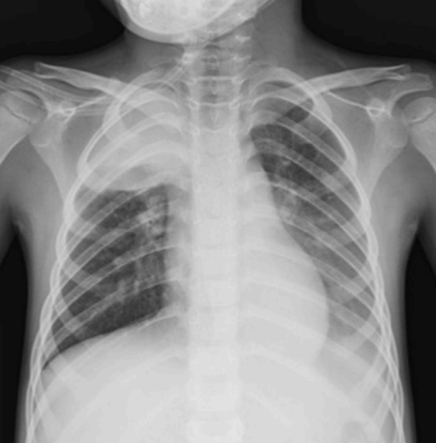
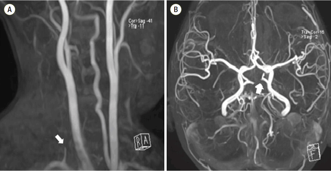
The patient’s chest radiogram demonstrated consolidation and atelectasis in the right upper and left lower lungs (Figure 1). The electrocardiography and echocardiography findings were normal. MRI performed at the previous hospital was suspicious for bilateral acute ischemic stroke in posterior circulation (Figure 2). The right vertebral and basilar arteries were not visualized on magnetic resonance angiography (MRA) (Figure 3).
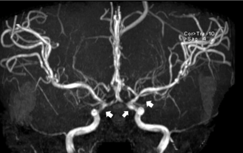
On the day of admission, mannitol and dexamethasone were continuously infused for IICP management. The antibiotic regimen was changed to vancomycin, cefotaxime, and levofloxacin and acyclovir was added. On the second hospital day after transfer to our facility, the patient’s seizures worsened despite increased doses of midazolam. He suddenly became apneic and intubation and mechanical ventilator support were required. Anti-inflammatory therapy included methylprednisolone at 30 mg/kg for 3 days and intravenous immunoglobulin (IVIG) at 0.5 g/kg for 4 days. Because of the deterioration in his neurologic condition, he underwent repeat brain MRI and MRA, which revealed further multifocal narrowing in the bilateral distal internal carotid arteries and progressive swelling of the right cerebellum with brain stem compression. To avert impending herniation, the patient underwent craniotomy, bilateral suboccipital craniectomy, and insertion of external ventricular drainage for IICP control. Analysis of the CSF showed a leukocyte count of 0/μL, glucose level of 105 mg/dl, and protein level of 13 mg/dl. The PCR of the CSF was negative for Mycoplasma. On the 5th day after the transfer to our hospital, the patient’s neurologic status was stable; however, the follow-up brain MRI and MRA revealed a slight interval progression of the T2 high signal intensity in the right pons and a multifocal narrowing in the proximal middle cerebral artery (Figure 4). As a result, enoxaparin was added to control the progression of the brain lesion. Seven days after admission, the consolidation was shown to be resolved on a follow-up chest radiography. His neurologic status improved, and he started breathing spontaneously, including spontaneous eye blinking and withdrawal of painful stimuli. He received bedside physiotherapy for rehabilitation. On the 13th day of admission, a tracheostomy was performed because his cough reflex remained weak. On the 25th hospital day, enoxaparin was changed to warfarin, and the patient was transferred to the general ward. Despite active rehabilitation, he remained bed-ridden when he was discharged to home 2 months later.
We report the case of a child with extensive and progressive acute cerebral artery occlusions after an MP infection, requiring decrompressive craniectomy. MP is an important pathogen of respiratory disease and has been reported in 10-40% of cases of community-acquired pneumonia in children [13]. Since the 2000s, a high prevalence of macrolide-resistant MP (MRMP) strains has been detected: as high as 90% in Asia, Europe, and the United States [13,14]. Tetracyclines and fluoroquinolones can be used as alternative to macrolides in the treatment of MRMP; however, their use in children is limited because of their adverse effects on bone and cartilage development [15]. In our case report, on the basis of the suspicion that the patient had an MRMP infection, clarithromycin was changed to levofloxacin; no adverse effects of the drug were observed.
The pathogenesis of refractory or severe MP infections is considered to be closely associated with an excessive immune response, such as a highly activated cellmediated immune response and the expression of proinflammatory cytokines such as interleukin (IL)-1, IL-2, IL-4, IL-6, interferon-gamma (IFN-γ) and tumor necrosis factor-alpha (TNF-α) [16]. Furthermore, several authors, including Miyashita et al. [17], determined that immunosuppressive therapy has a profound beneficial effect by reducing the immune-mediated pulmonary injury seen in MP infection. After the 5-year-old patient was transferred to our hospital, the antibiotics regimen was changed and he received high-dose methyl-prednisolone. The combination of the appropriate antibiotics and the immunemodulating therapies may have improved the pneumonia. As MRMP cases have been increasing rapidly in recent years worldwide, especially in younger children, appropriate treatment with antibiotics and immune-modulating agents should be considered, and further investigation are needed to assess for the adverse effects of drugs.
Various extrapulmonary manifestations have been reported in association with MP infection. Neurological impairments are the most common manifestations, including meningoencephalitis, acute disseminated encephalomyelopathy, transverse myelitis, seizures, peripheral nerve involvement, and stroke [1]. To date, a total of 12 children with MP-associated strokes have been reported in the literature [2-12]. Ten of the 12 cases involved an arterial occlusion in the anterior circulation of brain, and one case involved a posterior circulation stroke [10]. Occlusions of both the anterior and posterior circulation after an MP infection, like in our case, were reported in one 4-year-old patient, which resulted in death [5]. However, there are no previously reported cases of a rapidly progressing cerebral infarction, regardless of the specific brain regions, after an MP infection. Unlike the previously reported severe cases, we found that the stroke associated with the MP infection proceeded rapidly because of vascular narrowing and brain swelling; these findings were confirmed on consecutive MRI and MRA imaging of the brain.
The mechanisms of stroke associated with MP infections are well understood. Direct neurological invasion, hypercoagulable or thrombotic states, and vasculitis have all been suggested as possible etiologies for these strokes [1,5]. Assessing MP-specific antibodies and performing PCR of the CSF are useful for detecting direct central nervous system invasion. In our case, the PCR analyses of the CSF and brain tissue were negative for MP, and antibodies were not examined. A total of 6 of the 12 reported cases of stroke associated with MP infection had hypercoagulable and thrombotic conditions confirmed with laboratory tests [3,5-7,10,11]. Among these cases, one patient had a genetic defect in the MTHFR gene with hyperhomocysteinemia, which caused thrombosis, and one child had a sickle cell trait. Our patient was hypercoagulable with decreased protein S activity and an elevated D-dimer level. Although the heterozygous A1298C mutation of the MTHFR gene was discovered in our case, the causality between MTHFR gene polymorphism and cerebral infarction is weak. Compared with those in previous studies [18], our patient showed low levels of homocysteine and only one heterozygosity of C677T and A1298C mutations was found. The last possible mechanism is vasculitis, which results from direct invasion of the vascular wall with an autoimmune process or vascular inflammation by pro-inflammatory cytokines such as IL-6, IFN-γ, and TNF [19]. Brain biopsy is considered the gold standard for the diagnosis of central nervous system vasculitis; however, it is not always feasible. Our patient did not show elevation of acute-phase reactants, and compatible pathologic findings. However, we suspected that the anterior circulation infarction was due to vasculitis because multiple and bilateral stenotic vascular lesions were found on MRA. Furthermore, as both the neurologic condition and pneumonia were simultaneously deteriorated and improved, a systemic proinflammatory process was suggested to have affected the clinical course including progression of the cerebral infarction. Finally, in our case, the cerebral infarction appeared to be the result of the combined effects of hypercoagulability and vascular inflammatory injury.
The management of MP-associated stroke remains controversial. In the previously reported cases, the patients received antibiotics (macrolides in most cases), anti-inflammatory medications such as steroids and IVIG, or aspirin [2-12]. To date, there has been no consensus about the optimal treatment strategy. Among the 12 prior stroke cases, the patient with extensive cerebral involvement received antibiotics and low-dose aspirin based on the results of the hypercoagulability laboratory tests [5]. However, the patient did not receive anti-inflammatory medications, and ultimately died. Our patient received a combination of antibiotics, anticoagulants, and anti-inflammatory medications including methyl-prednisolone and IVIG. IVIG has not been used extensively in cerebral vasculitis; however, IVIG was infused for its anti-inflammatory effect through the modulation of cytokine antagonists and cytokine production, as previously reported [20]. We cannot specify which treatment affected the progression of the disease and improved his clinical condition, nor can we exclude the possibility that the cause of the stroke improved naturally. However, as several potential mechanisms could cause a stroke associated with MP infection, treatment with anti-inflammatory agents and antibiotics should be considered early, and should be based on the results of imaging and laboratory tests.
To summarize, we report a case of an extensive and rapidly progressive cerebral infarction after MP infection that possibly resulted from acquired hypercoagulability and cytokine-induced vascular inflammation. Clinicians should be cautious of the neurologic signs or symptoms when MP infections are especially severe and refractory, and multidisciplinary management may be necessary to improve the pulmonary and neurologic outcomes of MP infection that could otherwise proceed to death.
References
1. Tsiodras S, Kelesidis I, Kelesidis T, Stamboulis E, Giamarellou H. Central nervous system manifestations of Mycoplasma pneumoniae infections. J Infect. 2005; 51:343–54.

2. Ovetchkine P, Brugieres P, Seradj A, Reinert P, Cohen R. An 8-old boy with acute stroke and radiological signs of cerebral vasculitis after recent Mycoplasma pneumoniae infection. Scand J Infect Dis. 2002; 34:307–9.
3. Ryu JS, Kim HJ, Sung IY, Ko TS. Posterior cerebral artery occlusion after Mycoplasma pneumoniae infection associated with genetic defect of MTHFR C677T. J Child Neurol. 2009; 24:891–4.
4. Leonardi S, Pavone P, Rotolo N, La Rosa M. Stroke in two children with Mycoplasma pneumoniae infection. A causal or casual relationship? Pediatr Infect Dis J. 2005; 24:843–5.
5. Lee CY, Huang YY, Huang FL, Liu FC, Chen PY. Mycoplasma pneumoniae-associated cerebral infarction in a child. J Trop Pediatr. 2009; 55:272–5.

6. Fu M, Wong KS, Lam WW, Wong GW. Middle cerebral artery occlusion after recent Mycoplasma pneumoniae infection. J Neurol Sci. 1998; 157:113–5.

7. Tanir G, Aydemir C, Yilmaz D, Tuygun N. Internal carotid artery occlusion associated with Mycoplasma pneumoniae infection in a child. Turk J Pediatr. 2006; 48:166–71.
8. Parker P, Puck J, Fernandez F. Cerebral infarction associated with Mycoplasma pneumoniae. Pediatrics. 1981; 67:373–5.

9. Visudhiphan P, Chiemchanya S, Sirinavin S. Internal carotid artery occlusion associated with Mycoplasma pneumoniae infection. Pediatr Neurol. 1992; 8:237–9.

10. Antachopoulos C, Liakopoulou T, Palamidou F, Papathanassiou D, Youroukos S. Posterior cerebral artery occlusion associated with Mycoplasma pneumoniae infection. J Child Neurol. 2002; 17:55–7.

11. Kim GH, Seo WH, Je BK, Eun SH. Mycoplasma pneumoniae associated stroke in a 3-year-old girl. Korean J Pediatr. 2013; 56:411–5.
12. Papaevangelou V, Falaina V, Syriopoulou V, Theodordou M. Bell’s palsy associated with Mycoplasma pneumoniae infection. Pediatr Infect Dis J. 1999; 18:1024–26.

13. Kim EK, Youn YS, Rhim JW, Shin MS, Kang JH, Lee KY. Epidemiological comparison of three Mycoplasma pneumoniae pneumonia epidemics in a single hospital over 10 years. Korean J pediatr. 2015; 58:172–7.
14. Morozumi M, Iwata S, Hasegawa K, Chiba N, Takayanagi R, Matsubara K, et al. Increased macrolide resistance of Mycoplasma pneumoniae in pediatric patients with community-acquired pneumonia. Antimicrob Agents Chemother. 2008; 52:348–50.
15. Suzuki S, Yamazaki T, Narita M, Okazaki N, Suzuki I, Andoh T, et al. Clinical evaluation of macrolide-resistant Mycoplasma pneumoniae. Antimicrob Agents Chemother. 2006; 50:709–12.
16. Yang J, Hooper WC, Phillips DJ, Talkington DF. Cytokines in Mycoplasma pneumonia infection. Cytokine Growth Factor Rev. 2004; 15:157–68.
17. Miyashita N, Akaike H, Teranishi H, Ouchi K, Okimoto N. Macrolide-resistant Mycoplasma pneumoniae pneumonia in adolescents and adults: clinical findings, drug susceptibility, and therapeutic efficacy. Antimicrob Agents Chemother. 2013; 57:5181–5.

18. Macovei L, Gurzu A, Alexandrescu DM, Georgescu CA. Simultaneous tromboembolic events in a patient with heterozygous MTHFR mutation. Int Arch Med. 2015; 8.

19. Alba MA, Espígol-Frigolé G, Prieto-González S, Tavera-Bahillo I, García-Martínez A, Butjosa M, et al. Central nervous system vasculitis: still more questions than answers. Curr Neuropharmacol. 2011; 9:437–48.
20. Lukán N. Intravenous immunoglobulin therapy in
primary vasculitides. In : Amezcua-Guerra LM, editor. Advances in the diagnosis and treatment of vasculitis. 4th ed. Rijeka: InTech;2011. p. 149–66.
Figure 1.
Chest radiogram on the day of admission shows consolidation and atelectasis in the right upper and left lower lung.

Figure 2.
Magnetic resonance imaging (MRI) performed at the previous hospital on the day of admission. Axial section of diffusion-weighted MRI showing an area of restricted diffusion in the left thalamus (A), pons, and bilateral cerebellum (arrow) (B). Axial section of the apparent diffusion map showing a corresponding low apparent diffusion coefficient value in the left thalamus (arrow) (C), pons, and bilateral cerebellum (arrow) (D).


 PDF
PDF Citation
 Print
Print


 XML Download