

 PDF
PDF Citation
 Print
Print


Portal hypertension is a common complication of liver cirrhosis and porto-systemic collaterals significantly develops in advanced liver cirrhosis. Gastrointestinal bleeding from these collaterals is the most serious complication in liver cirrhosis. Although a common site of bleeding is esophageal and gastric varices, a significant bleeding from rectal varices has also been reported.[1,2] Various therapeutic modalities such as endoscopic variceal ligation (EVL), endoscopic injection sclerotherapy (EIS), transjugular intrahepatic portosystemic shunting (TIPS), percutaneous transvenous obliteration (PTO) and surgery have been suggested in patients with significant bleeding from rectal varices.[3] However, these definite hemostasis modalities are not readily available and require the experienced intervention radiologist or endoscopist. We report a case with massive rectal variceal bleeding in which Sengstaken-Blakemore (SB) tube was effective at ceasing the bleeding until the definite hemostasis was performed.
Case Report
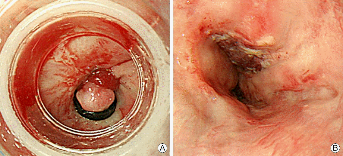
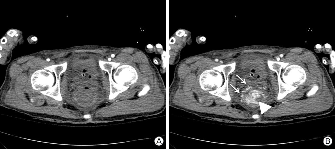
A 48-year-old man awaiting liver transplantation presented to the emergency department (ED) with hematochezia. He had liver cirrhosis associated with chronic hepatitis B infection and hepatocellular carcinoma treated with five times of transarterial chemoembolization. He had also received nine times of EVL for esophageal varices. Initial blood pressure was 87/51 mmHg and heart rate 67 per minute at ED. Initial hemoglobin level was 7.1 g/dl (8.9 g/dl at one week ago). Esophagogastroduodenoscopy (EGD) revealed esophageal varices with red color sign and EVL was performed for five esophageal varices (Fig. 1A). After EVL, patient’s blood pressure was stabilized and patient was admitted to general ward. Next morning, patient’s blood pressure decreased to 59/33 mmHg with massive hematochezia (about 650 ml) and patient was transferred to intensive care unit. After 4 packs of red blood cells (RBC) and 2 packs of fresh frozen plasma (FFP) were transfused, patient’s blood pressure was normalized. Follow-up EGD showed no evidence of active bleeding (Fig. 1B). After EGD, massive hematochezia persisted and massive transfusion (7 packs of RBC) was required to maintain blood pressure. The patient underwent three phase CT angiography, which demonstrated no contrast extravasation in arterial phase. However, contrast filling in rectal varices and extravasation to the rectum were observed in the portal phase (Fig. 2). Rectal varices were located within 10 cm from the anal verge.
Because it was holiday, intervention radiologist was called for the emergency PTO and several hours were required for the procedure. During 2 hours, about 3,500 ml of hematochezia was present and patient’s blood pressure decreased to 64/44 mmHg even after the transfusion of 12 packs of RBC and 9 packs of FFP. Therefore, we decided to apply SB tube to control bleeding mechanically. Because the length of the anal canal is about 4 cm and the proximal end of the gastric balloon of SB tube is about 13 cm from the tip, we inserted SB tube per anus and advanced more than 30 cm. Then, gastric balloon was inflated using 200 ml of air and the SB tube was retracted by applying 500 g of saline bag. Shortly after the application of SB tube, systolic blood pressure rose above 100 mmHg. Although additional 2 hours were required to perform the emergency PTO, 2 packs of RBC and FFP were enough to avoid hypotension. To describe PTO briefly, portal vein was accessed transhepatically guided by ultrasound and pigtail catheter was located at main portal vein. Rectal variceal angiography was obtained using Davis catheter advanced through inferior mesenteric vein. Rectal varix could not be fully visualized during the inflation of SB tube, which represented that SB tube could provide the successful hemostasis (Fig. 3A). After deflation of SB tube, contrast materials filled rectal varices and the extravasation to rectum could be observed (Fig. 3B and 3C). Rectal varices were obliterated by using glue-lipiodol mixture. After no further extravasation was confirmed, catheters were removed and transhepatic puncture tract was sealed by glue-lipiodol mixture and standard vascular coils. Patient showed no evidence of further bleeding and was discharged home after subsequent ward care. After 3 months, living donor liver transplantation was performed and the patient is alive for over 8 months.
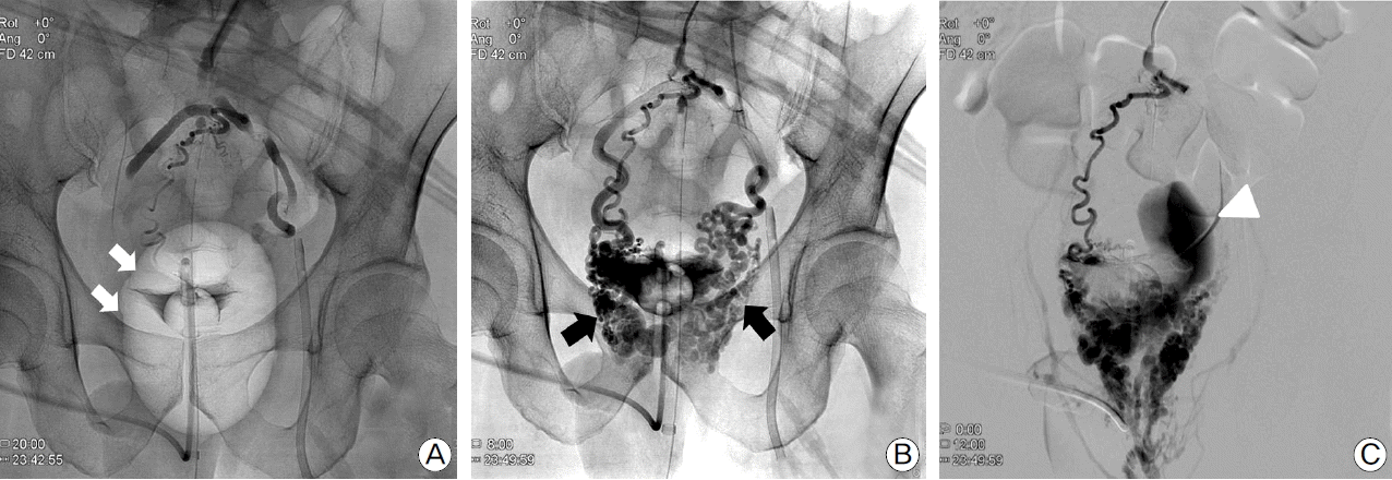 | Fig. 3.Venography demonstrates no contrast filling in rectal varices during the inflation of Sengstaken-Blakemore (SB) tube (white arrows), which represents that the effective hemostasis could be achieved (A). After deflation of SB tube, rectal varices (black arrows) are well visualized (B) and active extravasation of contrast materials (triangle) are observed (C).
|
Go to : 
Discussion
Variceal bleeding is common and most serious complications of liver cirrhosis. Esophageal varices are known to be detected in about 50% of cirrhosis patients and tend to bleed in about a third of patients.[4] Therefore, esophageal varix is the most common site of bleeding.
Ectopic varices including rectal varices accounts for only 2% to 5% of a gastrointestinal tract variceal bleeding.[5] However, variceal bleeding from ectopic varices can have a mortality rate as high as 40% because the therapeutic approach is more complicated than that in esophageal variceal bleeding.[5]
Initial medical management includes the administration of systemic vasopressin and octreotide in addition to the hemodynamic stabilization. For the hemostasis, portal decompression such as transjugular intrahepatic portosystemic shunt (TIPS) and endoscopic approach such as direct injection therapy and band ligation has been used.[6,7] Also, direct obliteration of varices by approaching through systemic or portal vein is widely used.[8,9]
As mentioned above, bleeding from rectal varices may become fatal because rectal blood flow is abundant and therapeutic interventions require experienced endoscopist or interventional radiologist.[10] Therefore, bridge hemostatic method which can be applied bedside is essential.
SB tube is a device originally invented for the management of esophageal variceal bleeding.[11] Although the modern endoscopic treatment has decreased the utility of SB tube, it is still preferred in emergencies when the endoscopist is not available or it is impossible to control bleeding.
There are some reasons why we have decided to apply SB tube in this case. First is that bleeding site could be clearly demonstrated by CT angiography. Second is that rectal varices were mainly located in the distal part of the rectum. The third is that venous bleeding has relatively lower pressure than that of arterial bleeding, which enables the balloon compression to be an effective hemostatic method. The last one is that only the gastric balloon of SB tube is expandable enough to fill up rectum. Generally, the inflation volume of gastric balloon of SB tube is 300 ml and the inflation pressure of esophageal balloon of SB tube is 30 mmHg. However, there is no recommended volume or pressure for the rectal ballooning. We have used 200 ml of air for the gastric balloon in this case and did not measure the pressure of the balloon. To measure the pressure of balloon for the guidance of proper volume would be better since over-expansion might have caused the rupture of rectum and under-expansion would be ineffective for the hemostasis.
In this case report, massive rectal bleeding could be successfully controlled using rectal application of SB tube. In addition, the effectiveness of SB tube to control rectal variceal bleeding could be objectively seen in variceal angiography. Therefore, we propose that SB tube can be considered in patients with massive rectal variceal bleeding and may give successful bridging hemostasis until the definitive hemostasis can be achieved.
Go to :
 XML Download