Journal List > Korean J Crit Care Med > v.29(3) > 1153863

Nam, Park, Lee, Lee, Lee, Park, Yoon, Lee, Lee, and Cho: Percutaneous Dilatational Tracheostomy in Critically Ill Patients Taking Antiplatelet Agents

Abstract

Background

Percutaneous dilatational tracheostomy (PDT) has been considered as an alternative to surgical tracheostomy in intensive care units (ICU), and is widely used for critically ill patients who need prolonged mechanical ventilation. Few studies have reported on PDT performed in critically ill patients taking antiplatelet agents. Our goals are to assess not only the feasibility and safety of PDT, but also bleeding complications in the patients receiving such therapy.

Methods

In a single institution, PDTs were performed by pulmonologists at the medical ICU bedside using the single tapered dilator technique and assisted by flexible bronchoscopy to confirm a secure puncture site. From March 2011 to February 2013, the patients’ demographic and clinical data, procedural parameters, outcomes and complications were analyzed and compared complications between patients taking antiplatelet agents and those not.

Results

PDTs were performed for 138 patients; the median age was 72 years, mean body mass index was 20.3 ± 4.8 kg/m2, and mean acute physiology and chronic health evaluation II score was 24.4 ± 9.4. Overall, the procedural success rate was 100% and the total procedural time was 25 ± 8.5 min. There were no periprocedural life-threatening complications, and no statistical difference in the incidence of bleeding complications between patients who had taken antiplatelet agents and those had not (p = 0.657).

Conclusions

PDT performed in critically ill patients taking antiplatelet agents was a feasible procedure and was implemented without additional bleeding complications.

Introduction

Percutaneous tracheostomy was first performed in 1955[1] after Seldinger’s earlier description of a needle placement over a guide wire for arterial cannulation.[2] After Ciaglia’s initial report of the dilatational technique in 1985,[3] percutaneous dilatational tracheostomy (PDT) has become a commonly performed procedure in intensive care units (ICUs). It has largely replaced conventional surgical tracheostomy in ICU patients, and provides benefits in terms of ease of performance, reduced complications, no need for additional transportation, and cost-effectiveness.[4,5] Because PDT is a bedside procedure, it avoids the inconvenience of long waiting lists for operating room scheduling. With PDT there is a shorter delay between the time a decision is made to perform a tracheostomy and the time of the actual procedure.[6] Currently, the single tapered dilator (STD) with hydrophilic coating method has now appears to be the single most common technique for performing percutaneous tracheostomy.[79]
In recent medical ICU, patients taking antiplatelet agents, which had become a crucial therapy for maintaining patency of vascular stents, increased due to cardiovascular or cerebrovascular comorbidities. Once the decision for PDT is made, it can be performed promptly at bedside without any significant delay associated discontinuing antiplatelet therapy. However, few studies have reported any evidence-based practical guidelines about whether stopping the agents or not before PDT performed in these patients until now.[10]
The aim of this study is the feasibility and safety of PDT performed in critically ill patients, including especially who had taking antiplatelet agents, whether or not there is a risk of bleeding.

Materials and Methods

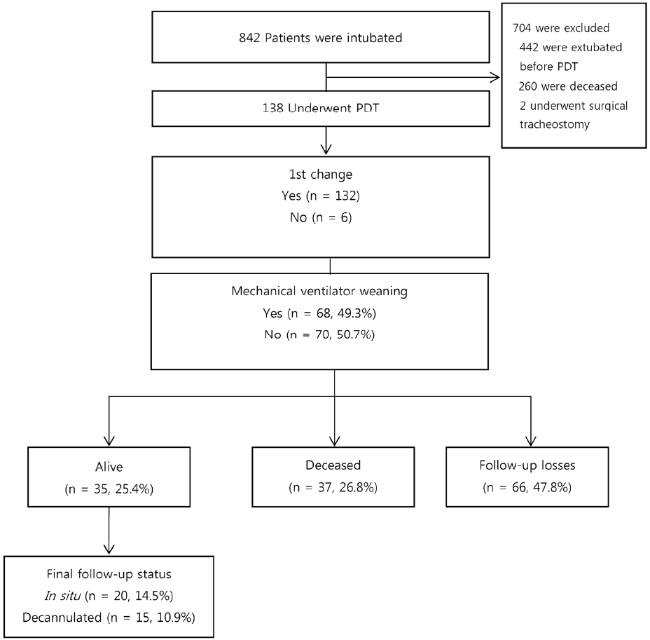
The study population (n = 138) was recruited from a medical ICU at a tertiary university hospital from March, 2011 to February, 2013 (Fig. 1). The patients’ demographic and clinical characteristics were retrospectively reviewed and procedural data were analyzed. We also analyzed immediate complications as the primary outcome, including bleeding complications associated with antiplatelet agent regimens; and the clinical outcomes of long-term follow-ups during 6 months were also reviewed. This study was was conducted in compliance with the Declaration of Helsinki and given an exemption from informed consent by the Institutional Review Board Committee of our hospital (B-1110/137-104).
During the PDT procedure, one bronchoscopist, two operators and one nurse were participated. It was performed by using the single tapered dilator (Ciaglia Blue Rhino Percutaneous Tracheotomy Introducer Kit; Cook Critical Care™, Bloomington, IL, USA) technique. Under fiberoptic bronchoscopic guidance, the PDT procedure was performed as follows. Routine local anesthesia with short duration of sedative and neuromuscular blocker was used in all patients with proper hemostasis of the skin and subcutaneous tissue using local electrocauterization. During the entire procedure, a mechanical ventilator was set on a volume-controlled modality with a 100% inspiratory fraction of oxygen. Vital signs were continuously monitored, and arterial blood gas analyses were obtained before and after the procedure. Asepsis of the skin was obtained with chlorhexidine 2% and povidone iodine 10%. The correct positioning of tracheostomy tube was obtained by the palpation of standard anatomical landmarks and bronchoscopic assistance.
Before and after the procedure, simple cervical spine view and chest radiography were checked. The duration of the procedure (from skin incision to tracheostomy tube placement) as well as any operative complications was noted after the time of tracheostomy placement.
Continuous data are presented as the mean ± standard deviation (SD) if normally distributed, and the median (range) if skewed. Proportions are presented as a number (%). Comparisons between groups were analyzed by using Student’s t-test or a chi-squared test, as appropriate. All statistical analyses were performed using IBM SPSS Statistics 20.0 (IBM, Armonk, NY, USA) with p < 0.05 considered significant.

Results

During the study period, 138 patients underwent PDT. Of the subjects, 94 were male (68.1%) with a median age of 72 years (17–92), and 81 (58.7%) of the patients were admitted to the ICU resulting from respiratory failure. PDT was performed 10.7 ± 8.5 days after endotracheal intubation. The baseline characteristics between antiplatelet groups are shown in Tables 1 and 2.
All patients underwent successful procedures; no patient was unable to receive a tracheostomy tube. Vertical incisions were used in 109 cases (79.0%), and the incision size was 1.7 ± 0.4 cm.
Over the year, the total procedural time was shortened from 33 min to 22 min. Times were reduced from 32 to 20 min for two attending physicians, and from 43 to 23 min for four fellows who had their first PDT experience. However, there are no statistical difference in procedural time between attending physicians and fellows (p = 0.571).
Immediate complications are described in Table 3. Overall complications occurred in 28 patients (20%); however, there were no periprocedural life-threatening complications and/or major bleeding (Table 3). There were minor bleedings in the anti-platelet group were only 2 (1.4%) patients. Noteworthy, there was no statistical difference in the incidence of bleeding complications between patients who had taken antiplatelet agents and those who had not (p = 0.657, Table 4).
Tracheostomy tubes were exchanged a week after PDT, and the mean follow-up duration was 120.6 days (Fig. 1). The final follow-up statuses were as follows: 66 follow-up losses (47.8%), 37 deceased (26.8%) and 35 alive (25.4%). Twenty (57.1 %) of the surviving patients were successfully weaned from their tracheostomy without any airway complications. The median days from PDT to decannulation (n = 20) was 62 (20–423).

Discussion

PDT is considered to be a minimally invasive procedure that can be performed in the ICU at the patient’s bedside. The time required for performing a bedside PDT is considerably shorter than that of open tracheostomy,[11] even performed by medical intensivisit or intensivist trainee,[12,13] and the other advantages of PDT are as follows: it minimizes scheduling difficulties associated with the operating room and anesthesiology staff, does not require transporting critically ill patients to the operating room, and reduces the overall cost of the tracheostomy. For these reasons, PDT is presently a standard technique in critical care medicine and is usually considered as having a low risk of complications.[14,15]
In this study, we elucidated that 138 PDTs performed in a medical ICU were a feasible and safe procedure, especially in 26 critically ill patients even taking antiplatelet agents without increasing periprocedural complications. There was no statistical difference in the incidence of bleeding complications between patients who had taken antiplatelet agents and those who had not (p = 0.657). There are several reasons for low incidence of bleeding complications in this study; First of all, when all PDTs are performed under bronchoscopy assistance, we could perform PDT more accurately. Second, the minimal amount of skin incision required, with practically less bleeding observed. Third, before the procedure, we checked meticulously the anterior neck area around puncture site on the chest CT and did palpation using fingers for avoid aberrant vascular structures crossing the midline. For this reason, in the recent study the use of ultrasound to evaluate cervical anatomy and to guide puncture in real-time has been advocated to improve safety and efficacy of PDT.[16,17]
In our study, the overall procedural success rate was 100%, and the mean total procedural time was 25 min; it was shortened as the operators became more experienced (33 min in 2011, 22 min in 2012). This result demonstrates that PDT procedures have educational merit, even for first-year fellows, based on non-surgical training experience from instructors such as pulmonologists. Undoubtedly, bronchoscopy assistance results in PDT being an easier procedure.
In our study population, there were two patients who can be safely performed with PDT even taking dual antiplatelet therapy. These patients suffered from cardiac arrest following myocardial infarction. After a primary angioplasty with stent placement, dual antiplatelet therapy was commenced. Because of the risk of in-stent thrombosis, PDT was performed without an interruption in antiplatelet therapy. No immediate or late hemorrhagic complications occurred during the procedure. Several studies have demonstrated the safety of this procedure in patients with congenital or acquired hemostasis diseases,[1820] but few studies have evaluated the safety of PDT in patients receiving dual antiplatelet therapy. However, no evidence-based guidelines clearly define the potential hazards of the procedure in these patients and large-scaled, randomized controlled studies should be needed to confirm our result.
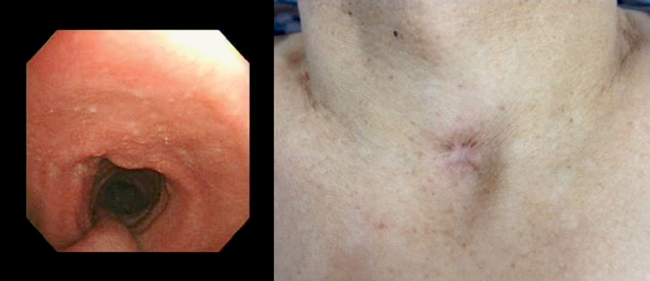
The overall rate of complications was 20.3%, which included minor bleeding, tracheal ring fracture, temporary desaturation, and unintended extubation; however, the patients’ prognoses in the ICU were not significant influences. Of the various complications, tracheal ring fracture could be considered a special issue related to PDT, which occurred immediately during the use of a single tapered dilator. In this study, we experienced 3 cases of tracheal ring fracture; inner diameter of tracheostomy tube was 7.5 mm. Nevertheless, there was no problem securing upper airway patency after long-term follow-up, regardless of whether decannulation was performed or not, there seen only mild scarring at the decannulated PDT site externally and on the endobronchial side inspected by bronchoscopy (Fig. 2).
However, the present study has several limitations. First, this study was conducted at a single center with not much PDT patient experience compared to abroad big hospitals. However, in Asia, and particularly in developing countries, there are few studies like this study. Second, while a study reporting on the associations of bleeding tendencies with the use of antiplatelet agents is new, it is a retrospective study, so a randomized, controlled trial is needed to confirm the findings. Third, in Korea, medical costs in the ICU are not properly reflected when a surgical tracheostomy is performed, considering the expenses incurred from the additional costs resulting from increased lengths of stays on the ICU. More multicenter studies about PDT as a treatment tool in accordance with the cost of complications, including comparative effectiveness are needed.
In conclusion, PDT performed in critically ill patients taking antiplatelet agents was a feasible and implementable procedure without additional bleeding complications.

Notes

No potential conflict of interest relevant to this article was reported.

ACKNOWLEDGMENTS

We would like to thank Soyeon Ahn, Ph.D., for dedicated statistical review of the study as an independent statistician.

References

1). Shelden CH, Pudenz RH, Freshwater DB, Crue BL. A new method for tracheotomy. J Neurosurg. 1955; 12:428–31.
crossref
2). Seldinger SI. Catheter replacement of the needle in percutaneous arteriography; a new technique. Acta radiol. 1953; 39:368–76.
crossref
3). Ciaglia P, Firsching R, Syniec C. Elective percutaneous dilatational tracheostomy. A new simple bedside procedure; preliminary report. Chest. 1985; 87:715–9.
4). Freeman BD, Isabella K, Cobb JP, Boyle WA 3rd, Schmieg RE Jr, Kolleff MH, et al. A prospective, randomized study comparing percutaneous with surgical tracheostomy in critically ill patients. Crit Care Med. 2001; 29:926–30.
crossref
5). Delaney A, Bagshaw SM, Nalos M. Percutaneous dilatational tracheostomy versus surgical tracheostomy in critically ill patients: a systematic review and meta-analysis. Crit Care. 2006; 10:R55.
6). Friedman Y, Fildes J, Mizock B, Samuel J, Patel S, Appavu S, et al. Comparison of percutaneous and surgical tracheostomies. Chest. 1996; 110:480–5.
crossref
7). Krishnan K, Elliot SC, Mallick A. The current practice of tracheostomy in the United Kingdom: a postal survey. Anaesthesia. 2005; 60:360–4.
crossref
8). Kluge S, Baumann HJ, Maier C, Klose H, Meyer A, Nierhaus A, et al. Tracheostomy in the intensive care unit: a nationwide survey. Anesth Analg. 2008; 107:1639–43.
crossref
9). Veenith T, Ganeshamoorthy S, Standley T, Carter J, Young P. Intensive care unit tracheostomy: a snapshot of UK practice. Int Arch Med. 2008; 1:21.
crossref
10). Abouzgheib W, Meena N, Jagtap P, Schorr C, Boujaoude Z, Bartter T. Percutaneous dilational tracheostomy in patients receiving antiplatelet therapy: is it safe? J Bronchology Interv Pulmonol. 2013; 20:322–5.
11). Massick DD, Powell DM, Price PD, Chang SL, Squires G, Forrest LA, et al. Quantification of the learning curve for percutaneous dilatational tracheotomy. Laryngoscope. 2000; 110(2 Pt 1):222–8.
crossref
12). Yoo H, Lim SY, Park CM, Suh GY, Jeon K. Safety and feasibility of percutaneous tracheostomy performed by medical intensivists. Korean J Crit Care Med. 2011; 26:261–6.
crossref
13). Lee D, Chung CR, Park SB, Ryu JA, Cho J, Yang JH, et al. Safety and Feasibility of Percutaneous Dilatational Tracheostomy Performed by Intensive Care Trainee. Korean J Crit Care Med. 2014; 29:64–9.
crossref
14). Grant CA, Dempsey G, Harrison J, Jones T. Tracheo-in-nominate artery fistula after percutaneous tracheostomy: three case reports and a clinical review. Br J Anaesth. 2006; 96:127–31.
crossref
15). Al-Ansari MA, Hijazi MH. Clinical review: percutaneous dilatational tracheostomy. Crit Care. 2006; 10:202.
16). Flint AC, Midde R, Rao VA, Lasman TE, Ho PT. Bedside ultrasound screening for pretracheal vascular structures may minimize the risks of percutaneous dilatational tracheostomy. Neurocrit Care. 2009; 11:372–6.
crossref
17). Rudas M, Seppelt I. Safety and efficacy of ultrasonography before and during percutaneous dilatational tracheostomy in adult patients: a systematic review. Crit Care Resusc. 2012; 14:297–301.
18). Al Dawood A, Haddad S, Arabi Y, Dabbagh O, Cook DJ. The safety of percutaneous tracheostomy in patients with coagulopathy or thrombocytopenia. Middle East J Anesthesiol. 2007; 19:37–49.
19). Kluge S, Meyer A, Kühnelt P, Baumann HJ, Kreymann G. Percutaneous tracheostomy is safe in patients with severe thrombocytopenia. Chest. 2004; 126:547–51.
crossref
20). Kost KM. Endoscopic percutaneous dilatational tracheotomy: a prospective evaluation of 500 consecutive cases. Laryngoscope. 2005; 115(10 Pt 2):1–30.
crossref

Fig. 1.
Long-term (6 months) follow-up results. PDT: percutaneous dilatational tracheostomy.
kjccm-2014-29-3-183f1.tif
Fig. 2.
Bronchoscopic findings and skin scarring after percutaneous dilatational tracheostomy decannulation.
kjccm-2014-29-3-183f2.tif
Table 1.
Baseline characteristics of the patients that underwent PDT
Characteristics Values
Number of patients 138
  Median age, yr (range) 72 (17–92)
  Male 94 (68.1)
  BMI, kg/m2 20.3 ± 4.8
  PEEP, cm H2O 4.6 ± 2.0
  FiO2 0.4 ± 0.2
  Duration of MV before PDT, d 10.7 ± 8.5
  ICU length of stay, d 30.8 ± 36.7
  Duration of mechanical ventilation, d 35.9 ± 52.4
  Acute Physiology and Chronic Health Evaluation II 24.4 ± 9.4
Causes of ICU admission
  Respiratory 81 (58.7)
  Neurologic 41 (29.7)
  Cardiologic 11 (8.0)
  Septic 5 (3.6)
Reasons for PDT
  Need for long-term free airway maintenance 84 (60.9)
  Prolonged mechnical ventilation 54 (39.1)
Periprocedural parameters
  Procedural success rate (%) 100
  Direction of incision
    Vertical 109 (79.0)
    Transverse 29 (21.0)
  Mean procedural time, min 25 ± 12
    1st yr (n = 41, range) 33 (5–64)
    2nd yr (n = 96, range) 22 (8–50)

Values are presented as mean ± SD or number (%) unless otherwise indicated. PDT: percutaneous dilatational tracheostomy; ICU: intensive care unit.

Table 2.
Baseline characteristics of the patients who underwent PDT whether taking antiplatelet agents or not
Total No antiplatelet group Antiplatelet group p value
Number of patients 138 112 26
Median age, yr (range) 72 (17–92) 70 (1–92) 77 (53–91) 0.31
PT INR 1.2 ± 0.2 1.2 ± 0.2 1.1 ± 0.1 0.84
Platelet count, ×103/μL 236 ± 117 237 ± 121 234 ± 99 0.90
Acute Physiology and Chronic Health Evaluation II 24.4 ± 9.4 23.6 ± 9.7 27.6 ± 7.3 0.051
Size of incision, cm 1.7 ± 0.4 1.7 ± 0.4 1.8 ± 0.4 0.177

Values are presented as number (%) or mean ± SD. PDT: percutaneous dilatational tracheostomy; PT INR: the international normalized ratio of prothrombin time.

Table 3.
Immediate PDT complications between two groups (n = 138)
Total No antiplatelet group Antiplatelet group p value
Number of patients 138 112 (81.2) 26 (18.8) 0.338
No complications 110 (79.7) 90 (65.2) 20 (14.6)
Complications 28 (20) 22 (16) 6 (4.2)
  Minor bleeding (3–5 small, soaked swabs) 10 (7.2) 8 (5.8) 2 (1.4)
  Others* 7 (5.1) 5 (3.6) 2 (1.4)
  Proximal tracheal ring fracture visible by bronchoscopy 4 (2.9) 4 (2.9) 0 (0)
  Oxygen desaturation (≤ 88%) 3 (2.2) 1 (0.7) 2 (1.4)
  Unintended extubation 2 (1.4) 2 (1.4) 0 (0)
  Subcutaneous emphysema 2 (1.4) 2 (1.4) 0 (0)

Values are presented as number (%).

* Hypotension 5, tachycardia 1, site hematoma 1. PDT: percutaneous dilatational tracheostomy.

Table 4.
Bleeding tendencies associated with antiplatelet agents
Minor bleeding* p value

No Yes
No antiplatelet group 103 (75) 9 (7) 0.657
Antiplatelet group 24 (17) 2 (1)

Values are presented as number (%).

* Minor bleeding was any documented bleeding that did not meet the definition of a hemorrhagic complication.

Antiplatelet group, n (%): Aspirin 12 (8.7), aspirin hold within 3 days 7 (5.1), clopidogrel 3 (2.2), aspirin with clopidogrel 2 (1.4), aspirin and clopidogrel hold within 3 days 2 (1.4)

TOOLS
Similar articles