Myocarditis is an inflammatory disease of the myocardium caused by different infectious and noninfectious triggers.[1] Myocarditis often results from viral infections such as parvovirus B19, Coxsackie B virus, human herpesvirus 6, or adenoviruses.[2] Numerous medications or toxins like antipsychotics, antibiotics, and ethanol also can cause myocarditis.[1] Recently, we encountered a case of acute fulminant myocarditis following a coffee diet program. These days, many people undergo a variety of diet. Therefore, it could be bound to have ramifications for public health, although this is the only case reported of its kind. So we report the case with a review of literatures.
CASE REPORT
A 41-years-old female visited emergency department for vomiting and restlessness. She had no past medical history except for a surgery for herniation of the intervertebral disc 5 years ago. She had no recent travel history or medication history. However, she was in emotional stress because she was divorced 2 months before the presentation. For 7 days before the presentation, she was on an extraordinary weight reduction diet program so called ‘coffee diet’. She reduced her daily meals by half and took several cups of grinded coffee powders with water. She even used it as a form of enema. From the day before the presentation, she had suffered excessive vomiting.
At presentation, her vital signs were checked as systolic blood pressure of 180 mmHg, diastolic blood pressure of 100 mmHg, pulse rate of 124 beats per minute, respiration rate of 28 times per minute, and body temperature of 36°C. The oxygen saturation was rapidly worsened from 90% to 60% in the first 30 minutes. Immediate endotracheal intubation was performed and mechanical ventilation was initiated. An arterial blood gas analysis (ABGA) revealed pH of 7.306 (reference value: 7.35–7.46), partial pressure of carbon dioxide (PaCO2) of 33.5 mmHg (reference value: 35–45), partial pressure of oxygen (PaO2) of 60 mmHg (reference value: 80–100), bicarbonate of 16.3 mM/L (reference value: 21–29), and oxygen saturation of 89% despite using a fraction of inspired oxygen (FiO2) of 1.0. A chest radiograph showed bilateral infiltration consistent with acute pulmonary edema. The blood pressure also rapidly dropped to 70/45 mmHg even though 0.4 µg/kg/min of norepinephrine and 20 µg/kg/min of dobutamine were infused. A transthoracic echocardiography showed severe global hypokinesia of the left ventricle (LV) with ejection fraction (EF) of 15%. A serum lactate was measured as 9.74 mM/L (reference value: 0–2). The initial cardiac enzymes were elevated as creatine kinase (CK) of 86 IU/L (reference value: 38–160), CK-MB of 5.21 ng/mL (reference value: 0–5), and troponin-T of 0.248 ng/mL (reference value: 0–0.1).
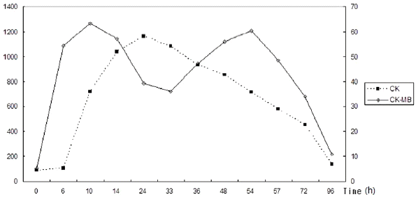
The blood pressure did not respond to vasopressors and inotropics, and the serum lactate was not reduced by medical treatment, thus we decided to apply mechanical circulatory and respiratory support using a veno-arterial (VA) extracorporeal membrane oxygenation (ECMO). A 24F venous catheter and a 21F arterial catheter were inserted each via the right femoral vein and the left femoral artery. We used the Emergency Bypass System (CAPIOX® EBS) of TERUMO® with revolutions per minute (RPM) of 2,500, and the ECMO flow was checked as 3.7 L/min. With ECMO support, the serum lactate became normalized in 6 hours. The initial blood chemistry showed serum blood urea nitrogen (BUN) of 14.8 mg/dl (reference value: 8–23) and serum creatinine of 1.9 mg/dl (reference value: 0.9–1.2). Although serum BUN/creatinine rose to 30.6/4.0 mg/dl in two days after the presentation, the urine output was preserved (over 25 ml/kg/h). Then the BUN and creatinine were gradually normalized during the following 2 weeks. Aspartate amino-transferase (AST) and alanine aminotransferase (ALT) were normal at presentation. AST (reference value: 10–40 IU/L)/ALT (reference value: 5–40 IU/L) reached peak level of 208/219 IU/L at day 2. The AST and ALT were normalized 5 days later. The serum CK reached peak level of 1170 IU/L at 23 hours after the presentation and gradually decreased thereafter, while serum CK-MB reached peak level of 63.29 ng/ml in 9 hours and remained at high level over the 48 hours after the presentation (Fig. 1). All serological tests for viruses (including coxsackie viruses, parvo viruses, Ebstein-Barr virus, cytomegalovirus, and herpes viruses) showed negative results and no autoantibodies were detected.
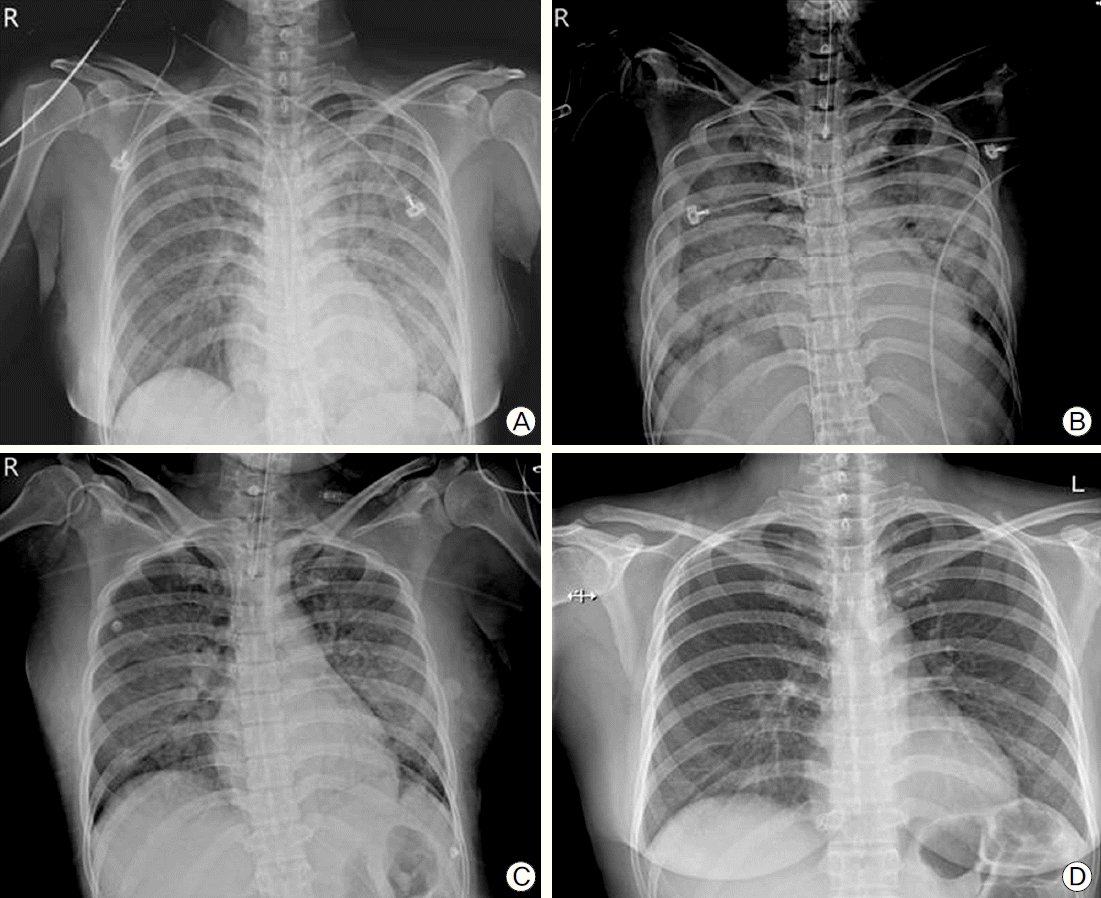
Nine days later, vital signs were kept stable and serum lactate was also normal even when the ECMO flow reached nearly zero. The patient was weaned from ECMO on the ninth day and from mechanical ventilation on the tenth day. A follow-up echocardiography at discharge showed improved systolic function of the LV with EF of 53% and normal cardiac chamber size (interventricular septal distance of 12 mm, LV end-diastolic distance of 50 mm, and LV end-systolic distance of 34 mm). However, the lateral and the anterior wall of the left ventricle still showed reduced contractility from mid-LV to apex. The serial chest radiograph showed pulmonary edema improving over time (Fig. 2). The patient was discharged with the prescription of angiotensin-converting enzyme inhibitor (ACEI) and furosemide on the 17th day.
 | Fig. 2.(A) A chest X-ray at presentation shows diffuse bilateral infiltration. (B) A chest X-ray taken on day 4 reveals aggravated bilateral infiltration and newly developed pleural effusion. (C) A chest X-ray on the day of ECMO-weaning (day 9) shows improved pulmonary edema and pleural effusion. (D) A chest X-ray taken on the day of discharge (day 17) shows normal X-ray findings. |
Three months later, a follow-up echocardiography showed normalized LV systolic function (EF 60%) without any regional wall motion abnormalities.
Go to : 
DISCUSSION
Myocarditis is usually caused by viral infections or post-viral immune-mediated responses.[1] Most commonly associated viruses are parvovirus B19, enteroviruses, adenoviruses and herpesviruses.[1] Myocarditis also can be triggered by nonviral infections such as Borrelia burgdorferi, Corynebacterium diphteriae, or Trypanosoma cruzi.[3] A number of medications like clozapine, penicillin, ampicillin, sulfonamides, tetracyclines or mesalamine are also associated with myocarditis.[1,4,5] Toxins, scorpion or spider bites also can trigger myocarditis.[6–8] To the best of our knowledge, this is the first case report of a myocarditis following a coffee diet program.
Why myocarditis occurred following an extensive coffee diet is currently inexplicable. The extensive dose of caffeine may explain the cardiac damage. The patient even used the diet materials as a form of enema. There is a case report of a patient who ingested 27 g of caffeine in a suicide attempt.[9] The patient experienced very severe cardiac arrhythmia and hypotension. Caffeine is supposed to act as an antagonist of adenosine at cell surface receptors, inhibit phosphodiesterase, and increase cytosolic calcium especially in very high concentrations (100 to 1000 μM/L).[10,11] There is also a study which showed that caffeine damages mitochondrial of the heart.[11] However, there is limited literature that examined the adverse effects of caffeine on heart, such as myocarditis. We do not know about the relationship between caffeine intoxication and myocarditis. In fact, the patient bought the ‘coffee diet materials’ from some kind of a black market. It is possible that the materials had been contaminated by unknown toxins or microorganisms and the amount of caffeine in the formulas is unknown.
There could be a controversy in the diagnosis of myocarditis for this patient. We did not perform endomyocardial biopsy (EMB). The World Health Organization (WHO)/International Society and Federation of Cardiology (ISFC) defined myocarditis as inflammatory disease of the heart muscle, diagnosed by established histological, immunological, and immunohistochemical criteria.[12] However, the clinical presentation of acute congestive heart failure, echocardiographic changes, elevation of cardiac enzymes, negative serologic markers for viruses, no evidence of other immunological disorders, and complete recovery of the LV systolic function suggest myocarditis as the most possible diagnosis for this patient.
The treatment of myocarditis includes usual medications for heart failure (ACEI, angiotensin-II receptor blockers, diuretics, beta-blockers, aldosterone antagonists and inotropics), calcium channel blockers, nonsteroidal anti-inflammatory drugs, and colchicine. For patients with cardiogenic shock with acute fulminant myocarditis, mechanical circulatory support may be required. In a study, mechanical circulatory support rescued 68% of patients with fulminant myocarditis.[13] For a refractory myocarditis, heart transplantation can be an option.
This is a very rare case of life-threatening myocarditis following a coffee diet. However, it has a potential to become a great heath care problem, because such a diet program is widely used by many people and there are a large number of unauthorized products available in the market.
Go to : 


 PDF
PDF Citation
 Print
Print


 XML Download