Journal List > Cancer Res Treat > v.49(1) > 1153460

Li, Wei, Yao, and Wang: Clinicopathological Features of Low-Grade Thyroid-like Nasopharyngeal Papillary Adenocarcinoma

Abstract

Purpose

Primary low-grade thyroid-like papillary adenocarcinomas are extremely rare neoplasms that generally originate in the nasopharynx. Here, we describe a novel case of a 15-year-old Chinese girl who was diagnosed with low-grade thyroid-like papillary adenocarcinoma, including a brief review of the literature to reveal the clinicopathological features of low-grade thyroid-like nasopharyngeal papillary adenocarcinoma.

Materials and Methods

Immunohistochemistry was used to evaluate the expression of pan-cytokeratin (CKpan), cytokeratin (CK) 7, thyroid transcription factor 1 (TTF-1), vimentin, epithelial membrane antigen (EMA), thyroglobulin, CD15, S100, P40, CK20, CDX-2, glial fibrillary acidic protein (GFAP), and Ki-67. Additionally, in situ hybridization investigation was utilized to identify the presence of small Epstein-Barr virus (EBV)–encoded RNA.

Results

Histopathological analysis revealed florid proliferation of papillary structures lined by columnar epithelial cells with fibrovascular cores. Immunohistochemically, the neoplastic cells were positive for CKpan, CK7, TTF-1, vimentin, and EMA, but negative for thyroglobulin, CD15, S100, P40, CK20, CDX-2, and GFAP. The Ki-67–labeling index reached 5% in the most concentrated spot. In situ hybridization for EBV was negative.

Conclusion

Due to the distinct rarity of low-grade thyroid-like papillary adenocarcinomaswith a favorable clinical outcome, a nationwide effort to raise public awareness of this neoplasm is required.

Introduction

As a common head and neck cancer, nasopharyngeal carcinomas are generally either keratinizing or nonkeratinizing squamous cell carcinomas. Primary nasopharyngeal adenocarcinomas (NPACs) are rare neoplasms, constituting only 0.38% to 0.48% of all malignant nasopharyngeal neoplasms [1,2]. Thyroid-like low-grade nasopharyngeal papillary adenocarcinoma (TL-LGNPPA) is a small minority of conventional nasopharyngeal adenocarcinoma that exhibits papillary growth and abnormal expression of thyroid transcription factor 1 (TTF-1). To the best of our knowledge, only thirteen cases have been reported in the English-language literature and eleven in the Chinese-language literature to date (Table 1). In this article, we present a case of primary TL-LGNPPA and an analysis of its clinicopathological features.

Materials and Methods

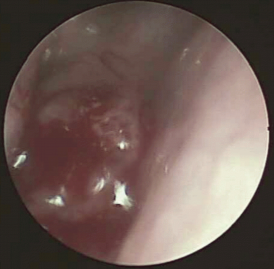
A 15-year-old Chinese girl presented with a complaint of rhinorrhoea and nasal congestion with a duration of 1 month. On clinical examination, no thyroid tumor or other physical abnormalities were found. Nasal endoscopy identified a pedunculated polypoid mass with smooth surface that measured approximately 2.5×2 cm arising from the posterior nasal septum (Fig. 1). A biopsy of the dominant portion of the mass was performed. Pathological analysis revealed an adenocarcinoma with papillary structure. Therefore, complete excision of the tumor was performed through an endoscope. Follow-up data showed no signs of local recurrence at up to 2 years after complete surgical removal.
The tissue was fixed in formalin and embedded in paraffin, after which 4-μm thin sections were cut and stained with hematoxylin and eosin. Immunohistochemical staining was performed using commercially available antibodies to the following antigens: pan-cytokeratin (CKpan), epithelial membrane antigen (EMA), vimentin, cytokeratin (CK) 7, CD15, thyroid transcription factor 1 (TTF-1), Ki-67, P40, S100, glial fibrillary acidic protein (GFAP), CK20, CDX-2, and thyroglobulin. At the same time, in situ hybridization for the presence of small Epstein-Barr virus (EBV)–encoded RNA was performed to identify the association between this tumor and EBV. All protocols were employed according to the manufacturers’ recommendations (Table 2).

Results

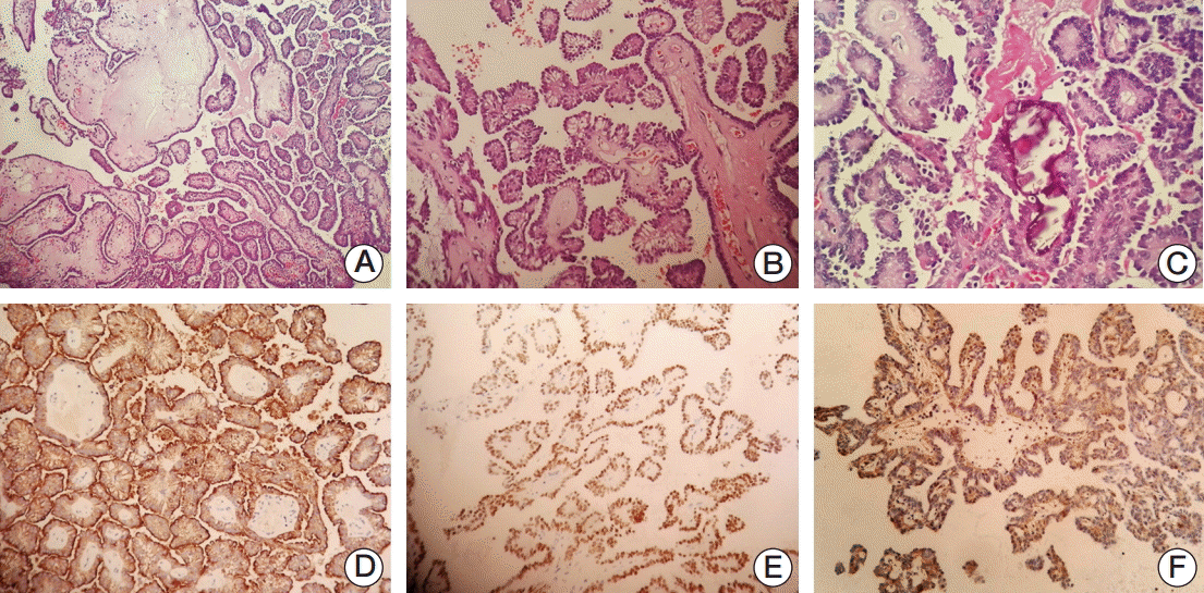
Histological examination of the biopsy specimen revealed a tumor that showed papillary configuration with hyalinized fibrovascular cores, similar to thyroid papillary carcinoma (Fig. 2A and B). The papillae were complex and tightly packed, and most were lined with cuboidal or columnar epithelia containing overlapping round to ovoid nuclei that displayed fine chromatin and mildly eosinophilic cytoplasm (Fig. 2C). Only minor degrees of pleomorphism and hyperchromatism were observed. Small intra-nuclear cytoplasmic inclusions were present focally, although neither nuclear grooving nor ground glass nuclei were found. In small parts of the tumor, psammoma bodies were exhibited (Fig. 2C). No necrosis or mitotic activity was discerned and no continuity between the tumor epithelia and normal nasopharyngeal mucosa or mucous glands was identified.
Immunoperoxidase studies showed that neoplastic cells were positive for CK7, vimentin, TTF-1, CKpan, and EMA (Fig. 2D-F). There was no immunoreactivity for thyroglobulin, CD15, S100, P40, CK20, CDX-2, and GFAP. In the most concentrated spot, the Ki-67–labeling index reached 5%. Negative results were revealed by in situ hybridization investigation of EBV.

Discussion

NPAC contains two subtypes: conventional/surface origin-type and salivary gland-type. The former often originates from nasopharyngeal surface mucosa and presents with papillary configuration. The latter includes polymorphous low-grade adenocarcinoma, mucoepidermoid adenocarcinoma, and adenoid cystic carcinoma [12]. In 1988, Wenig et al. [20] first described thyroid-like papillary adenocarcinoma of the nasopharynx and proposed that these papillary adenocarcinomas should been regarded as a distinct entity from conventional adenocarcinomas in this region based on their indolent clinical behavior and low-grade histological features. Nasopharyngeal papillary adenocarcinoma (NPPA) was enrolled in the World Health Organization classification system of malignant epithelial tumors of the nasopharynx in 2005 [21].
Papillary configuration and aberrant TTF-1 expression are the distinguishing features of TL-LGNPPA, mimicking papillary thyroid carcinoma (PTC). TL-LGNPPA is an extremely rare neoplasm. To the best of our knowledge, few cases have been reported [3-19]. Clinical features of the published cases are listed in Table 1. The median age is 35 years in patients, ranging from 9 to 68 years. No difference was observed between the ratio of males to females. Instances of TL-LGNPPA were usually localized in the roof of the nasopharynx and posterior edge of the nasal septum. The excellent prognosis was obvious, with local excision being performed in all reported cases, and no local recurrence or metastasis reported.
Histologically, these neoplasms frequently exhibit papillary architecture lined by moderately pleomorphic columnar epithelial cells with fibrovascular cores, overlapping nuclei with clear optically chromatin, and psammoma bodies. These features are generally ascribed to PTC as well. However, the biphasic pattern of low-grade NPPA with a spindle cell component has been described in three case reports [7,9,12]. In this case, no obvious spindle cell component was observed. The biphasic pattern of TL-LGNPPA should be further investigated if a large number of cases can be collected.
Immunohistochemically, the positive expression of TTF-1 was the most characteristic feature of TL-LGNPPA. As a homeodomain containing transcription factor coded by NKX2-1, TTF-1 is usually found in lung, thyroid, and central nervous system tissue [22,23]. Nevertheless, the positive expression of TTF-1 was also observed in other organs, including endometrium, colon, and breast [24]. Although the etiology of TTF-1 positive staining in TL-LGNPPA is still not clear, three mechanisms to explain this phenomenon were proposed in a recent case report. First, TL-LGNPPA may develop from ectopic thyroid tissue. Second, a gene rearrangement that affects TTF-1/NKX2-1 may result in abnormal expression of TTF-1. Finally, genetic instability and reprogramming of the cancer cells can cause dis-differentiation and lead to deregulation of TTF-1/NKX2-1 [12]. However, these presumptions are poorly evidenced owing to the extreme rarity of this tumor at the present time.
There are limited options available for differential diagnosis of papillary lesions in the nasopharynx. Because TL-LGNPPA display a striking resemblance to PTC, it is important to exclude nasopharyngeal metastasis from papillary adenocarcinoma of the thyroid gland for accurate diagnosis, treatment, and prognosis of the patient. Immunostaining for thyroglobulin and CD15 are critical and highly recommended to distinguish these two entities [25]. However, a case of TL-LGNPPA with focal expression of thyroglobulin was recently reported [11]. Polymorphous low-grade papillary adenocarcinoma (PLGA) is more aggressive and positive for vimentin and S100-protein. To date, positivity for TTF-1 has never been reported in PLGA [12]. Papillary variants of the intestinal type of adenocarcinoma (ITAC) show more nuclear atypia and commonly display mucinous differentiation. Moreover, these are often positive for CK20 and CDX2 [26]. Acinic cell carcinomas (ACC) with a papillary component are frequently cystic and variably positive for S100-protein and vimentin according to the range of differentiation (acinar to intercalated ducts) [9]. Extraventricular choroid plexus papillomas (CPP) at unusual localization must be taken into consideration, most of them was positive for S100 and negative for EMA, part of them was positive for GFAP. In our report, negative staining for thyroglobulin, CD15, S100, GFAP, CK20, and CDX-2 was revealed; therefore, PTC, PLGA, ITAC, ACC, and CPP should not be considered for accurate diagnosis.
It has been well documented that tumors originating from the nasopharyngeal epithelium were associated with the EBV. Investigation of in situ hybridization for EBV revealed negative results, which is concordant with reports by Wu et al. [4] and Fu et al. [5]. A close relationship between TL-LGNPPA and EBV was not confirmed; however, further investigation is needed because of the rarity of this neoplasm.
To date, no case of lymphatic metastasis or recurrence has been reported, indicating excellent prognosis for patients with thyroid-like papillary adenocarcinoma [8]. Generally, surgical excision is adequate for the treatment of TL-LGNPPA [20]. When surgical excision is not feasible or positive surgical margins have been observed, radiotherapy can be employed as an adjuvant treatment [20,27].

Conclusion

In conclusion, we present here a novel case of TL-LGNPPA with a review of its main clinicopathological features. Due to the rarity of this neoplasm and favorable clinical outcome, clinicians should pay more attention to accurate diagnosis of this entity, as well as its treatment and clinical prognosis.

Notes

Conflict of interest relevant to this article was not reported.

References

1. He JH, Zong YS, Luo RZ, Liang XM, Wu QL, Liang YJ. Clinicopathological characteristics of primary nasopharyngeal adenocarcinoma. Ai Zheng. 2003; 22:753–7.
2. McGuire LJ, Lee JC. The histopathologic diagnosis of nasopharyngeal carcinoma. Ear Nose Throat J. 1990; 69:229–36.
3. Carrizo F, Luna MA. Thyroid transcription factor-1 expression in thyroid-like nasopharyngeal papillary adenocarcinoma: report of 2 cases. Ann Diagn Pathol. 2005; 9:189–92.
crossref
4. Wu PY, Huang CC, Chen HK, Chien CY. Adult thyroid-like low-grade nasopharyngeal papillary adenocarcinoma with thyroid transcription factor-1 expression. Otolaryngol Head Neck Surg. 2007; 137:837–8.
crossref
5. Fu CH, Chang KP, Ueng SH, Wu CC, Hao SP. Primary thyroid-like papillary adenocarcinoma of the nasopharynx. Auris Nasus Larynx. 2008; 35:579–82.
crossref
6. Bansal A, Pradeep KE, Gumparthy KP. An unusual case of low-grade tubulopapillary adenocarcinoma of the sinonasal tract. World J Surg Oncol. 2008; 6:54.
crossref
7. Ohe C, Sakaida N, Tadokoro C, Fukui H, Asako M, Tomoda K, et al. Thyroid-like low-grade nasopharyngeal papillary adenocarcinoma: report of two cases. Pathol Int. 2010; 60:107–11.
crossref
8. Sillings CN, Weathers DR, Delgaudio JM. Thyroid-like papillary adenocarcinoma of the nasopharynx: a case report in a 19-year-old male. Oral Surg Oral Med Oral Pathol Oral Radiol Endod. 2010; 110:e25.
crossref
9. Petersson F, Pang B, Loke D, Hao L, Yan B. Biphasic low-grade nasopharyngeal papillary adenocarcinoma with a prominent spindle cell component: report of a case localized to the posterior nasal septum. Head Neck Pathol. 2011; 5:306–13.
crossref
10. Huang CH, Chang YL, Wang CP, Wu HP. Positive immunostaining of thyroid transcription factor-1 in primary nasopharyngeal papillary adenocarcinoma. J Formos Med Assoc. 2015; 114:473–4.
crossref
11. Ozer S, Kayahan B, Cabbarzade C, Bugdayci M, Kosemehmetoglu K, Yucel OT. Thyroid-like papillary adenocarcinoma of the nasopharynx with focal thyroglobulin expression. Pathology. 2013; 45:622–4.
crossref
12. Oishi N, Kondo T, Nakazawa T, Mochizuki K, Kasai K, Inoue T, et al. Thyroid-like low-grade nasopharyngeal papillary adenocarcinoma: case report and literature review. Pathol Res Pract. 2014; 210:1142–5.
crossref
13. Ozturk K, Midilli R, Veral A, Ertan Y, Karci B. Primary thyroid-like papillary adenocarcinoma of the nasal septum: a case report. Ear Nose Throat J. 2015; 94:E19–21.
crossref
14. Wu R, Liu H. Clinicopathological features of low-grade nasopharyngeal papillary adenocarcinoma. Zhonghua Bing Li Xue Za Zhi. 2014; 43:613–7.
15. Zhang X, Xu X, Qian K, Liu Y, Wang L, Li M, et al. Low-grade thyroid-like papillary of the nasopharynx with squamous differentiation: report of a case. Lin Chuang Yu Shi Yan Bing Li Xue Za Zhi. 2015; 31:1199–200.
16. Kang Z, Ou Y, Yao L, Qu L, Zheng Z, Yu Y. Low-grade nasopharyngeal papillary adenocarcinoma: report of two cases and literature review. Lin Chuang Yu Shi Yan Bing Li Xue Za Zhi. 2015; 31:1165–7.
17. Li JF, Ye Q, Hong B, Gao X, Xu KL. Thyroid-like low-grade nasopharyngeal papillary adenocarcinoma: report of a case. Zhonghua Bing Li Xue Za Zhi. 2011; 40:638–9.
18. Han S, Yang F, Yuan C, Zhang R. Clinicopathological observation of 1 cases of low grade nasopharyngeal papillary adenocarcinoma. Lin Chuang Yu Shi Yan Bing Li Xue Za Zhi. 2013; 29:328–30.
19. Chen Z, Zhuo M, Zheng X. Thyroid-like low-grade nasopharyngeal papillary adenocarcinoma: one case report. Lin Chung Er Bi Yan Hou Tou Jing Wai Ke Za Zhi. 2014; 28:1266–7.
20. Wenig BM, Hyams VJ, Heffner DK. Nasopharyngeal papillary adenocarcinoma: a clinicopathologic study of a low-grade carcinoma. Am J Surg Pathol. 1988; 12:946–53.
21. Thompson L. World Health Organization classification of tumours: pathology and genetics of head and neck tumours. Ear Nose Throat J. 2006; 85:74.
crossref
22. Bingle CD. Thyroid transcription factor-1. Int J Biochem Cell Biol. 1997; 29:1471–3.
crossref
23. Katoh R, Kawaoi A, Miyagi E, Li X, Suzuki K, Nakamura Y, et al. Thyroid transcription factor-1 in normal, hyperplastic, and neoplastic follicular thyroid cells examined by immunohistochemistry and nonradioactive in situ hybridization. Mod Pathol. 2000; 13:570–6.
crossref
24. Ordonez NG. Value of thyroid transcription factor-1 immunostaining in tumor diagnosis: a review and update. Appl Immunohistochem Mol Morphol. 2012; 20:429–44.
25. Imamura Y. CD15 (C3D-1) immunoreactivity in normal, benign and malignant thyroid lesions. Appl Immunohistochem. 1998; 6:181–6.
crossref
26. Ortiz-Rey JA, Alvarez C, San Miguel P, Iglesias B, Anton I. Expression of CDX2, cytokeratins 7 and 20 in sinonasal intestinal-type adenocarcinoma. Appl Immunohistochem Mol Morphol. 2005; 13:142–6.
crossref
27. Wang CP, Chang YL, Chen CT, Yang TH, Lou PJ. Photodynamic therapy with topical 5-aminolevulinic acid as a post-operative adjuvant therapy for an incompletely resected primary nasopharyngeal papillary adenocarcinoma: a case report. Lasers Surg Med. 2006; 38:435–8.
crossref

Fig. 1.
Nasal endoscopy shows a pedunculated polypoid mass originating in the posterior nasal septum.
crt-2016-195f1.tif
Fig. 2.
(A) The tumor displayed papillary structures with hyalinized fibrovascular cores (H&E staining, ×100). (B) Most papillae were lined with cuboidal or columnar epithelia (H&E staining, ×200). (C) Overlapping round to ovoid nuclei that displayed fine chromatin and psammoma bodies were exhibited (H&E staining, ×400). (D) Strong positive staining of cytokeratin 7 was displayed (immunohistochemical [IHC] staining, ×200). (E) Nuclear staining for thyroid transcription factor 1 was shown (IHC staining, ×200). (F) Positivity of vimentin was evident (IHC staining, ×200).
crt-2016-195f2.tif
Table 1.
Clinical summary of reported thyroid-like low-grade nasopharyngeal papillary adenocarcinoma
No. Source Age (yr) Sex Location Size (cm) Macroscopic appearance Follow-up Recurrence
1 Carrizo and Luna [3] 9 M Right nasopharyngeal wall 2.0 Submucosal mass 2 yr N
2 Carrizo and Luna [3] 13 M Roof of the nasopharynx 1.5 NA 15 yr N
3 Wu et al. [4] 36 F Left nasopharynx 1 Pedunculated polypoid mass 3 yr N
4 Fu et al. [5] 68 M Roof of the nasopharynx NA Pedunculated tumor 1 yr N
5 Bansal et al. [6] 32 M Posterior end of the left nasal septum NA Pedunculated well-circumscribed lesion 2 yr N
6 Ohe et al. [7] 25 M Roof of the pharynx 0.8 Pedunculated mass 13 mo N
7 Ohe et al. [7] 41 M Posterior roof of the nasopharynx 0.5 Pedunculated mass 9 mo N
8 Sillings et al. [8] 19 M Posterior superior free edge of the nasal septum 1.5 Pedunculated mass NA N
9 Petersson et al. [9] 39 F Posterior edge of bony septum 1 Polypoid mass NA N
10 Huang et al. [10] 36 F Roof of the nasopharynx NA Pedunculated tumor 31 mo N
11 Ozer et al. [11] 17 F Posterior nasopharyngeal wall 2.7×2.2 Bilobulated mass 1 yr N
12 Oishi et al. [12] 47 F Posterior edge of the left nasal septum 2 Pedunculated mass 19 mo N
13 Ozturk et al. [13] 24 F Posterior septum 3.0×2.5 Polypoid mass 4 yr N
14 Wu and Liu [14] 31 M Posterior of the nasopharynx 0.6×0.5×0.6 Polypoid mass 9 yr and 8 mo N
15 Wu and Liu [14] 49 M Pharyngeal recess of the nasopharynx 1.5×1 NA 4 yr and 7 mo N
16 Wu and Liu [14] 62 M Posterior nasal septum 0.5×0.5×0.3 NA 1 yr and 4 mo N
17 Wu and Liu [14] 42 F Roof of the nasopharynx 1×1×0.5 NA 7 mo N
18 Wu and Liu [14] 51 M Posterior nasal septum 1×1×0.8 Pedunculated papillary mass 1 mo N
19 Zhang et al. [15] 57 F Nasopharynx 2×1.5×1.5 Papillary mass 9 mo N
20 Kang et al. [16] 25 M Nasopharynx NA Polypoid mass 1 yr N
21 Kang et al. [16] 36 M Posterior nasal septum 0.8×0.8×0.8 Polypoid mass 18 mo N
22 Li et al. [17] 26 F Roof of the nasopharynx 1.5×1.3×0.4 Polypoid mass 8 mo N
23 Han et al. [18] 40 F Roof of the right posterior nasopharynx 2×1 Pedunculated polypoid mass 1 yr N
24 Chen et al. [19] 42 F Roof of the nasopharynx 0.5×0.5 Polypoid mass 1 yr N
25 Present case 15 F Posterior nasal septum 2.5×2 Pedunculated polypoid mass with smooth surface 24 mo N

M, male; N, no; NA, not acquired; F, female.

Table 2.
Summary of primary antibodies and results of immunohistochemistry
Antibody Source Dilution Result
CK Ascend Bio, Guangzhou, China 1:100 +
EMA Ascend Bio, Guangzhou, China 1:100 +
Vim Ascend Bio, Guangzhou, China 1:100 +
CK7 Ascend Bio, Guangzhou, China 1:100 +
CD15 Ascend Bio, Guangzhou, China 1:200
TTF-1 Ascend Bio, Guangzhou, China 1:400 +
Ki-67 Ascend Bio, Guangzhou, China 1:200 5% in the most concentrated spot
P40 ZSGB Bio, Beijing, China 1:200 -
S-100 Dako, Glostrup, Denmark 1:3,200 -
GFAP Dako, Glostrup, Denmark 1:1,600 -
CK20 Ascend Bio, Guangzhou, China 1:200 -
CDX2 Ascend Bio, Guangzhou, China 1:400 -
Thyroglobulin Ascend Bio, Guangzhou, China 1:800 -
EBER Triplex International Bio, Fuzhou, China RTU -

CK, cytokeratin; EMA, epithelial membrane antigen; Vim, vimentin; TTF-1, thyroid transcription factor-1; GFAP, glial fibrillary acidic protein; CK, cytokeratin; EBER, Epstein-Barr virus–encoded RNA; RTU, ready to use.

TOOLS
Similar articles