This article has been
cited by other articles in ScienceCentral.
Abstract
Histiocytosis is an uncommon disease characterized by excessive accumulation of histiocytes. Here, we report a rare case of non-Langerhans-cell histiocytosis in a 51-year-old woman who presented with severe symptoms of pericardial effusion. Radiologic investigation also detected multiple bone (lower limbs, vertebrae, ribs, and ilium) lesions. Resected pericardium showed abundant mono- or multi-nucleated non-foamy histiocytes (CD68+/CD163+/S-100+/CD1α−/langerin−) in a fibroinflammatory background. The histiocytes demonstrated emperipolesis of lymphocytes, a hallmark feature of Rosai-Dorfman disease (RDD). However, molecular analysis revealed a BRAF V600E mutation of the proliferating histiocytes, highlighting the neoplastic features frequently observed in another non-Langerhans-cell histiocytosis known as Erdheim-Chester Disease (ECD). We consider this case to be a unique presentation of ECD harboring some RDD-like cells with emperipolesis, but not a case of RDD with a BRAF mutation concerning its clinical manifestation (involvement of the heart and bones) and neoplastic features.
Go to :

Keywords: Histiocytosis, Erdheim-Chester disease, Rosai-Dorfman disease, Emperipolesis
Introduction
Histiocytosis refers to a group of rare diseases characterized by the abnormal accumulation of macrophages, dendritic cells or monocyte-derived cells [
1]. Recently, a revised classification schema divided histiocytosis into five groups: (1) Langerhans-related, (2) cutaneous and muco-cutaneous, (3) malignant histiocytoses, (4) Rosai-Dorfman disease (RDD), and (5) haemophagocytic lymphohistiocytosis and macrophage activation syndrome [
1]. Erdheim-Chester disease (ECD) and RDD are common histiocytic disorders affecting adult bone and/or heart.
ECD, a member of the “Langhans-related” group according to the latest revised classification, is a histiocytosis characterized by foamy histiocytes (CD68
+/CD163
+/S-100
−/+/CD1α
−/langerin
−) accompanied by lymphocytes, Touton giant cells, and dense fibrosis [
2]. The radiographic features of symmetric diaphyseal and metaphyseal osteosclerosis in the lower limbs are mostly present in patients with ECD. Other classic extraskeletal organs involved include the cardiovascular, pulmonary, neurological, and endocrine systems [
3,
4]. RDD, which is also known as sinus histiocytosis with massive lymphadenopathy, is diagnosed on the basis of abundant pale cytoplasm in the histiocytes (CD68
+/CD163
+/S-100
+/CD1a
–/langerin
–) and the presence of lymphocytes and plasma cells infiltrating the background. Emperipolesis of histiocytes is a hallmark feature of RDD that has not yet been observed in ECD [
5]. In contrast to ECD, which is characterized by its neoplastic features and often harbors a
BRAF mutation, RDD is mostly defined as a reactive disorder of histiocytosis [
6].
Here, we present an extraordinary case of non-Langerhans-cell histiocytosis (CD68+/CD163+/S-100+/CD1a–/langerin–) involving the heart and skeleton in which emperipolesis and BRAF mutation was confirmed. We consider this case the first report of an atypical ECD with emperipolesis rather than RDD with a BRAF mutation. This study was approved by the Ethics Committee of Tongji Hospital, Tongji Medical College, Huazhong University of Science and Technology, Wuhan, China. Written informed consent was obtained from the patient.
Go to :

Case Report
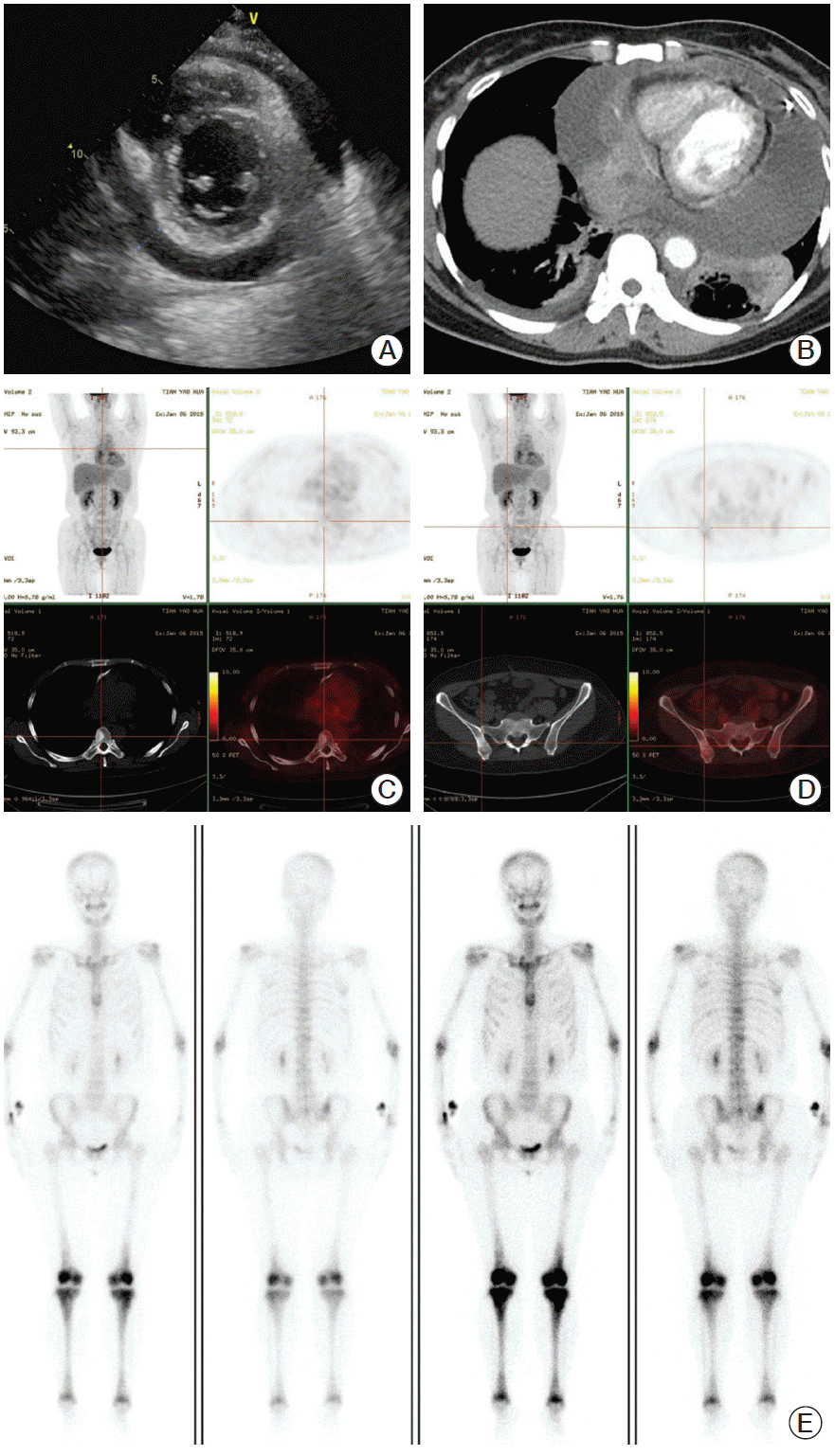
A 51-year-old woman was referred to our hospital because of shortness of breath, chest pain, recurrent edema of the lower extremities, and night sweats without fever or vomiting. Ultrasound and a chest enhanced computed tomography scan showed liquid density in the pericardial cavity (
Fig. 1A and
B) and soft tissue density in the pericardial wall (
Fig. 1B). Positron emission tomography showed radiotracer uptake in the wall of the pericardium and multiple bones, including the vertebrae (
Fig. 1C), ilium (
Fig. 1D), ribs, and lower limbs. Analysis using
99mTc bone scintigraphy also demonstrated significant symmetrical radiotracer uptake in the distal ends of the femurs and the proximal and distal tibia, in addition to the vertebrae, ribs, and ilium (
Fig. 1E). To relieve the symptoms caused by the pericardial effusion, pericardial fenestration and pericardiectomy were performed. A fragment of the hard and grey-white pericardial tissue measuring 7 cm×2.5 cm×0.4 cm was excised and submitted for pathological examination. A biopsy sample from the bone lesion could not be obtained due to patient refusal.
 | Fig. 1.(A) Ultrasound shows a massive circumferential pericardial effusion. (B) Computerized tomography shows pericardial effusion and pericardial soft tissue density. (C, D) Computerized tomography and positron emission tomography shows radiotracer uptake in the thoracic vertebra (C) and ilium (D). (E) Electrical capacitance tomography demonstrates symmetrical radiotracer uptake in the distal ends of the femurs and the proximal and distal tibia, as well as the ribs and vertebrae. 
|
The surgical samples were fixed in 4% buffered formalin, embedded in paraffin, cut into 4 μm sections and stained with hematoxylin and eosin (H&E) for histological evaluation. Immunohistochemical staining was performed on paraffin-embedded sections using a three-step Avidin-Biotin Complex staining method (VECTASTAIN ABC kit, Vector Laboratories, Burlingame, CA). The primary antibodies included langerin (1:100, clone 12D6, Novocastra, Newcastle upon Tyne, UK), CD1α (1:100, clone O10, Dako, Glosrup, Denmark), S-100 (1:1,000, clone Z0311, Dako), CD68 (1:50, clone KP-1, Dako), CD163 (1:100, clone Ber-MAC3, Dako), smooth muscle actin (SMA; 1:400, clone M0879, Dako), desmin (1:200, clone D33, Dako), and pan-cytokeratin (pCK; 1:300, Dako). Diaminobenzidine was used as chromogen. Nuclei were stained with Mayer’s hematoxylin. Appropriate positive controls were included. For molecular analysis, genomic DNA was extracted from formalin-fixed, paraffin-embedded tumor specimens using the TIANamp Genomic DNA Kit (Tiangen Biotech Co., Ltd., Beijing, China) according to the manufacturer’s instructions. The BRAF mutation was detected with the AmoyDx Human BRAF V600E Mutation Detection kit using a BioRad CFX96 Real-Time PCR Machine (BioRad Laboratories, Inc., Berkeley, CA). Briefly, 4.7 μL DNA was added to 35.3 μL polymerase chain reaction (PCR) master mix containing PCR primers, fluorescent probes, PCR buffer, and Taq DNA polymerase. The PCR conditions were as follows: 5-minute denaturation at 95°C, followed by 15 cycles of 95°C for 25 seconds, 64°C for 20 seconds, 72°C for 20 seconds, and then 31 cycles of 95°C for 25 seconds, 60°C for 35 seconds, and 72°C for 20 seconds. The fluorescent signal was collected from the FAM and HEX channels. The results were analyzed according to the instructions in the user manual.
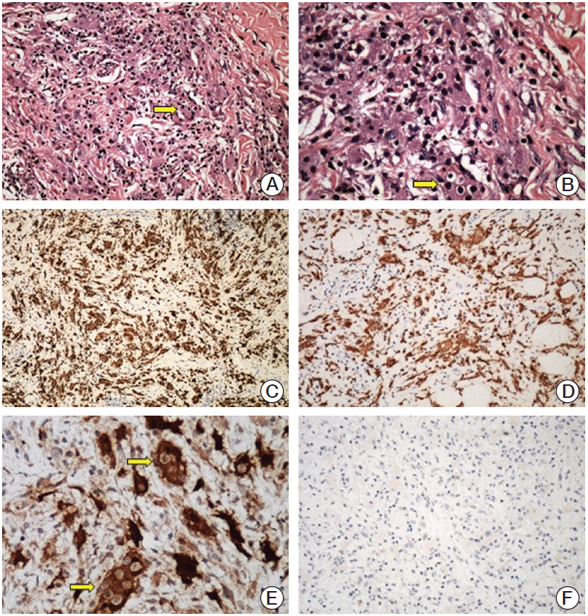
Microscopic examination of the H&E-stained pericardial lesion revealed an exuberant histiocytic and chronic fibroinflammatory process. As shown in
Fig. 2A and
B, the inflammatory cells were primarily composed of lymphocytes, plasma cells, sparse neutrophils, and eosinophils. Intermixed with the infla mmatory cells and fibrotic collagen fibers were scattered mononucleated or multinucleated histiocytes characterized by round or oval vesicular or hyperchromatic nuclei containing distinct nucleoli and abundant amphophilic cytoplasm. Under higher magnification, some of the histiocytes exhibited emperipolesis (
Fig. 2B). Immunohistochemically, these histiocytes showed positive staining of CD68 (
Fig. 2C), CD163 (
Fig. 2D), and S-100 (
Fig. 2E) and negative staining of Langerin (
Fig. 2F), CD1α, and other relevant markers (SMA, desmin, and pCK). The positive S-100 staining highlighted the emperipolesis of the histiocytes (
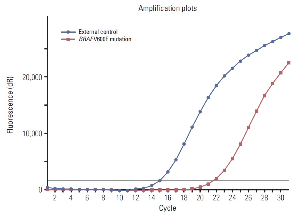
Fig. 2E). Reverse transcription polymerase chain reaction was performed, and a
BRAF V600E mutation was detected (
Fig. 3).
 | Fig. 2.(A) The lesion shows infiltration of non-foamy histocytes (arrow) in a marked fibroinflammatory background (H&E staining, ×100). (B) The lesion shows granular histiocytes in a fibroinflammatory background. Emperipolesis (arrow) shows engulfed intact lymphocytes inside the cytoplasm of non-foamy histiocytes (H&E staining, ×200). (C, D) Positive immunostaining of CD68 (C, ×100) and CD163 (D, ×100) in non-foamy histiocytes in a diffuse cytoplasmic pattern. (E) The histiocytes show strong cytoplasmic and nuclear staining for S-100. Engulfed lymphocytes (arrows) are well demonstrated in some histiocytes as emperipolesis (×200). (F) The histiocytes are negative for Langerin (×100). 
|
 | Fig. 3.
BRAF–polymerase chain reaction detecting V600E mutation: red amplification curve, BRAF V600E mutation; blue amplification curve, external positive control. 
|
Based on the overall findings, a diagnosis of atypical ECD with emperipolesis was considered. After diagnosis, the patient underwent interferon injections subcutaneously for 7 months. During the follow-up period, the patient indicated that her quality of life had improved because she could now get out of bed and was functioning well in her daily life, but no radiologic investigation was performed.
Go to :

Discussion
Histiocytosis refers to a heterogeneous group of diseases characterized by proliferation of cells thought to be derived from dendritic cells, macrophages, or monocyte derived cells. Differentiation between the subtypes relies on the morphology, immunohistochemistry, molecular analysis, and the clinical setting. The major differential diagnosis in this case included Langerhans cell histiocytosis (LCH), RDD, and ECD.
In the present case, LCH was easily excluded because (1) the lesional histiocytes did not show intranuclear pseudoinclusions, prominent nuclear indentations, or grooves; (2) the histiocytes were CD1α and langerin negative, which is inconsistent with the diagnostic immunophenotype for LCH (S-100
+/CD1α
+/langerin
+); and (3) LCH in adults mostly involves bones (52%), lungs (40%), or skin (7%) [
7]. Cardiovascular involvement has only been reported in one case based on radiographic findings and pathologic examination [
8].
Based on the histopathological features, the morphological characteristics observed in this case were consistent with those of RDD. The lesion was characterized by cytoplasm rich, proliferative mononucleated or multinucleated histiocytes in a fibroinflammatory background. Furthermore, the immunophenotype of the cells was identical to that found in RDD (CD68
+/S-100
+/CD1α
−/langerin
−). Moreover, the histiocytes demonstrated emperipolesis, a conventional hallmark feature of RDD, which is facilitated by positive staining of S-100 protein. However, these findings are inconsistent with the typical histopathological features of ECD, which is generally characterized by xanthomatous infiltration of lipid-laden foamy histiocytes (CD68
+/S-100
−/+/CD1α
−/langerin
−) within a variably fibrous stroma with admixed chronic inflammatory cells and Touton giant cells [
4]. It has been reported that, in the extraskeletal ECD, histiocytes have a less or non-foamy appearance, but display a granular appearance in a fibrous stroma without the presence of Touton giant cells [
4]. This atypical morphological feature of extraskeletal ECD may make differential diagnosis of ECD and RDD challenging if emperipolesis is not taken into consideration. Emperipolesis is an uncommon physiological or pathological process in which the engulfed hematopoietic or inflammatory cells exist in another living cell, are viable, and can exit at any time. To date, emperipolesis has been considered one of the diagnostic features of RDD, as well as some hematolymphoid disorders (lymphoma, leukemia, myeloproliferative disorders, etc.) and non-hematological malignant tumors (giant cell tumors of the lung, neuroblastoma, etc.) [
5]. However, emperipolesis has never been observed in ECD and LCH. Apparently, the histopathological features of the pericardial lesion are more closely related to RDD.
With regard to its clinical manifestation, besides lymphadenopathy, various extranodal involvement of RDD has also been reported, such as skin, soft tissue, respiratory tract, bone, eye, and retro-orbital tissue, as an isolated lesion or a concomitant lesion of lymphadenopathy [
9]. However, involvement of the cardiovascular system is extremely rare. To date, there have only been eight adult cases of RDD with heart lesions reported. All of the patients with RDD were diagnosed between the ages of 40 and 69 years based on incidental findings on chest radiographs or because of complaints of chest pain with dyspnea. In these patients, the heart lesion was either isolated or concomitant with lymphadenopathy, with no evidence of bone osteosclerosis [
10-
13]. During follow-up, the patients displayed no signs of disease progression without any treatment, with the exception of one patient who died following the development of pericardial and bilateral pleural effusions [
10-
13]. However, in this case, the patient presented with lesions located in the pericardium and in multiple bones. Osteosclerosis of long bones, especially the distal ends of the femurs and the proximal and distal tibia, and pericardial lesions are the most common radiographic findings in ECD [
14]. Pericardial thickening and effusion occur in approximately 40% of patients with ECD who have bone lesions [
4]. In contrast to the indolent and self-limited clinical course of RDD without therapy, interferon α (IFN-α) and pegylated IFN-α are the recommended first-line therapy for ECD. In this case, IFN-α therapy led to significant symptomatic relief during the follow-up. Based on this patient's clinical course, ECD is the more likely diagnosis.
Currently, RDD is regarded as a reactive/non-neoplastic disorder of the histiocytes, and no genetic alterations have been identified to date [
15]. Conversely, the recent discovery of
BRAF V600E (54%), NRAS (18%), and phosphoinositide 3-kinase (13%) mutations confirm the clonal features associated with ECD [
15].
BRAF/RAS/PI3KCA mutations result in activation of the RAS-ERK pathway, a process that may play an important role in the pathogenesis of ECD. Our case demonstrated an apparent
BRAF V600E mutation, supporting the neoplastic features that are more consistent with ECD.
With regard to the distinct morphological features, immunohistochemical profile, molecular abnormalities, and the clinical setting, this case represents a rare non-LCH histiocytosis characterized by emperipolesis and a BRAF mutation involving the heart and multiple bones. Additional cases are needed to determine whether this case is an atypical ECD with emperipolesis, an RDD with a BRAF mutation, or a new entity of non-LCH between RDD and ECD. We consider this to be a unique case of ECD with atypical morphology, especially the presence of RDD-like cells with emperipolesis.
Go to :

Notes
Go to :

ACKNOWLEDGMENTS
This project was supported by grants from the Natural Science Foundation of China (31271040).
Go to :

References
1. Emile JF, Abla O, Fraitag S, Horne A, Haroche J, Donadieu J, et al. Revised classification of histiocytoses and neoplasms of the macrophage-dendritic cell lineages. Blood. 2016; 127:2672–81.

2. Blombery P, Wong SQ, Lade S, Prince HM. Erdheim-Chester disease harboring the BRAF V600E mutation. J Clin Oncol. 2012; 30:e331.

3. Diamond EL, Abdel-Wahab O, Pentsova E, Borsu L, Chiu A, Teruya-Feldstein J, et al. Detection of an NRAS mutation in Erdheim-Chester disease. Blood. 2013; 122:1089–91.

4. Diamond EL, Dagna L, Hyman DM, Cavalli G, Janku F, Estrada-Veras J, et al. Consensus guidelines for the diagnosis and clinical management of Erdheim-Chester disease. Blood. 2014; 124:483–92.

5. Rastogi V, Sharma R, Misra SR, Yadav L, Sharma V. Emperipolesis: a review. J Clin Diagn Res. 2014; 8:ZM01–2.
6. Haroche J, Arnaud L, Amoura Z. Erdheim-Chester disease. Curr Opin Rheumatol. 2012; 24:53–9.

7. Satter EK, High WA. Langerhans cell histiocytosis: a review of the current recommendations of the Histiocyte Society. Pediatr Dermatol. 2008; 25:291–5.

8. Chen CY, Wu MH, Huang SF, Chen SJ, Lu MY. Langerhans' cell histiocytosis presenting with a para-aortic lesion and heart failure. J Formos Med Assoc. 2001; 100:127–30.
9. Foucar E, Rosai J, Dorfman R. Sinus histiocytosis with massive lymphadenopathy (Rosai-Dorfman disease): review of the entity. Semin Diagn Pathol. 1990; 7:19–73.
10. Sarraj A, Zarra KV, Jimenez Borreguero LJ, Caballero P, Nuche JM. Isolated cardiac involvement of Rosai-Dorfman disease. Ann Thorac Surg. 2012; 94:2118–20.

11. Maleszewski JJ, Hristov AC, Halushka MK, Miller DV. Extranodal Rosai-Dorfman disease involving the heart: report of two cases. Cardiovasc Pathol. 2010; 19:380–4.

12. Chen J, Tang H, Li B, Xiu Q. Rosai-Dorfman disease of multiple organs, including the epicardium: an unusual case with poor prognosis. Heart Lung. 2011; 40:168–71.

13. Bi Y, Huo Z, Meng Y, Wu H, Yan J, Zhou Y, et al. Extranodal Rosai-Dorfman disease involving the right atrium in a 60-year-old male. Diagn Pathol. 2014; 9:115.

14. Lim J, Kim KH, Suh KJ, Yoh KA, Moon JY, Kim JE, et al. A Unique case of Erdheim-Chester disease with axial skeleton, lymph node, and bone marrow involvement. Cancer Res Treat. 2016; 48:415–21.

15. Haroche J, Charlotte F, Arnaud L, von Deimling A, Helias-Rodzewicz Z, Hervier B, et al. High prevalence of BRAF V600E mutations in Erdheim-Chester disease but not in other non-Langerhans cell histiocytoses. Blood. 2012; 120:2700–3.

Go to :


 PDF
PDF Citation
 Print
Print


 XML Download