Introduction
Angiogenesis is a fundamental event in breast cancer growth and metastasis [
1], and therefore has been a major target in development of new agents for breast cancer. A number of types of antiangiogenic agents have been investigated in phase III trials in patients with advanced breast cancer [
2]. Of these, oral small-molecule multikinase agents, which inhibit diverse angiogenesis targets, including vascular endothelial growth factor receptor (VEGFR) and platelet-derived growth factor receptor (PDGFR), have been widely investigated, although their role in management of breast cancer has yet to be established.
TSU-68 (orantinib), a novel oral antiangiogenic multikinase agent, selectively inhibits VEGFR-2, PDGFR, and fibroblast growth factor receptor (FGFR) [
3]. Based on promising results from multiple single-arm phase II trials of TSU-68 in patients with breast cancer [
4,
5], we previously conducted a randomized phase II trial comparing the combination of TSU-68 and docetaxel with docetaxel monotherapy in patients with metastatic breast cancer in whom anthracycline therapy had failed [
6].
In the era of targeted therapy, the role of biomarkers has become increasingly important for selection of patients who will benefit most, as the magnitude of benefit from each agent varies among patients. A number of antiangiogenic agents have been approved for management of various types of cancers, although the role of biomarkers remains to be clearly demonstrated for these agents. However, previous studies have suggested the potential value of soluble plasma proteins as pharmacodynamic and predictive biomarkers for antiangiogenic agents [
7]. Based on these findings, an exploratory translational study was incorporated into a randomized phase II trial of TSU-68. Candidate markers related to angiogenesis, apoptosis, and inflammation were analyzed for the pharmacodynamics of TSU-68 and prediction of clinical outcomes in breast cancer patients treated with TSU-68–containing chemotherapy. Here, we report on the results of this biomarker analysis of TSU-68 in patients with metastatic breast cancer.
Go to :

Materials and Methods
1. Patients, treatment, and outcome
This study was an exploratory analysis of a multicenter randomized phase II trial comparing TSU-68 plus docetaxel with docetaxel alone in patients with metastatic breast cancer who had previously received therapy with an anthracycline-containing regimen. Eligible patients were randomized at a 1:1 ratio to receive either docetaxel 60 mg/m
2 on day 1 plus TSU-68 400 mg/m
2 on days 1-21 every 3 weeks or docetaxel 60 mg/m
2 alone on day 1 every 3 weeks. The primary endpoint was progression-free survival (PFS) by independent review. The details of this study have been published elsewhere [
6].
In brief, between November 2006 and December 2007, 77 patients were eligible for analysis (38 for TSU-68 plus docetaxel and 39 for docetaxel alone). The median PFS was 6.8 months (95% confidence interval [CI], 5.4 to 12.5 months) in the TSU-68 plus docetaxel group and 8.1 months (95% CI, 4.0 to 13.7 months) in the docetaxel-alone group (hazard ratio, 1.0; 95% CI, 0.6 to 1.8; p=0.95). No significant differences in the overall response rates and overall survival were observed between groups (p=0.29 and p=0.42, respectively). In subgroup analyses for prespecified patient groups (anthracycline sensitive/resistant, prior taxanes/non-prior taxanes, estrogen receptor positive/negative, progesterone receptor positive/negative, HER2 positive/negative, triple negative), no differences in PFS was observed across all subgroups between arms. This study was approved by the institutional review boards of each participating institution and conducted in accordance with the Declaration of Helsinki and Good Clinical Practice guidelines. All participants provided written informed consent before enrollment.
2. Plasma collection and enzyme-linked immunosorbent assay assays
Blood samples were collected at baseline and prior to the start of each cycle for evaluation of potential TSU-68 biomarkers. Plasma levels of angiogenesis-related factors (vascular endothelial growth factor [VEGF], platelet-derived growth factor [PDGF]-AA, PDGF-AB, PDGF-BB, and fibroblast growth factor [FGF]), apoptosis markers (caspase-cleaved fragment of cytokeratin 18 [M30]), and inflammatory markers (C-reactive protein [CRP] and interleukin [IL]-6) were measured using an enzyme-linked immunosorbent assay according to the manufacturer’s instructions (R&D Systems, Minneapolis, MN).
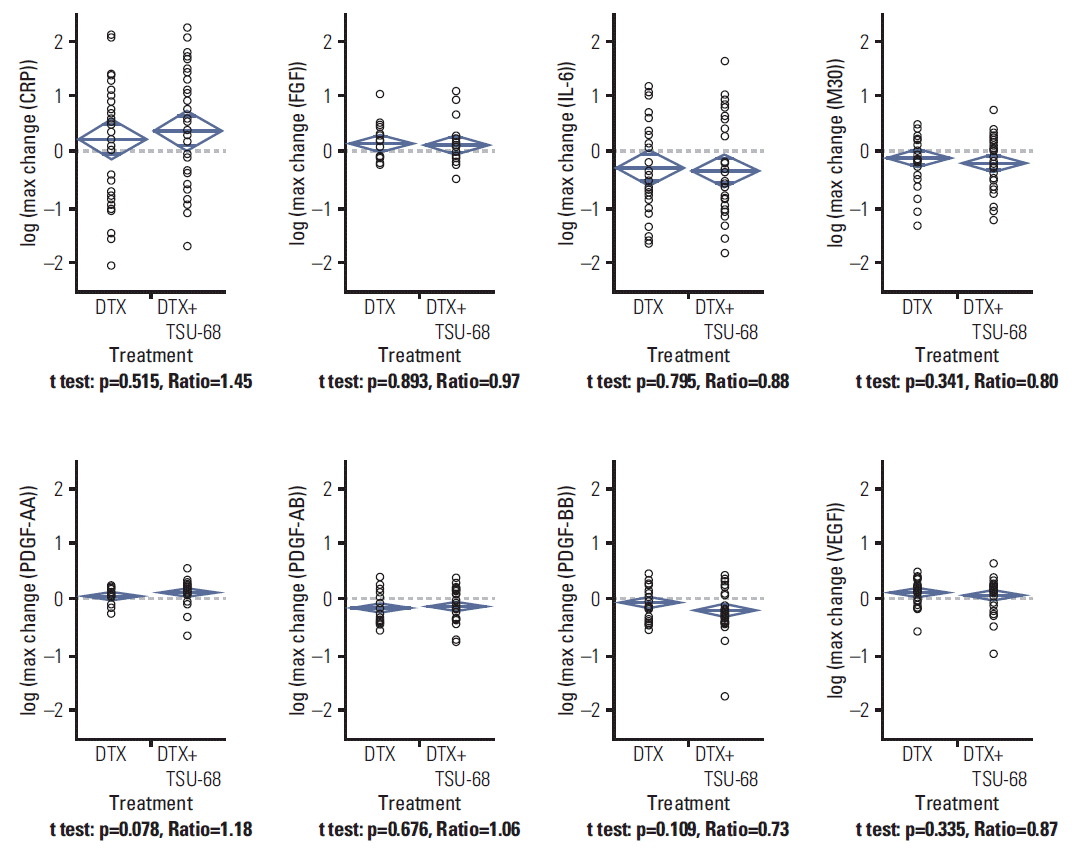
The pharmacodynamics of TSU-68 was assessed by estimation of fold changes in plasma biomarker concentration from baseline to each cycle of the study treatment and their maximal changes during the entire study course. For correlative analysis of candidate plasma biomarkers, baseline plasma concentration and fold changes from baseline to the end of the first cycle of the study treatment were dichotomized according to the median values. In the patient subgroup categorized by baseline biomarker levels, PFSs were compared between the two arms to evaluate the impact of baseline biomarker levels on the benefit of TSU-68. Analysis of fold changes in plasma biomarker concentrations from baseline across study treatment cycles was performed to determine the relationship with PFS in each treatment arm.
3. Statistical analysis
The chi-square test or Fisher exact test was used to determine the association between categorical variables, as appropriate. Log-transformation of the plasma biomarker concentration was performed for comparison. Comparisons of the means of biomarker-related variables between treatment groups and within groups were performed using the Student’s t test and Wilcoxon rank sum tests, respectively. The Kaplan-Meier method was used for estimation of the probability of survival, with comparison using a log-rank test. PFS was defined as the time between randomization and disease progression or death from any cause, whichever occurred first. Correlation analyses in terms of PFS were performed using baseline biomarker levels, and fold changes after the first cycle of study treatment. A p-value of < 0.05 was considered statistically significant, and all analyses were performed using SPSS ver. 18.0 (SPSS Inc., Chicago, IL).
Go to :

Discussion
In our current study, we found that the activity of TSU-68, which inhibits VEGFR-2, PDGFR, and FGFR, might be affected by baseline plasma PDGF-AA levels. The dynamics of circulating soluble proteins that reflect angiogenesis and tumor microenvironmental status showed correlation with PFS in patients treated with TSU-68, but not in those who received cytotoxic chemotherapy alone. Our findings have implications for the clinical development of TSU-68 and the identification of potential biomarkers for TSU-68.
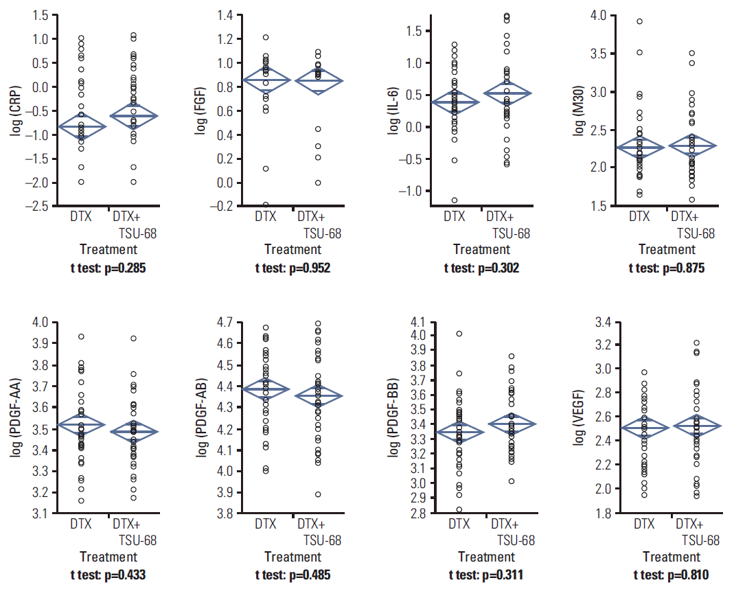
The combination of TSU-68 and docetaxel induced significant increases in the levels of CRP, PDGF-AA, PDGF-AB, and VEGF at the end of the first cycle compared to those at baseline, while VEGF was the only marker showing a significant increase with docetaxel. However, in comparison of the maximal magnitude of changes in biomarkers during the serial measurements, no significant difference in any biomarker was observed between the two groups. Previous studies have reported conflicting results regarding the pharmacodynamic effects of TSU-68 [
4,
5,
8]. In several studies, no significant changes in angiogenesis-related plasma proteins were observed after treatment with TSU-68 [
4,
5]. Although a previous pharmacodynamic analysis of colorectal cancer patients treated with TSU-68 in combination with fluoropyrimidine and oxaliplatin reported a significant decrease in the PDGF series (PDGF-AA, AB, and BB) [
8], it is unlikely that this result reflects the pharmacodynamic features of TSU-68 itself. Although it is possible that docetaxel confounded the effect of TSU-68 in our study, due to the randomized design of this trial, we were able to evaluate the pharmacodynamic impact of the addition of TSU-68 to docetaxel monotherapy. The limited impact of TSU-68 on pharmacodynamic markers in many trials, including ours, suggests that the current dose and schedule for TSU-68 may not be optimal to induce significant target inhibition and clinical efficacy.
In our primary analysis of this randomized phase II trial, TSU-68 plus docetaxel did not demonstrate superiority to docetaxel alone in terms of PFS [
6]. In addition, when patients were categorized by clinical factors, including hormone receptors, HER-2 overexpression, and resistance to previous therapy, there was no subgroup benefit from combination therapy.
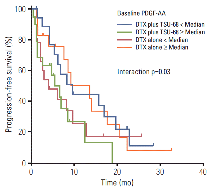
In our subanalysis of biomarker for TSU-68, we found that the efficacy of TSU-68 may depend on the pretreatment plasma concentration of PDGF-AA. The addition of TSU-68 appears to be associated with improved PFS in patients with low baseline PDGF-AA levels. However, a detrimental effect of TSU-68 was observed in patients with high baseline PDGF-AA levels, which contrasts with the expectation that PDGFR inhibition by TSU-68 may be more effective in patients with PDGF overexpression. This finding seems to be in line with the results of a previous randomized phase II study that compared TSU-68 with observation in patients with hepatocellular carcinoma [
9]. Although the result was not statistically significant, patients who received TSU-68 had a numerically longer PFS than those included in the observation arm in the subgroup with low baseline PDGF-AA levels, whereas this trend was reversed in patients with high baseline PDGF-AA levels.
Until recently, investigation of the predictive role of circulating dimeric PDGF isoforms for antiangiogenic agents has been limited. Dimeric PDGF isoforms primarily induce paracrine stimulation of stromal fibroblasts and perivascular cells and also activate autocrine signaling for the proliferation of tumors [
10]. However, the mechanism of the interaction between PDGF-AA and TSU-68 efficacy is unclear and will have to be defined in future studies. In many clinical trials for unselected patients with various types of cancer, TSU-68 has not demonstrated a significant improvement in clinical outcomes [
6,
9,
11]. Although the implication of PDGF-AA as a predictive factor for TSU-68 should be validated in a large patient population, our results indicate that further investigations of TSU-68 for patients with relatively low expression of PDGF-AA will be necessary.
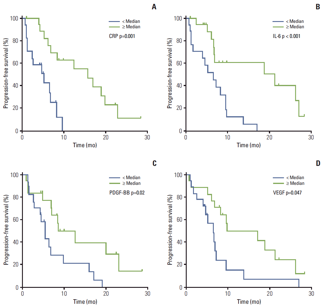
Monitoring of the dynamics of circulating protein biomarkers may provide the opportunity to predict clinical outcomes as well as achieve a better understanding of the mechanism of action of investigational drugs. In our current study, the dynamics of CRP, IL-6, PDGF-BB, and VEGF levels showed significant association with PFS in patients treated with TSU-68 plus docetaxel. This result confirms that the activity of TSU-68 is driven by the inhibition of angiogenesis and by its impact on the tumor microenvironment. The relationship between the dynamics of plasma biomarkers and PFS was not seen in patients who received docetaxel monotherapy, thus the significance of our finding appears quite robust. A greater increase above the median in plasma VEGF (a median fold increase of 1.1) and PDGF-BB (1.1) following TSU-68 plus docetaxel was associated with better PFS. Given that inhibition of VEGFR and PDGFR results in a reactive increase in plasma VEGF and PDGF-BB [
4,
12], this result may indicate that stronger VEGFR and PDGFR inhibition leads to a better PFS with TSU-68 treatment.
IL-6 is a proinflammatory marker and CRP is a well-known indicator of inflammatory status [
13,
14]. Patients with fold changes above the median in CRP (the median of 1.5) and IL-6 (0.7) after treatment with TSU-68 had improved clinical outcomes in terms of PFS compared with those who showed less increased or decreased CRP and greater decrease of IL-6, respectively. Increases in these inflammatory markers can be regarded as a consequence of therapy-induced inflammation [
13]. Death of cancer cells from anticancer treatment induces the release of necrotic products, leading to stimulation of cytokine-producing inflammatory cells. Currently, it is unclear whether therapy-induced inflammation is beneficial or harmful in cancer treatment [
13]. The inflammation may stimulate residual cancer cells and help in regrowth via the activation of prosurvival genes; however, inflammation may also lead to activation of adaptive anti-tumor immune responses due to the increased presentation of tumor antigens. Further investigations will be needed in order to elucidate the effects of the dynamics of these biomarkers, which reflect the status of the tumor microenvironment, on the efficacy of antiangiogenic agents.
There are several limitations to our current study. Although the randomized design of this trial enabled comparisons of the impact of candidate biomarkers for TSU-68, our analysis was based on a small number of patients, limiting the ability of the multivariate analysis to adjust for potential confounding effects.
Go to :


 PDF
PDF Citation
 Print
Print


 XML Download