Abstract
Purpose
Prostate specific antigen is not reliable in diagnosing prostate cancer (PCa), making the identification of novel, precise diagnostic biomarkers important. Since chemokines are associated with more aggressive disease and poor prognosis in diverse malignancies, we aimed to investigate the diagnostic relevance of chemokines in PCa.
Materials and Methods
Preoperative and early postoperative serum samples were obtained from 39 consecutive PCa patients undergoing radical prostatectomy. Serum from 15 healthy volunteers served as controls. Concentrations of CXCL12, CXCL13, CX3CL1, CCL2, CCL5, and CCL20 were measured in serum by Luminex. The expression activity of CXCR3, CXCR4, CXCR5, CXCR7, CXCL12, CXCL13, CX3CR1, CXCL1, CCR2, CCR5, CCR6, CCR7, CCL2, and CCL5 mRNA was assessed in tumor and adjacent normal tissue of prostatectomy specimens by quantitative real-time polymerase chain reaction. The associations of these chemokines with clinical and histological parameters were tested.
Results
The gene expression activity of CCL2 and CCR6 was significantly higher in tumor tissue compared to adjacent normal tissue. CCL2 was also significantly higher in the blood samples of PCa patients, compared to controls. CCL5, CCL20, and CX3CL1 were lower in patient serum, compared to controls. CCR2 tissue mRNA was negatively correlated with the Gleason score and grading.
Since serum prostate specific antigen (PSA) was introduced as an early detection marker of prostate cancer (PCa) in the 1980s, several trials involving novel and more precise biomarkers have failed to provide new markers with definitive clinical relevance and applicability. Considering the low sensitivity of PSA and the importance of distinguishing latent from clinically significant PCa to optimize treatment, the need for specific diagnostic markers is apparent. Recent investigations have pointed to caveolin-1 [1], exosomes [2], and PCA-3 [3] as diagnostic markers. However, their clinical applicability is still to be determined.
This pilot study was designed to investigate the potential of chemokines as biomarkers for PCa. Chemokines are low molecular weight secretory proteins divided into four groups (CXC, CC, C, and the CX3C family [4]) according to the arrangement of conserved cysteine (C) residues of the mature proteins, which interact as ligands (CL) with their corresponding receptors (CR). Due to their proinflammatory properties, chemokines regulate chemotaxis and metabolic activity of leukocytes circulating in the body and therefore significantly affect normal development, angiogenesis and atherosclerosis [4,5]. They are involved in many aspects of oncogenesis, including regulation of cancer cell growth, dissemination and host-tumor response [6,7]. Hence, various tumor types differentially trigger a complex chemokine network determining the qualitative and quantitative degree of immunocellular infiltration [8]. Alterations of the chemokine expression level are significantly associated with more aggressive disease and poorer prognosis in diverse malignancies [9,10]. However, their role in the tumorigenesis of PCa is unknown. Preliminary work pointed out the potential of CXC chemokines as prognostic biomarkers for PCa [11]. To assess the diagnostic relevance of chemokines in PCa, the expression of a broad panel of chemokines in tumor tissue and their serum levels in patients with PCa was investigated and associations tested with clinical and pathological parameters.
The local medical ethics committee approved the study. Patients undergoing radical prostatectomy for biopsyproven PCa between January 2008 and October 2010 in the Department of Urology, Goethe University, Frankfurt, Germany, were included in the study after signing informed consent for utilization of biomaterials and further scientific assessment. The tumor stage and grade was determined according to the sixth edition of the TNM classification [12]. Tumors were graded with the Gleason score [13] as well. Clinical parameters, including the patient’s age and preoperative and postoperative PSA values, were documented.
Ten milliliters of peripheral blood was drawn from patients the day before surgery and 5 to 7 days after surgery. As controls, 10 mL blood was drawn from 15 healthy, male, age-matched volunteers. Blood samples were allowed to coagulate and then centrifuged at 3,000 rpm at 4°C for 10 minutes. The serum supernatant was stored at –80°C.
The human cytokine multiplex bead assay kit (Millipore, Darmstadt, Germany) was used to determine CXCL12, CXCL13, CX3CL1, CCL2, CCL5, and CCL20 in serum, according to the manufacturer’s instructions. The filter plate was pre-wetted with 200 μL of washing solution. Anti-chemokine antibody coated beads (25 μL) were pipetted into each well. The plates were washed using a vacuum manifold. Samples (serum) or standards (25 μL) were then added to the wells and incubated with the beads for 2 hours. After washing, biotin conjugated detector antibody was added and incubated for 1 hour, followed by a 30-minute incubation with R-phycoerythrin conjugated streptavidin. All incubation steps were performed at room temperature in the dark. Analysis was carried out with a Luminex 100 (Gurce, Nivelles, Belgium) and expressed as pg/mL.
Immediately after resection, tissue was transferred to the Department of Pathology and reviewed by a pathologist. Tumor tissue, as well as adjacent normal tissue, was fresh-frozen in liquid nitrogen.
Total RNA from solid tumor tissue of patients with PCa and adjacent normal tissue was extracted using RNeasy kit (Qiagen, Hilden, Germany), according to the manufacturer’s instructions. Solid tissues were sliced into small pieces and transferred to a Precellys tube pre-filled with 1.4-mm ceramic beads (Bertin Technologies, Saint-Quentin-en-Yvelines Cedex, France) containing lysis buffer. Cells were lysed under rapid agitation at 6,500 rpm for 1 minute in three cycles using a Precellys 24 homogenizer (Bertin Technologies). cDNA was synthesized from 1 μg of total RNA with oligo(dT) primer using an AffinityScript QPCR cDNA synthesis kit (Stratagene, Santa Clara, CA), according to the manufacturer’s instructions. The amount and quality of RNA were analyzed using Nanovue (GE Healthcare, Piscataway, NJ) and bioanalyzer (Agilent Technologies, Palo Alto, CA), respectively.
Real-time quantitative PCR (qRT-PCR) was performed using RT2 SYBR-Green/RoxqPCR master mix and gene specific primers for CL and CR (both from Sabioscience, Madison, WI) (Table 1), according to the manufacturer’s protocol. Gene amplification was performed using MX3005P (Stratagene). One hundred nanograms of cDNA was used in the qPCR reaction in duplicate. The dissociation curve was checked at the end of each PCR reaction. Glyceraldehyde 3-phosphate dehydrogenase and β-actin were used as housekeeping gene and for normalization. Gene expression (fold change) was calculated using the ΔΔCt method, after normalizing the tumor sample with a corresponding normal sample.
All experiments were performed three to six times. Statistical significance of differences between parameters within the same group was tested by the Wilcoxon matched pairs test and in different groups by the Wilcoxon Mann-Whitney test. Subgroup analyses employed the Kruskal-Wallis test with the Iman-Conover method (Bonferroni-Holm correction). Correlation between two parameters was evaluated by the analysis of Spearman’s coefficient. Linear regression was assessed by Pearson’s linear regression test. The statistical program applied was BiAS for Windows (ver. 9.11, Dr. rer. nat. Hanns Ackermann, Epsilon Publishers, Frankfurt, Germany). Null hypotheses of no difference were rejected if p-values were less than 0.05. Continuous data are presented as mean±standard deviation.
The study population consisted of 39 patients (Table 2) with a mean age at tumor diagnosis of 68.9±5.6 years. None of the patients had evident clinical signs of infection or acute or chronic inflammation at the time of surgery. Histologically, all tumors were conventional acinar adenocarcinomas. In four patients the final histopathological analysis revealed positive margins (R1). Limited lymph node metastasis was detected in two other patients (N1). Perineural infiltration (Pn1) was found in 10 patients. All patients with microscopic residual disease (R1) and five other patients (2× pT3a, 2× pT3b and 1× pT2c with biochemical relapse after surgery) received radiation therapy. The mean follow-up time was 18.1±15.1 months. At the study endpoint, all patients were alive without signs of biochemical or clinical recurrence.
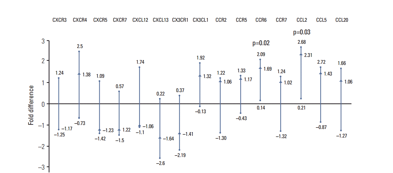
Using qRT-PCR, significantly higher expression of CCL2 and CCR6 in tumor tissue was observed, compared to normal adjacent tissue (Fig. 1). The expression of CXCR4, CX3CR1, CCR2, CCR5, CCR7, CCL5, and CCL20 was higher and that of CXCR3, CXCR5, CXCR7, CXCL12, CXCL13, and CX3CL1 lower in the tumor tissue without reaching significance.
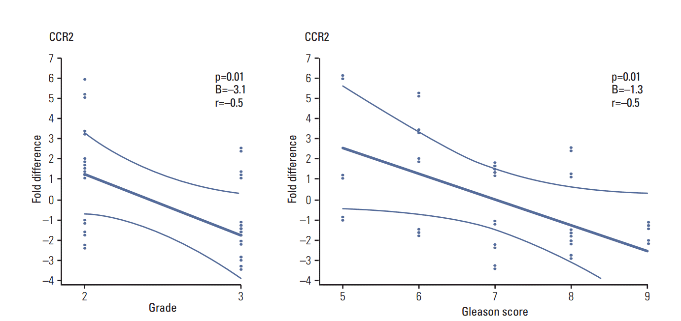
The association of gene expression of the chemokines in the tumor tissue was tested with clinical and oncologic parameters (Fig. 2). CCR2 revealed a significant negative linear correlation with the Gleason score and grading. No association was found between the other investigated chemokines and T stage, Gleason score, grade, R1- and Pn1-status, preoperative and postoperative PSA or biochemical relapse.
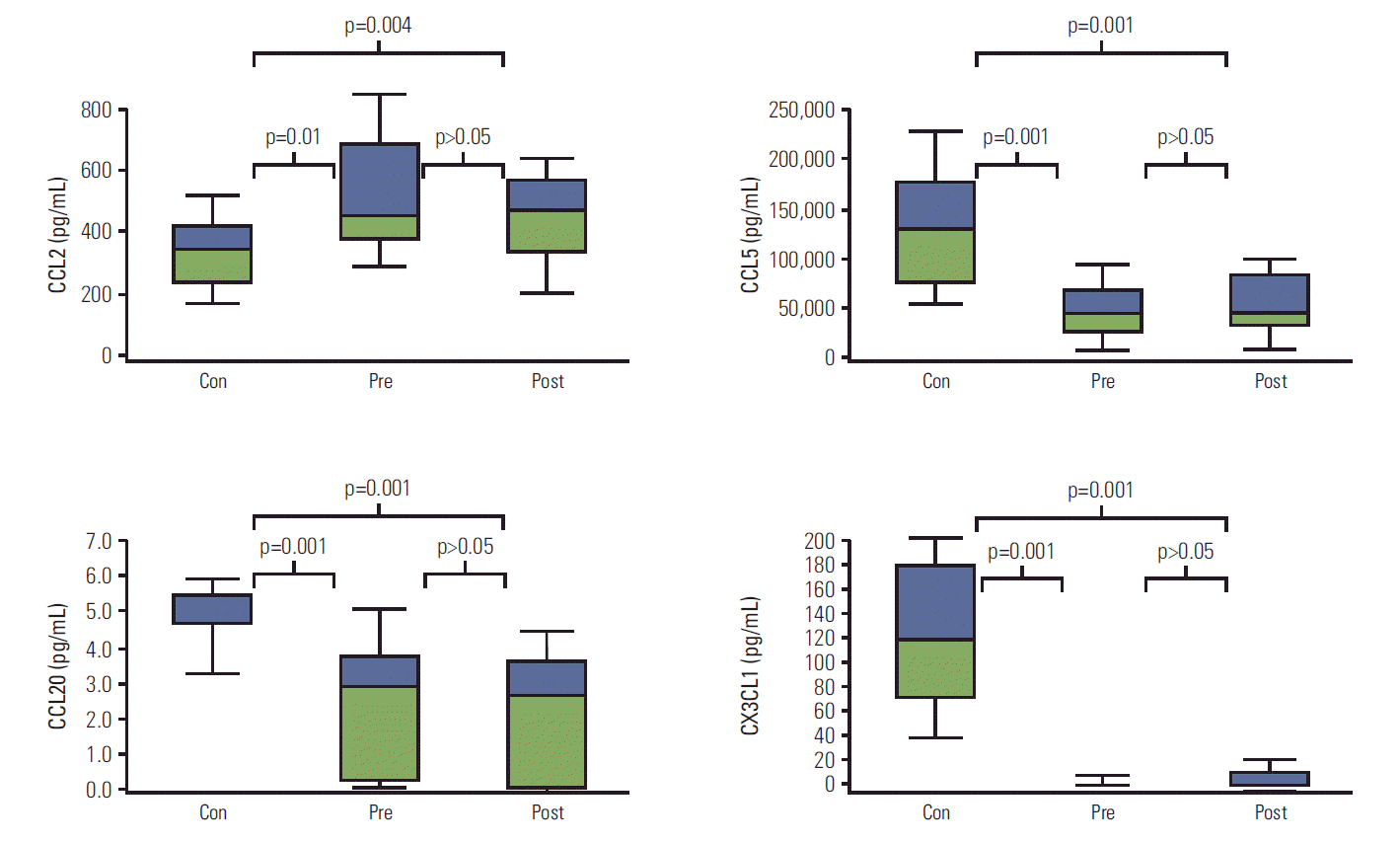
The chemokine concentration in serum was determined with Luminex (Fig. 3). CCL2 was significantly over-expressed both preoperatively and postoperatively in PCa patients, compared to controls. Significantly diminished concentrations of CCL5 in preoperative and postoperative samples were detected, compared to controls. The concentration of CCL20 increased significantly in the control group, compared to both preoperative and postoperative serum concentration in PCa patients. The serum concentration of CX3CL1 also increased significantly in the controls compared to that in patient serum, both preoperatively and postoperatively. The level of CXCL12 and CXCL13 did not significantly differ among any of the groups.
The association of chemokine serum concentrations with T stage, Gleason score, grade, R1- and Pn1-status, preoperative PSA as well as biochemical relapse were then tested. CCL2 was significantly negatively correlated with the preoperative PSA value (Spearman’s correlation coefficient rho, –0.48; p=0.035). No associations among these parameters and the other chemokines were apparent.
Chemokines are small secreted chemotactic cytokines that coordinate immunological machinery by promoting leukocyte migration and cross-communication among different cell types of the immune system [14]. Due to similarities between leukocyte trafficking and cancer cell dissemination, the role of chemokines in tumorigenesis is drawing increasing attention [15]. Recent studies regarding different tumor entities point to significant involvement of chemokines in facilitating tumor promotion and dissemination [16]. The present data explore the potential of chemokines as biomarkers in patients with PCa.
Based on the evaluation of 15 members of the chemokine family, evidence is provided here that the gene activity of particular molecules is altered in PCa, compared to normal tissue. CCL2 and CCR6 mRNA were overexpressed in the tumor specimens, compared to adjacent normal tissue. CCL2, but not CCR6, was also significantly augmented in serum from tumor patients compared to healthy controls, indicating a critical role of CCL2 in neoplastic progression. A meta-analysis of gene-expression data has identified CCL2 as a primary driver in PCa development [17]. Immunohistochemistry and immunofluorescence analysis of the chemokine expression profile reveal CCL2 to be predominantly expressed at the tumor site, more than for other chemokines [18]. Tissue microarray analysis shows that levels of CCL2 expression in PCa tissue are greater than in non-neoplastic epithelia [19]. Therefore, CCL2 could serve as a specific diagnostic marker, closely associated with PCa, even though CCL2 did not reach control values postoperatively. A possible explanation is that immunologic processes are still active five to seven days after surgical intervention. Studies incorporating blood sampling at later post-surgical dates should, therefore, more clearly define the specific diagnostic value of CCL2. One problem in employing CCL2 to diagnose PCa is the lack of association with tumor stage and grade, meaning that CCL2 may not allow evaluation of tumor dissemination. Shirotake et al. [20] found differences in CCL2 levels only between very early localized, and advanced castration resistant PCa. In contrast, the report from Lu et al. [19] pointed to a significant relationship between CCL2 level and Gleason score. Different analytic techniques (microarray) or a selected patient cohort (Asian) may account for this discrepancy. Obviously, further patient studies are necessary to clarify the diagnostic value of CCL2 for PCa.
CCL5, CCL20, and CX3CL1 were all reduced in patient sera, whereas no differences were seen in the gene analyses. Therefore, PCa progression seems accompanied by alterations in CCL5, CCL20, and CX3CL1 protein release but not in transcriptional modification. Data concerning this issue are sparse. In a recent report, CCL5 serum level was not altered in PCa patients, compared to controls. However, this study was limited to PCa with serum PSA values of < 10 ng/mL [21], which cannot be applied to our cohort with 41% of patients with a preoperative PSA > 10 ng/mL. Immunohistochemistry of 80 PCa cases revealed absent or weak CCL20 expression in 61%, whereas strong staining was evident in only 10% of patients [22]. This may corroborate our CCL20 data, although serum probes were not analyzed in this study [22]. CX3CL1 is down-regulated in high-risk PCa patients who develop biochemical recurrence following prostatectomy [23]. The authors of this case-control study recommend CX3CL1 as a potential indicator of future recurrence.
CXCL12 was not different between PCa patients and controls. CXCL12 is suggested to facilitate tumor cell recruitment to the bone marrow and a high CXCL12 expression is associated with lymph node metastatic prostate carcinoma compared with non–lymph-node metastatic cancer in an immunohistochemical analysis [24]. Semiquantitative PCR evaluation points to an enhanced CXCL12 mRNA expression level in prostate tumor, compared to adjacent 'normal' tissue [11]. Serum CXCL12 levels are significantly higher for men who were biopsy positive compared to those who were biopsy negative for PCa [25].
In contrast, Agarwal et al. [21], who performed multiplex enzyme-linked immunosorbent assay assays on 272 men, found no association between CXCL12 and PCa status [21]. These results agree with the present investigation. A final assessment of the role of CXCL12 in PCa is difficult since different techniques have been applied in relevant studies. More research is required to better interpret the clinical significance of CXCL12.
In conclusion, particular chemokines have been shown to be related to PCa. The CCL2 changes dominate other chemokine changes, since PCa related CCL2 alterations were found at both gene and protein level. However, CCR6, CCL5, CCL20, and CX3CL1 might be further candidates pointing to neoplastic transformation. Further large-volume biomarker studies are required to expand and validate the preliminary findings of this pilot study.
ACKNOWLEDGMENTS
We would like to thank Karen Nelson for critically reading the manuscript and Dr. Hanns Ackermann for statistical supervision. Grant from the “Heinrich und Erna Schaufler-Stiftung” (received 2012, principal investigator: Igor Tsaur).
References
1. Gumulec J, Sochor J, Hlavna M, Sztalmachova M, Krizkova S, Babula P, et al. Caveolin-1 as a potential high-risk prostate cancer biomarker. Oncol Rep. 2012; 27:831–41.
2. Principe S, Jones EE, Kim Y, Sinha A, Nyalwidhe JO, Brooks J, et al. In-depth proteomic analyses of exosomes isolated from expressed prostatic secretions in urine. Proteomics. 2013; 13:1667–71.

3. Shen M, Chen W, Yu K, Chen Z, Zhou W, Lin X, et al. The diagnostic value of PCA3 gene-based analysis of urine sediments after digital rectal examination for prostate cancer in a Chinese population. Exp Mol Pathol. 2011; 90:97–100.

4. Bonecchi R, Galliera E, Borroni EM, Corsi MM, Locati M, Mantovani A. Chemokines and chemokine receptors: an overview. Front Biosci (Landmark Ed). 2009; 14:540–51.

5. Lin TH, Liu HH, Tsai TH, Chen CC, Hsieh TF, Lee SS, et al. CCL2 increases alphavbeta3 integrin expression and subsequently promotes prostate cancer migration. Biochim Biophys Acta. 2013; 1830:4917–27.
6. Craig MJ, Loberg RD. CCL2 (monocyte chemoattractant protein-1) in cancer bone metastases. Cancer Metastasis Rev. 2006; 25:611–9.

7. Homey B, Muller A, Zlotnik A. Chemokines: agents for the immunotherapy of cancer? Nat Rev Immunol. 2002; 2:175–84.

8. Kulbe H, Levinson NR, Balkwill F, Wilson JL. The chemokine network in cancer--much more than directing cell movement. Int J Dev Biol. 2004; 48:489–96.
10. Strieter RM, Belperio JA, Phillips RJ, Keane MP. CXC chemokines in angiogenesis of cancer. Semin Cancer Biol. 2004; 14:195–200.

11. Wedel SA, Raditchev IN, Jones J, Juengel E, Engl T, Jonas D, et al. CXC chemokine mRNA expression as a potential diagnostic tool in prostate cancer. Mol Med Rep. 2008; 1:257.
12. Wittekind C, Meyer HJ, Bootz F. UICC: TNM Klassifikation maligner Tumoren. 6. Auflage. Berlin: Springer;2002.
13. Epstein JI, Allsbrook WC Jr, Amin MB, Egevad LL; ISUP Grading Committee. The 2005 International Society of Urological Pathology (ISUP) Consensus Conference on Gleason Grading of Prostatic Carcinoma. Am J Surg Pathol. 2005; 29:1228–42.

14. Waugh DJ, Wilson C, Seaton A, Maxwell PJ. Multi-faceted roles for CXC-chemokines in prostate cancer progression. Front Biosci. 2008; 13:4595–604.

15. Koizumi K, Hojo S, Akashi T, Yasumoto K, Saiki I. Chemokine receptors in cancer metastasis and cancer cell-derived chemokines in host immune response. Cancer Sci. 2007; 98:1652–8.

16. Tsaur I, Noack A, Waaga-Gasser AM, Makarevic J, Schmitt L, Kurosch M, et al. Chemokines involved in tumor promotion and dissemination in patients with renal cell cancer. Cancer Biomark. 2011; 10:195–204.

17. Gorlov IP, Sircar K, Zhao H, Maity SN, Navone NM, Gorlova OY, et al. Prioritizing genes associated with prostate cancer development. BMC Cancer. 2010; 10:599.

18. Izhak L, Wildbaum G, Weinberg U, Shaked Y, Alami J, Dumont D, et al. Predominant expression of CCL2 at the tumor site of prostate cancer patients directs a selective loss of immunological tolerance to CCL2 that could be amplified in a beneficial manner. J Immunol. 2010; 184:1092–101.

19. Lu Y, Cai Z, Galson DL, Xiao G, Liu Y, George DE, et al. Monocyte chemotactic protein-1 (MCP-1) acts as a paracrine and autocrine factor for prostate cancer growth and invasion. Prostate. 2006; 66:1311–8.

20. Shirotake S, Miyajima A, Kosaka T, Tanaka N, Kikuchi E, Mikami S, et al. Regulation of monocyte chemoattractant protein-1 through angiotensin II type 1 receptor in prostate cancer. Am J Pathol. 2012; 180:1008–16.

21. Agarwal M, He C, Siddiqui J, Wei JT, Macoska JA. CCL11 (eotaxin-1): a new diagnostic serum marker for prostate cancer. Prostate. 2013; 73:573–81.

22. Ghadjar P, Loddenkemper C, Coupland SE, Stroux A, Noutsias M, Thiel E, et al. Chemokine receptor CCR6 expression level and aggressiveness of prostate cancer. J Cancer Res Clin Oncol. 2008; 134:1181–9.

23. Blum DL, Koyama T, M'Koma AE, Iturregui JM, Martinez-Ferrer M, Uwamariya C, et al. Chemokine markers predict biochemical recurrence of prostate cancer following prostatectomy. Clin Cancer Res. 2008; 14:7790–7.

Fig. 1.
Analysis of chemokine gene activity obtained with real-time quantitative polymerase chain reaction in tumor tissue compared to normal adjacent tissue in patients with prostate cancer. Vertical line, Tukey confidence interval for Hodges-Lehmann estimator; ●, lower and upper limit of the Tukey confidence interval; ▲, Hodges-Lehmann estimator of the median fold difference.

Fig. 2.
Results from Pearson’s regression test for chemokine expression in prostate cancer samples and tumor grade or Gleason score. Presented as regression line (thick) and confidence borders (thin lines) with confidence p=0.95. p, p-value of the regression test; B, regression coefficient; r, Pearson’s correlation coefficient. Chemokine expression as fold difference obtained with real-time quantitative polymerase chain reaction.

Fig. 3.
Chemokine concentration obtained with Luminex. Chemokines with significant differences between at least two samples are presented. Box-plot: Con, control serum; Pre, preoperative serum; Post, postoperative serum. Box: lower line, quartile Q1 (25%-quantile); middle line, median; upper line, Q3 (75%-quantile); aerials, extreme values; horizontal bracket, respective p-value of the concentration difference in Wilcoxon-matched-pairs or Wilcoxon-Mann-Whitney test.

Table 1.
Gene specific primers used for qRT-PCR with catalogue numbers
Table 2.
Clinical and histopathological characteristics of the study cohort


 PDF
PDF Citation
 Print
Print


 XML Download