Introduction
Case Report
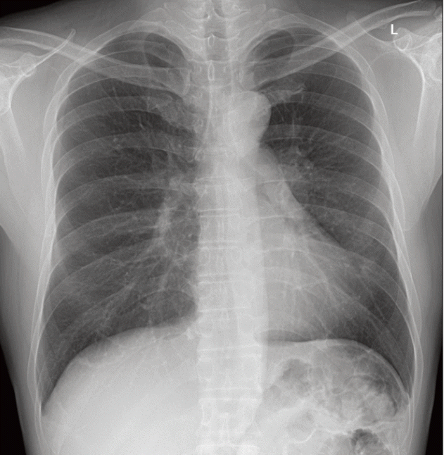 | Fig. 1.Initial chest radiography. The endobronchial mass with left upper lobar bronchial narrowing was overlooked on the initial chest radiography. |
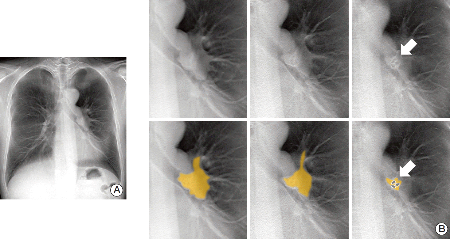 | Fig. 2.Digital tomosynthesis (DTS). (A) DTS reveals a lobulating contoured endobronchial lesion nearly obstructing the left main bronchus. (B) The tumor extends to the left upper lobar bronchus and the left lower lobar bronchus. However, the superior segmental bronchus is not obliterated on the last image with patent three holes (arrows). The extent and margins of the endobronchial tumor are well demarcated on serial anterior-to-posterior coronal images from the DTS. The yellow overlay and gray patent bronchi are demonstrated in the lower images. |
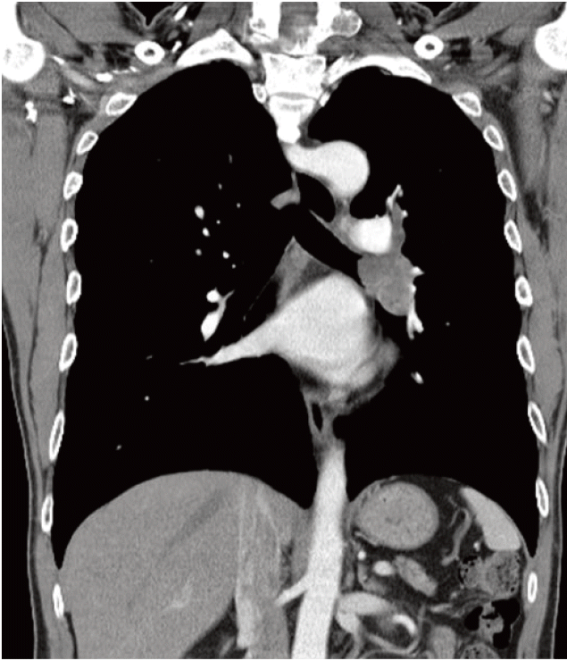 | Fig. 3.On the computed tomography images, a mildly enhanced lesion with finger-like appearance was noted along the left main bronchus extending to the upper and lower lobar bronchi, as suspected on digital tomosynthesis. |


 PDF
PDF Citation
 Print
Print


 XML Download