Introduction
Materials and Methods
1. Cell culture and TGF-β treatment
2. Western blot analysis
3. RNA extraction and Northern blot analysis
4. Actinomycin D chase experiment (mRNA stability assay)
5. Reverse transcription PCR and real-time PCR
6. Transient transfection and luciferase assays
7. Smad2, Smad3, and TTP knockdown using small interfering RNA (siRNA)
8. Statistic analysis
Results
1. TGF-β blocks spontaneous induction of COX-2 expression in A549 cells
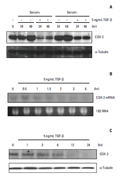
 | Fig. 1.Expression of cyclooxygenase 2 (COX-2) mRNA and protein is suppressed by transforming growth factor β (TGF-β) treatment in A549 cells. (A) COX-2 protein levels were spontaneously elevated after cell seeding. This increase was more marked with serum starvation. TGF-β blocked the induction of COX-2 expression in the presence or absence of serum. A549 cells were incubated with or without 5 ng/mL TGF-β in the presence or absence of serum. Total cellular proteins were isolated at the indicated times, after which Western blotting was performed with anti–COX-2 or anti– α-tubulin antibodies. (B) COX-2 mRNA expression was down-regulated by TGF-β. The cells were treated with 5 ng/mL TGF-β for 1.5 to 2 hours, after which the total RNA was isolated at the indicated times and subjected to Northern blot analysis (20 μg per lane) to measure COX-2 mRNA expression. (C) COX-2 protein levels were down-regulated by TGF-β in A549 cells. The cells were treated with 5 ng/mL TGF-β. Total cellular proteins were isolated at the indicated times and analyzed by Western blotting as described above. |
2. TGF-β does not affect COX-2 promoter activity in A549 cells
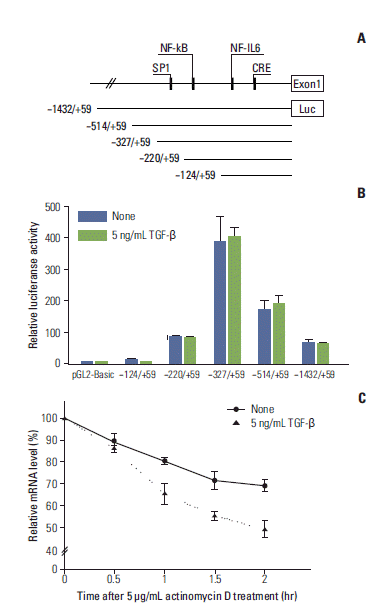
 | Fig. 2.Transforming growth factor β (TGF-β) has no effect on cyclooxygenase 2 (COX-2) promoter activity, but affects COX-2 mRNA half-life. (A, B) Cells were transfected with a pGL2 basic control vector or the indicated reporter constructs, then treated with or without 5 ng/mL TGF-β for 6 hours. Levels of luciferase activity were measured as described in the Materials and Methods section. Data were normalized relative to β-galactosidase activity. No significant difference between the luciferase activities of TGF-β–treated and untreated cells was observed. (C) Cells were stimulated with TGF-β (5 ng/mL) for 1 hour, then treated with actinomycin D (5 μg/mL). The cells were harvested at the indicated times after actinomycin D was added. Total RNA was isolated, and real-time polymerase chain reaction was performed to measure the expression of COX-2 and glyceraldehyde 3-phosphate dehydrogenase (GAPDH). The levels of COX-2 mRNA were normalized relative to GAPDH mRNA and presented as a percentage of that found in the untreated control. NF-kB, nuclear factor kB; IL-6, interleukin 6; CRE, cAMP response element. |
3. COX-2 mRNA is post-transcriptionally regulated via destabilization by TGF-β
4. COX-2 expression is down-regulated by TGF-β in a Smad3-dependent manner
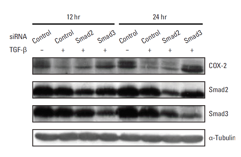
 | Fig. 3.Smad3 knockdown restores the transforming growth factor β (TGF-β)–mediated down-regulation of cyclooxygenase 2 (COX-2) expression. A549 cells were transiently transfected with 80 nM of Smad2- or Smad3-specific siRNA. After transfection, the cells were treated with vehicle or 5 ng/mL of TGF-β for 12 or 24 hours, after which cell lysates were obtained for Western blot analysis. Total cell proteins (70 μg) were subjected to Western blotting and the membranes were probed with the indicated antibodies. |
5. TTP expression is rapidly and transiently induced by TGF-β
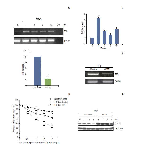
 | Fig. 4.Tristetraprolin (TTP)-mediated cyclooxygenase 2 (COX-2) mRNA destabilization is induced by transforming growth factor β (TGF-β). (A, B) Total RNA was recovered from A549 cells treated with TGF-β (5 ng/mL) for different periods of time and Reverse transcribed. Reverse transcription–polymerase chain reaction (RT-PCR) and real-time PCR were then performed using primers specific for TTP, β-actin, and glyceraldehyde 3-phosphate dehydrogenase (GAPDH) as described in the Materials and Methods section. The columns represent the mean of three independent experiments and are shown with error bars (±SE). *p < 0.001. (C) A549 cells were transfected with control or TTP-specific siRNA. After 24 hours of transfection, the cells were treated with TGF-β for 1 hour. RT-PCR and quantitative real-time PCR were used to measure the levels of TTP mRNA. Columns, the mean of three independent experiments; Bars, ±SE; *p < 0.001. (D) Cells transfected with control or TTP-specific siRNA were stimulated with or without TGF-β for 1 hour, after which actinomycin D (5 μg/mL) was added. Total RNA was isolated at different times, and COX-2 mRNA levels were quantified by real-time PCR. COX-2 mRNA expression was analyzed as described for Fig. 2C. (E) Cells transfected with control or TTP-specific siRNA were stimulated with TGF-β for the indicated times. Total cell proteins were then recovered and analyzed by Western blotting as described in the Materials and Methods. |


 PDF
PDF Citation
 Print
Print


 XML Download