Abstract
Notes
Ethics Statement
This study was approved by the Institutional Review Board of Samsung Medical Center with a waiver of informed consent (IRB No. 2021-03-125).
Availability of Data and Material
The datasets generated or analyzed during the study are available from the corresponding author on reasonable request.
Author Contributions
Conceptualization: YLO. Data curation: JL. Formal analysis: JL. Investigation: JL, YC, IH, KHC, YLO. Methodology: JL, YC. Resources: YLO. Supervision: YLO. Validation: YLO, JL. Visualization: JL, YC. Writing—original draft: JL. Writing—review & editing: YLO, YC. Approval of final manuscript: all authors.
References
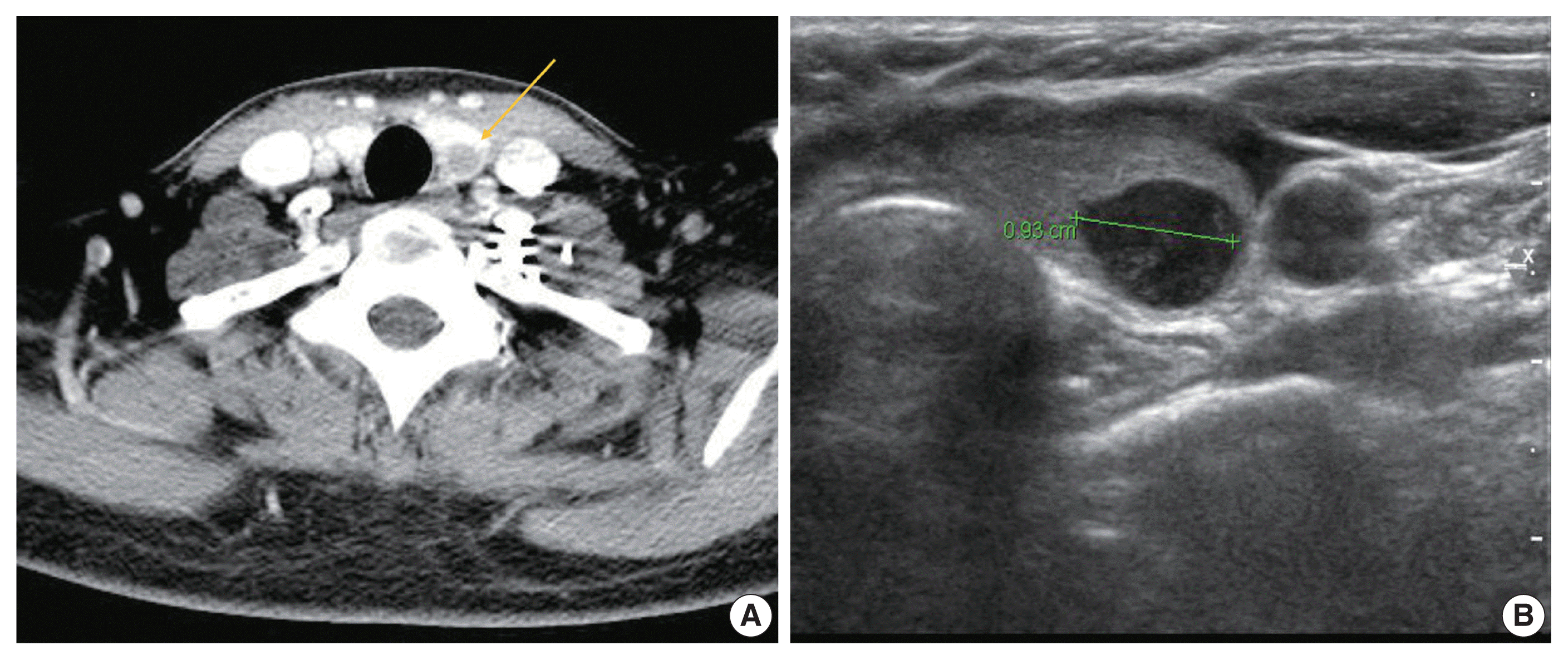
Fig. 1

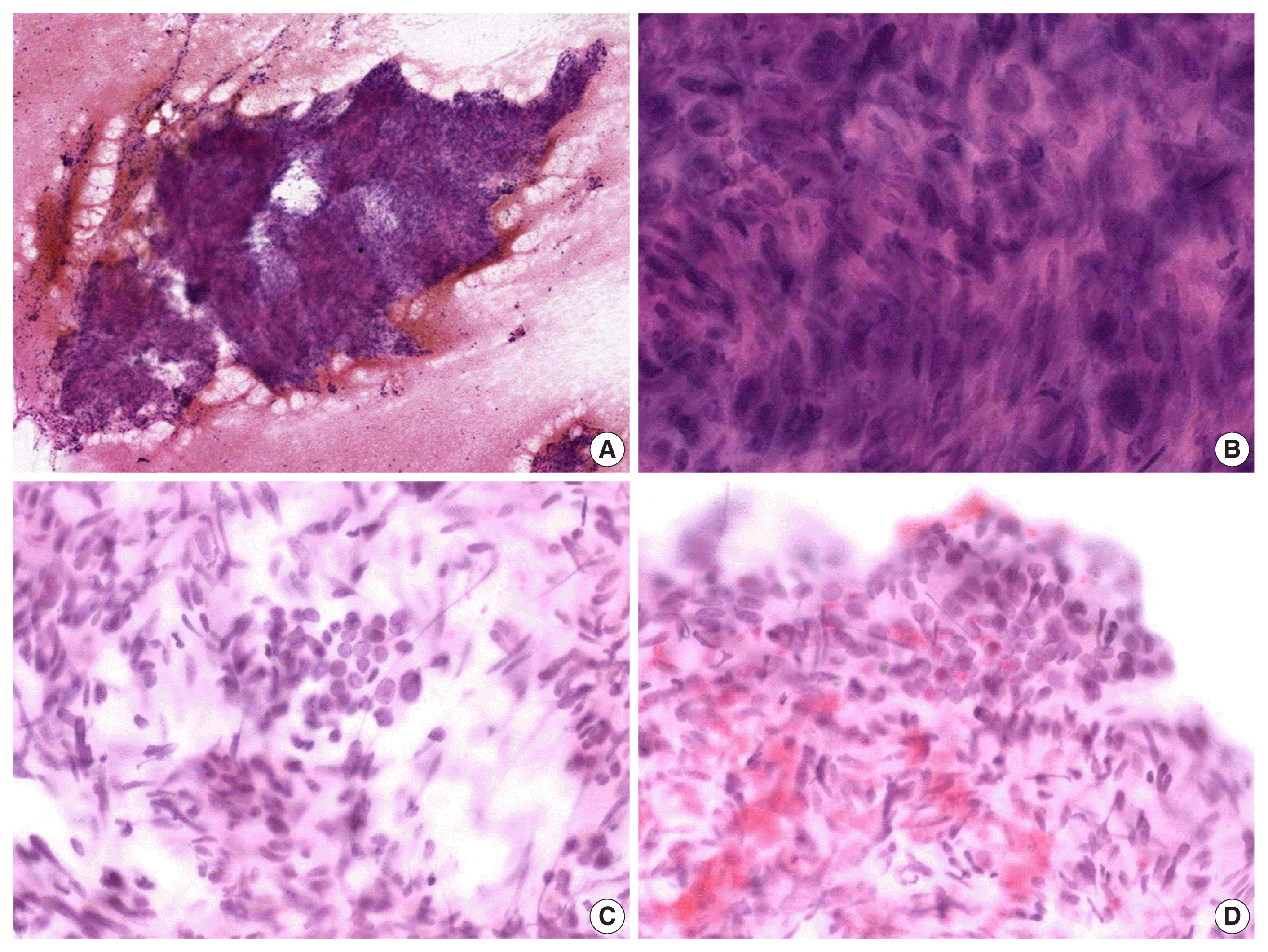
Fig. 2

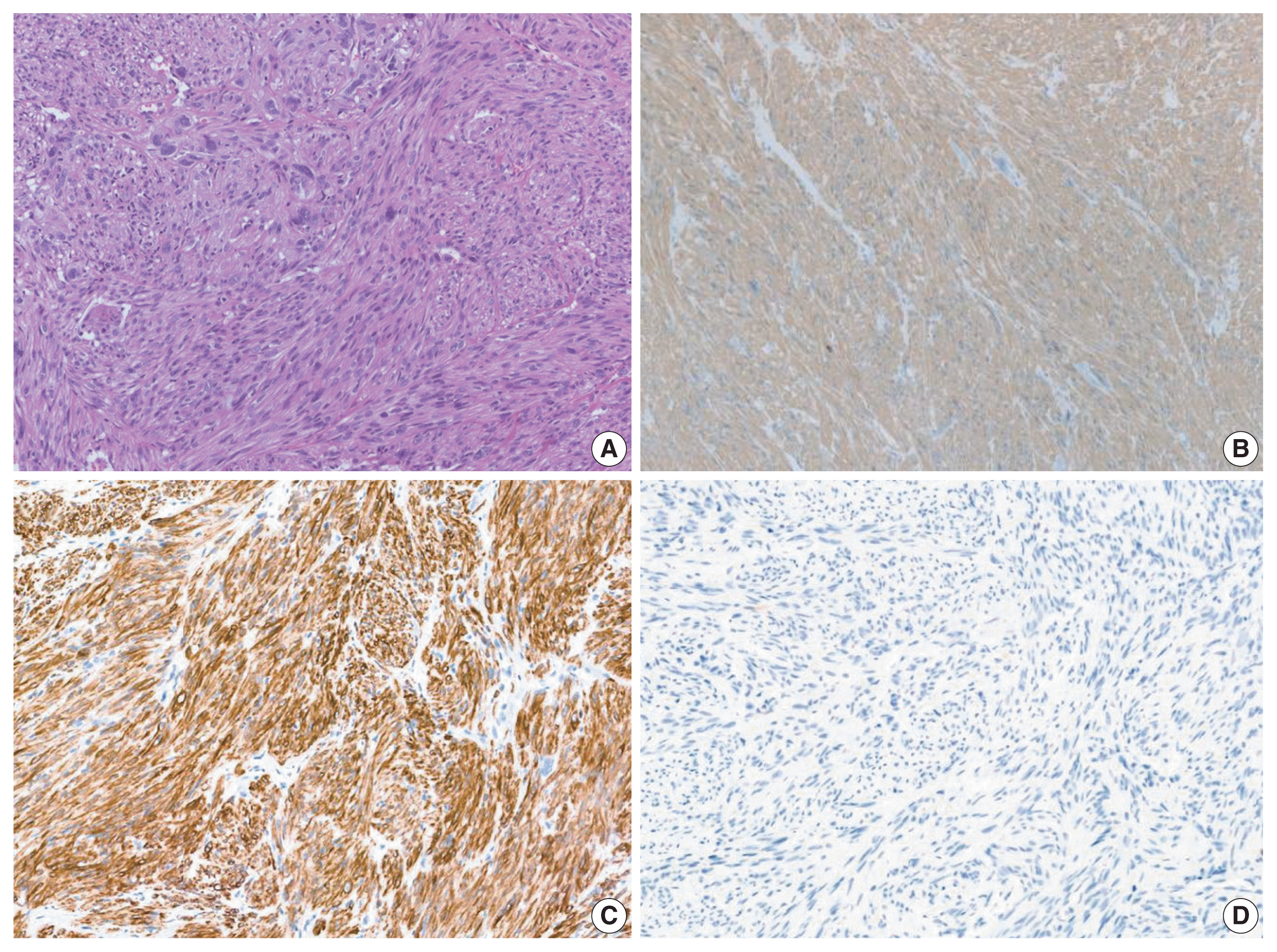
Fig. 3

Table 1
| Author | Age (yr) | Sex | Origin | Chief complaint | Location/size | Cytology | Pathology | Vimentin | Desmin | SMA | CD34 | S100 | Cytokeratin | Thyroglobulin |
|---|---|---|---|---|---|---|---|---|---|---|---|---|---|---|
| Gross and Horton (1975) [5] | 46 | M | Great saphenous vein | Painless swelling in neck | N/M | - | - | - | - | - | - | - | - | - |
| Cruickshank (1988) (as cited in Vankalakunti et al. [6]) | 30 | F | Uterus | Diffuse thyroid swelling | N/M | - | - | - | - | - | - | - | - | - |
| Gattuso et al. (1989) [2] | N/M | N/M | Retroperitoneal | N/M | Left thyroid/3 cm | - | - | - | - | - | - | - | - | - |
| Bode-Lesniewska et al. (1994) [7] | 69 | F | Duodenum | N/M | 5 cm | - | - | - | - | - | - | - | - | - |
| Michelow (1995) (as cited in Vankalakunt et al. [6]) | N/M | N/M | Uterus | Thyroid nodular swelling | N/M | - | - | - | - | - | - | - | - | - |
| Nakhjavani (1997) (as cited in Vankalakunti et al. [6]) | N/M | N/M | Uterus | N/M | N/M | - | - | - | - | - | - | - | - | - |
| Wang et al. (1998) [8] | 63 | F | Left leg | Rapidly progressive enlargement of the neck | N/M | - | - | - | - | - | - | - | - | - |
| Chen et al. 1999) [9] | N/M | N/M | Stomach | N/M | Unilateral | Anaplastic, malignant spindle cell tumor | N/D | - | - | - | - | - | - | - |
| Leath et al. (2002) [10] | 5th decade | F | Uterus | Postmenopausal bleeding | Diffuse and numerous/up to 2.9 cm | N/D | Smooth muscle cells with atypia and mitotic figures | N/D | Strong positive | Focal positive | N/D | N/D | N/D | N/D |
| D’Andrea et al. (2003) [11] | 60 | F | Pulmonary artery | Dyspnea and hemoptysis | Both thyroid/up to 4.7 cm | N/D | N/D | - | - | - | - | - | - | - |
| Deng et al. (2005) [12] | N/M | N/M | Leg | N/M | N/M | - | - | - | - | - | - | - | - | - |
| Giannikaki et al. (2006) [13] | 54 | F | Uterus | Single “cold” nodule in the right thyroid | Right thyroid/0.12 cm co-existing with PTC | N/D | Spindle cells with prominent nuclear atypia and mitoses | N/D | Positive | Positive | N/D | N/D | Negative | N/D |
| Eloy et al. (2007) [14] | 54 | F | Uterus | Rapidly enlarging and painful left neck mass | Left thyroid/2.5 cm | Consistent with a sarcomatous lesion | Large cells with hypercellularity, nuclear atypia, and pleomorphism with atypical mitoses | Positive | Negative | Negative | N/D | Negative | Negative | Negative |
| Nemenqani et al. (2010) [1] | 86 | F | Pelvic soft tissue | Right thyroid swelling | Right thyroid | Hypercellular spindle cell proliferation arranged in sheets mixed with stroma; marked pleomorphism; large hyperchromatic nuclei; numerous mitoses | Numerous mitoses and bizarre nuclei | Positive | Negative | Positive | Negative | Negative | Negative | Negative |
| Young et al. (2011) [15] | 55 | F | Lung | Cough | N/M | N/D | Fascicular pattern of spindle-shaped cells with increased cellularity and hyperchromatic, bizarre, malformed, and megakaryocytic nuclei | Positive | Focal positive | Positive | Negative | Negative | N/D | N/D |
| Gauthe et al. (2017) [16] | 47 | F | Uterus | Fast growing thyroid nodule | N/M | N/D | N/D | N/D | N/D | N/D | N/D | N/D | N/D | N/D |
| Golovko et al. (2020) [17] | 65 | F | Vagina | Multinodular toxic goiter | Right thyroid | Suggestive of the benign thyroid lesion | Consistent with metastatic vaginal leiomyosarcoma | N/D | N/D | N/D | N/D | N/D | N/D | N/D |
| Irizarry-Villafane et al. (2020) [18] | 47 | F | Uterus | Neck discomfort | Left upper lobe/2.4 cm | Marked cellularity of atypical spindle cells, dyscohesive, and in tissue aggregates with some binucleated cells | Spindle cell tumor with frequent mitotic figures | N/D | Positive | Positive | N/D | N/D | Negative | Negative |


 PDF
PDF Citation
 Print
Print


 XML Download