Journal List > J Pathol Transl Med > v.54(4) > 1152463

Kim, Kim, Kim, Bae, Kim, and Jung: Highly prevalent BRAF V600E and low-frequency TERT promoter mutations underlie papillary thyroid carcinoma in Koreans

Abstract

Background

The presence of telomerase reverse transcriptase (TERT) promoter mutations have been associated with a poor prognosis in patients with papillary thyroid carcinomas (PTC). The frequency of TERT promoter mutations varies widely depending on the population and the nature of the study.

Methods

Data were prospectively collected in 724 consecutive patients who underwent thyroidectomy for PTC from 2018 to 2019. Molecular testing for BRAF V600E and TERT promoter mutations was performed in all cases.

Results

TERT promoter alterations in two hotspots (C228T and C250T) and C216T were found in 16 (2.2%) and 4 (0.6%) of all PTCs, respectively. The hotspot mutations were significantly associated with older age at diagnosis, larger tumor size, extrathyroidal extension, higher pathologic T category, lateral lymph node metastasis, and higher American Thyroid Association recurrence risk. The patients with C216T variant were younger and had a lower American Thyroid Association recurrence risk than those with hotspot mutations. Concurrent BRAF V600E was found in 19 of 20 cases with TERT promoter mutations. Of 518 microcarcinomas measuring ≤1.0 cm in size, hotspot mutations and C216T variants were detected in five (1.0%) and three (0.6%) cases, respectively.

Conclusions

Our study indicates a low frequency of TERT promoter mutations in Korean patients with PTC and supports previous findings that TERT promoter mutations are more common in older patients with unfavorable clinicopathologic features and BRAF V600E. TERT promoter mutations in patients with microcarcinoma are uncommon and may have a limited role in risk stratification. The C216T variant seems to have no clinicopathologic effect on PTC.

The incidence of thyroid cancer has dramatically increased over the past several decades [1,2]. The highest incidence of thyroid cancer in the world has been observed in Korea [3,4]. The increase in the incidence of thyroid cancer was responsible for the increase in papillary thyroid carcinoma (PTC), which accounts for over 95% of all thyroid cancer cases in Korea [3,5,6]. Despite the increased incidence of thyroid cancer, the thyroid cancer mortality rate has not changed significantly over the last three decades [3,5]. A multicenter cohort study reported a disease-specific 10-year survival rate of 98% in Korean patients with well-differentiated thyroid carcinoma [6].
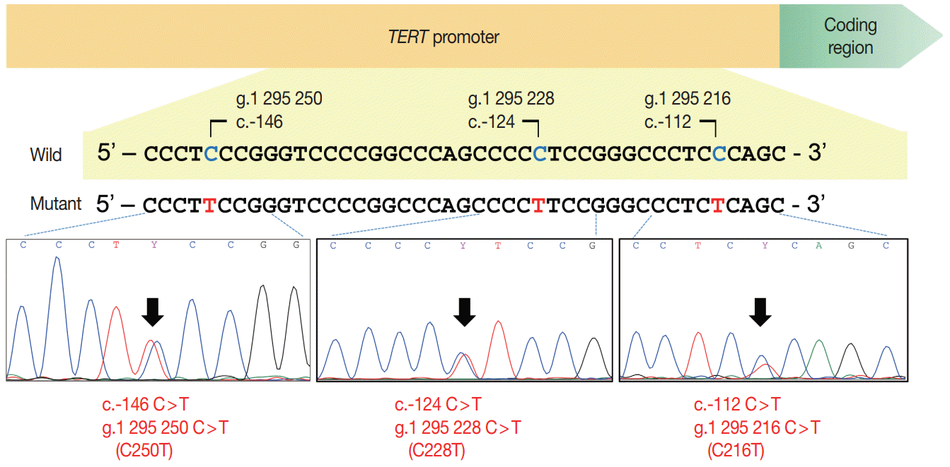
Independent prognostic factors related to survival in patients with PTC include elements of cancer staging, such as patient age at diagnosis, tumor size, extensive extrathyroidal extension, and distant metastasis [7]. There have been many studies demonstrating the prognostic value of molecular markers for tumor recurrence and survival. Telomerase reverse transcriptase (TERT) promoter mutation is one of the most evident molecular factors related to poor prognosis of patients with PTC [8-11]. The cancer-specific TERT promoter mutations occur in two mutually exclusive hotspots in chromosome 5, g.1 295 228 C>T (C228T) and g.1 295 250 C>T (C250T) which correspond to 124 bp (c.–124C>T) and 146 bp (c.–146C>T), upstream from the translation start codon of the TERT gene promoter sequence [10-16]. The pooled prevalence of TERT promoter mutations in PTC was 11.3% (95% confidence interval, 9.3 to 13.5) in a previous meta-analysis of 13 studies [14]. However, the retrospective data may overestimate the mutation frequency because of potential patient selection bias. Patients with microcarcinoma ≤ 1.0 cm were more easily excluded from the molecular studies [8-10,15,17-19]. Furthermore, old archival paraffin blocks may have suboptimal DNA quality that results in molecular test failures and analytical errors.
The BRAF V600E mutation is the most common genetic alteration in PTC and remains controversial as an independent prognostic factor [7]. The coexistence of BRAF V600E and TERT promoter mutations, however, could more accurately indicate the highest mortality risk for patients with PTC [8,19].
The present study aimed to evaluate the real-world frequency of TERT promoter mutations in prospectively-collected consecutive cases of PTC and assess the relationship between TERT promoter mutations and clinicopathological features in Korean patients with PTC and a high frequency of BRAF V600E mutations.

MATERIALS AND METHODS

Patients

We reviewed the prospectively collected data from 724 consecutive patients who underwent thyroidectomy for PTC and molecular testing at Seoul St. Mary’s Hospital of the Catholic University of Korea from 2018 to 2019. Molecular tests for BRAF and TERT promoter mutations were performed in all patients who agreed to allow molecular analysis of their surgical specimens. In cases of multifocal PTCs, the largest tumor was defined as the primary lesion and was chosen for evaluation. The histologic variants of PTC were classified following the diagnostic criteria and terminology of the World Health Organization [7]. The tall cell variant was defined using 30% of tall cell area as a criterion. The PTCs were further classified as classic PTC with tall cell features if it harbored between 10%–30% tall cells and as classic PTC if it contained less than 10% of tall cell area and any well-formed papillae. Cancer staging was done using the 8th edition of the American Joint Committee on Cancer (AJCC) staging system [20]. Minimal extrathyroidal extension was defined as extrathyroidal invasion that was restricted to the perithyroidal soft tissues detected only on microscopic examination (including microscopic strap muscle invasion). When strap muscle invasion was found on preoperative imaging and/or at the time of surgery, the case was considered as gross extrathyroidal extension. Risk stratification of patients for tumor recurrence was done using the 2015 American Thyroid Association (ATA) guidelines [21].

Mutational analyses for TERT promoter and BRAF V600E mutations
Genomic DNA was extracted from 10 μm-thick formalin-fixed paraffin-embedded (FFPE) tissue blocks using a Maxwell 16 FFPE Tissue LEV Purification Kit (Promega, Fitchburg, WI, USA). Tumor areas were manually dissected with a scalpel under a microscope.
The TERT promoter was amplified using the nested polymerase chain reaction (PCR) method. The first-round 235-bp PCR amplicon was amplified using forward 5'-AGTGGATTCGCGGGCACAGA-3' and reverse 5'-CAGCGCTGCCTGAA ACTC-3' primers. Then, the second-round 163-bp PCR amplicon was amplified using forward 5'-GTCCTGCCCCTTCACCTT3' and reverse 5'-CAGCGCTGCCTGAAACTC-3' primers. Bidirectional Sanger sequencing was performed in both directions using the same primers that were used for the second-round PCR. BRAF V600E mutation was analyzed using the real-time PCR clamping technology of PNAClampTM BRAF kit (Panagene, Daejeon, Korea) [22]. Each test had a positive control of mutation-holding human genomic DNA and a negative control of distilled water.

Statistical analysis

Categorical variables were analyzed using the Pearson’s chi-square, Fisher exact test, or linear-by-linear association when appropriate. Continuous variables were compared using the Student’s t-test or Mann-Whitney test when appropriate. The statistical significance threshold was defined as a p-value less than 0.05. All statistical analyses were done using SPSS Statistics program, ver. 21.0 (IBM Corp., Armonk, NY, USA).

RESULTS

Demographic and clinicopathologic characteristics

Table 1 summarizes the baseline clinicopathologic characteristics of the 724 patients with PTC. The median age of the patients at the time of diagnosis was 46 years (interquartile range [IQR], 36 to 56 years). The female to male ratio was 2.7:1. The median tumor size was 0.7 cm (IQR, 0.5 to 1.1 cm). The proportion of microcarcinomas (≤1.0 cm in size) was 71.5% (518/724). Lobectomy was done in 504 (69.6%) and total thyroidectomy in 191 patients (26.4%). The numbers of patients with minimal and gross extrathyroidal extension were 405 (55.9%) and 41 (5.7%), respectively. Cervical lymph node metastases were found in 409 patients (56.5%).

Frequency of TERT promoter and BRAF V600E mutations

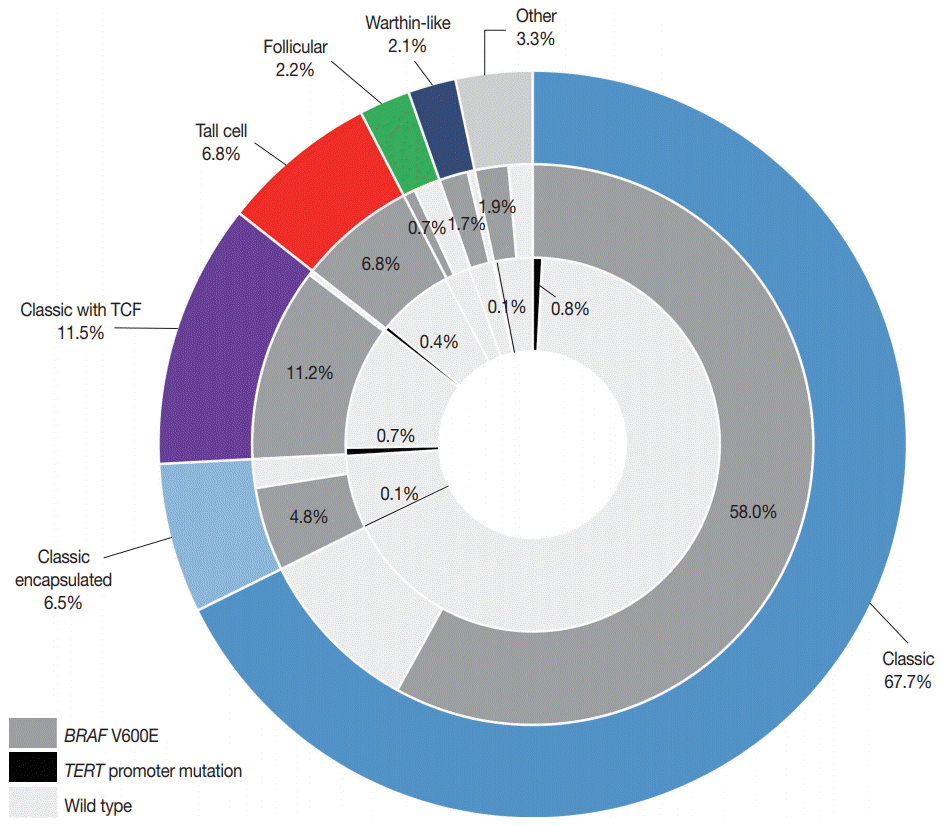
Hotspot-point mutations (C228T and C250T) in the TERT promoter were found in 16 (2.2%) patients: 14 with C228T and two with C250T (Table 2, Fig. 1). Four cases had the TERT promoter variant of g.1 295 216 C>T (c.–112C>T) (hereafter C216T) (Fig. 1). The BRAF V600E mutation was found in 616 (85.1%) patients. Of 20 PTCs with TERT promoter aberrations, 19 had coexisting BRAF V600E (Table 3, Fig. 2). Fig. 2 summarizes the distribution of histologic variants of PTC and mutational profiles according to the variants. There was no correlation between TERT promoter mutations and histologic variants.

Clinicopathologic features of patients with TERT promoter mutation

Hotspot mutations in the TERT promoter were significantly associated with age ≥55 years (p<.001), tumor size >1.0 cm (p=.001), extrathyroidal extension (p=.032), lateral lymph node metastasis (p=.041), and higher ATA recurrence risk (p<.001) (Table 2). Compared with patients with hotspot mutations, those with C216T variant were younger (p=.032) and had a lower rate of high ATA recurrence risk (p=.014). There were no significant differences in the clinicopathologic features between the patients with wild-type TERT promoter mutations and those with C216T variant of the TERT promoter (Table 2). Table 3 shows the detailed clinicopathologic features of the patients with a TERT promoter mutation.

DISCUSSION

The strength of this study stems from the prospectively collected data encompassing all consecutive patients treated for PTC with thyroid surgery. In our study, almost three-quarters of patients with PTC underwent thyroid surgery before the age of 55 years (73.3%) and had microcarcinomas (71.5%). Gross extrathyroidal extension was found in only 41 patients (5.7%). No case developed synchronous distant metastasis. Therefore, it stands to reason that the vast majority (86.0%) of patients with PTC were assigned to stage I by the 8th edition of the AJCC staging system. Although BRAF V600E mutations were highly prevalent in our study cohort, the frequency of hotspot mutations in the TERT promoter was 2.2%, which is far lower than that reported in previous studies for PTC (pooled mean prevalence of 11.3%) [14,23]. These results indicate that most PTC tumors in the current study should have indolent behavior.
In our study, additional benefits were gained by including microcarcinomas in the molecular analysis. Hotspot mutations of the TERT promoter were found in five of 518 papillary microcarcinomas (1.0%). Minimal extrathyroidal extension, found in three of the five patients with hotspot mutations, did not affect the pathologic T category. Patient age ranged from 39 to 84 years. Although the frequency of TERT promoter mutations is lower than that of previous studies, these findings are in line with a previous Italian study showing no correlation with unfavorable outcomes [24]. The Italian study showed that TERT promoter mutations were found in 4.7% of papillary microcarcinomas and were not associated with poor clinical features [24]. As active surveillance is one of the treatment options for low-risk papillary microcarcinomas, the identification of TERT promoter mutations may facilitate decision-making on appropriate candidates for active surveillance [21,25]. One Japanese study reported that no TERT promoter mutations were found in 25 patients selected from 1,252 patients with low-risk papillary microcarcinoma who were managed with active surveillance [25]. These results, however, need to be validated in further larger studies.
Since most studies reported only pathogenic hotspot mutations, little is known about the prevalence and functional role of the TERT promoter C216T variant in human cancers. The C216T variant was found in four cases of our study cohort and has been previously reported in two lung adenocarcinomas [26] and one esophageal squamous cell carcinoma [27]. In our study, all four patients with the C216T were younger (range, 29 to 55 years) than those with hotspot mutations and had no unfavorable clinicopathologic features. Therefore, we suggest that the TERT promoter C216T variant may be a non-pathogenic DNA polymorphism in PTC.
Many studies have shown synergistic effects of concurrent BRAF V600E and TERT promoter mutations on the poor prognosis and mortality risk of patients with PTC [8,11,17-19,23]. The C228T and C250T mutations of the TERT promoter generate an 11-bp binding motif (5'-CCCCTTCCGGG-3') for E-twentysix (ETS) transcription factors [13]. Mitogen-activated protein kinase pathway activation by the BRAF V600E mutation upregulates ETS transcription factors, which results in increased TERT mRNA expression by the binding of the mutated TERT promoter to ETS [28]. In our study, all 14 patients with the TERT promoter C228T mutation had a concurrent BRAF V600E mutation. In Korean patients with PTC and a high prevalence of the BRAF V600E, further studies are needed to validate the prognostic utility of risk stratification of patients with PTC by combining BRAF V600E and TERT promoter mutations.
In summary, this study demonstrated that the TERT promoter mutation frequency was 2.2% in prospectively collected patients, and the presently reported frequency is lower than that reported in previous studies. TERT promoter mutations were more common in older patients with unfavorable clinicopathologic features and a BRAF V600E mutation. Although they were observed less frequently than in those with larger tumors, TERT promoter mutations also occurred in patients with microcarcinoma and low-risk clinicopathologic features. The C216T variant was found in 0.6% of all PTCs and may be a non-pathogenic DNA polymorphism.

Notes

Ethics Statement

This study was approved by the Institutional Review Board of Seoul St. Mary’s Hospital, the Catholic University of Korea (KC16SISI0709). Informed consent was obtained from each patient.

Author contributions

Conceptualization: CKJ. Data curation: SYK, TK, KK, JSB, JSK, CKJ. Formal analysis: SYK, TK, CKJ. Funding acquisition: CKJ. Investigation: SYK, TK, CKJ. Methodology: SYK, TK, CKJ. Project administration: SYK, CKJ. Resources: SYK, KK, JSB, JSK, CKJ. Software: SYK, CKJ. Supervision: CKJ. Validation: KK, JSB, JSK, CKJ. Visualization: SYK, KK, JSB, JSK, CKJ. Writing—original draft: SYK, CKJ. Writing—review & editing: SYK, TK, KK, JSB, JSK, CKJ. Approval of final manuscript: all authors.

Conflicts of Interest

C.K.J. is the editor-in-chief of the Journal of Pathology and Translational Medicine and was not involved in the editorial evaluation or decision to publish this article. All remaining authors declare that they have no potential conflicts of interest.

Funding

This study was supported by a grant (2017R1D1A1B03029597) from the Basic Science Research Program through the National Research Foundation of Korea.

References

1. Vaccarella S, Franceschi S, Bray F, Wild CP, Plummer M, Dal Maso L. Worldwide thyroid-cancer epidemic? The increasing impact of overdiagnosis. N Engl J Med. 2016; 375:614–7.
crossref
2. Ahn HS, Kim HJ, Kim KH, et al. Thyroid cancer screening in South Korea increases detection of papillary cancers with no impact on other subtypes or thyroid cancer mortality. Thyroid. 2016; 26:1535–40.
crossref
3. Ahn HS, Kim HJ, Welch HG. Korea's thyroid-cancer “epidemic”: screening and overdiagnosis. N Engl J Med. 2014; 371:1765–7.
4. Jung KW, Won YJ, Kong HJ, Lee ES. Cancer statistics in Korea: incidence, mortality, survival, and prevalence in 2016. Cancer Res Treat. 2019; 51:417–30.
crossref
5. Park S, Oh CM, Cho H, et al. Association between screening and the thyroid cancer “epidemic” in South Korea: evidence from a nationwide study. BMJ. 2016; 355:i5745.
crossref
6. Jeon MJ, Kim WG, Kim TH, et al. Disease-specific mortality of differentiated thyroid cancer patients in Korea: a multicenter cohort study. Endocrinol Metab (Seoul). 2017; 32:434–41.
crossref
7. Lloyd RV, Osamura RY, Klöppel G, Rosai J. WHO classification of tumours of endocrine organs. 4th ed. Lyon: International Agency for Research on Cancer (IARC);2017. p. 65–91.
8. Liu R, Bishop J, Zhu G, Zhang T, Ladenson PW, Xing M. Mortality risk stratification by combining BRAF V600E and TERT promoter mutations in papillary thyroid cancer: genetic duet of BRAF and TERT promoter mutations in thyroid cancer mortality. JAMA Oncol. 2017; 3:202–8.
9. Kim TH, Kim YE, Ahn S, et al. TERT promoter mutations and long-term survival in patients with thyroid cancer. Endocr Relat Cancer. 2016; 23:813–23.
10. Liu X, Qu S, Liu R, et al. TERT promoter mutations and their association with BRAF V600E mutation and aggressive clinicopathological characteristics of thyroid cancer. J Clin Endocrinol Metab. 2014; 99:E1130–6.
11. Melo M, da Rocha AG, Vinagre J, et al. TERT promoter mutations are a major indicator of poor outcome in differentiated thyroid carcinomas. J Clin Endocrinol Metab. 2014; 99:E754–65.
12. Horn S, Figl A, Rachakonda PS, et al. TERT promoter mutations in familial and sporadic melanoma. Science. 2013; 339:959–61.
13. Huang FW, Hodis E, Xu MJ, Kryukov GV, Chin L, Garraway LA. Highly recurrent TERT promoter mutations in human melanoma. Science. 2013; 339:957–9.
14. Bae JS, Kim Y, Jeon S, et al. Clinical utility of TERT promoter mutations and ALK rearrangement in thyroid cancer patients with a high prevalence of the BRAF V600E mutation. Diagn Pathol. 2016; 11:21.
crossref
15. Xing M, Liu R, Liu X, et al. BRAF V600E and TERT promoter mutations cooperatively identify the most aggressive papillary thyroid cancer with highest recurrence. J Clin Oncol. 2014; 32:2718–26.
16. Jung CK, Kim Y, Jeon S, Jo K, Lee S, Bae JS. Clinical utility of EZH1 mutations in the diagnosis of follicular-patterned thyroid tumors. Hum Pathol. 2018; 81:9–17.
17. Landa I, Ganly I, Chan TA, et al. Frequent somatic TERT promoter mutations in thyroid cancer: higher prevalence in advanced forms of the disease. J Clin Endocrinol Metab. 2013; 98:E1562–6.
18. Liu X, Bishop J, Shan Y, et al. Highly prevalent TERT promoter mutations in aggressive thyroid cancers. Endocr Relat Cancer. 2013; 20:603–10.
19. Song YS, Lim JA, Choi H, et al. Prognostic effects of TERT promoter mutations are enhanced by coexistence with BRAF or RAS mutations and strengthen the risk prediction by the ATA or TNM staging system in differentiated thyroid cancer patients. Cancer. 2016; 122:1370–9.
20. Amin MB, Edge S, Greene F, et al. AJCC cancer staging manual. 8th ed. New York: Springer;2017. p. 873–90.
21. Haugen BR, Alexander EK, Bible KC, et al. 2015 American Thyroid Association management guidelines for adult patients with thyroid nodules and differentiated thyroid cancer: the American Thyroid Association Guidelines Task Force on Thyroid Nodules and Differentiated Thyroid Cancer. Thyroid. 2016; 26:1–133.
22. Jeong D, Jeong Y, Lee S, et al. Detection of BRAF(V600E) mutations in papillary thyroid carcinomas by peptide nucleic acid clamp real-time PCR: a comparison with direct sequencing. Korean J Pathol. 2012; 46:61–7.
23. Yang J, Gong Y, Yan S, Chen H, Qin S, Gong R. Association between TERT promoter mutations and clinical behaviors in differentiated thyroid carcinoma: a systematic review and meta-analysis. Endocrine. 2020; 67:44–57.
24. de Biase D, Gandolfi G, Ragazzi M, et al. TERT promoter mutations in papillary thyroid microcarcinomas. Thyroid. 2015; 25:1013–9.
25. Yabuta T, Matsuse M, Hirokawa M, Yamashita S, Mitsutake N, Miyauchi A. TERT promoter mutations were not found in papillary thyroid microcarcinomas that showed disease progression on active surveillance. Thyroid. 2017; 27:1206–7.
26. Ma X, Gong R, Wang R, et al. Recurrent TERT promoter mutations in non-small cell lung cancers. Lung Cancer. 2014; 86:369–73.
27. Zhao Y, Gao Y, Chen Z, Hu X, Zhou F, He J. Low frequency of TERT promoter somatic mutation in 313 sporadic esophageal squamous cell carcinomas. Int J Cancer. 2014; 134:493–4.
28. Song YS, Yoo SK, Kim HH, et al. Interaction of BRAF-induced ETS factors with mutant TERT promoter in papillary thyroid cancer. Endocr Relat Cancer. 2019; 26:629–41.

Fig. 1.
Schematic figure of the telomerase reverse transcriptase (TERT) promoter region and sequencing electropherograms of two hotspot mutations (C228T and C250T) and a C216T variant in the TERT promoter. The hotspot mutations resulted from a cytosine-to-thymine transition at genomic loci Chr5:1,295,228 (C228T) and 1,295,250 (C250T), respectively. The C216T variant is a cytosine-to-thymine transition at the 1,295,216 position of Chr5.
jptm-2020-05-12f1.tif
Fig. 2.
A pie chart depicting a portion of BRAF V600E and telomerase reverse transcriptase (TERT) promoter mutations (C228T and C250T) in relation to histologic variants of papillary thyroid carcinoma (n=724). The middle and inner circles show the frequency of BRAF V600E and TERT promoter mutations, respectively. The other variants included eight diffuse sclerosing, eight oncocytic, five solid, three hobnail, and one cribriform-morular variant. TCF, tall cell features.
jptm-2020-05-12f2.tif
Table 1.
Baseline characteristics
Characteristic No. (%) (n = 724)
Age at diagnosis (yr) 45.9 ± 13.0
 < 55 531 (73.3)
 ≥ 55 193 (26.7)
Sex
 Female 528 (72.9)
 Male 196 (27.1)
Tumor size (cm)
 ≤ 1.0 518 (71.5)
 > 1.0 206 (28.5)
Surgical procedure
 Lobectomy 504 (69.6)
 Total thyroidectomy 191 (26.4)
 Isthmusectomy 29 (4.0)
Histologic types
 Classic 490 (67.7)
 Classic with tall cell features 83 (11.5)
 Classic encapsulated 46 (6.4)
 Tall cell variant 49 (6.8)
 Warthin-like variant 15 (2.1)
 Infiltrative follicular variant 10 (1.4)
 Invasive encapsulated follicular variant 6 (0.8)
 Diffuse sclerosing variant 8 (1.1)
 Oncocytic variant 8 (1.1)
 Solid variant 5 (0.7)
 Hobnail variant 3 (0.4)
 Cribriform-morular variant 1 (0.1)
Extrathyroidal extensiona
 Absent 278 (38.4)
 Minimal (microscopic) 405 (55.9)
 Gross (strap muscle invasion, pT3b) 30 (4.1)
 Gross (tracheal, esophageal or recurrent laryngeal nerve invasion, pT4a) 11 (1.5)
Pathologic T categorya
 pT1 651 (89.9)
 pT2 30 (4.1)
 pT3 32 (4.4)
 pT4 11 (1.5)
Lymph node metastasisa
 Absent (pN0) 315 (43.5)
 Central lymph node (pN1a) 346 (47.8)
 Lateral lymph node (pN1b) 63 (8.7)
ATA recurrence risk
 Low risk 241 (33.3)
 Intermediate risk 358 (49.4)
 High risk 125 (17.3)
AJCC cancer staginga
 Stage I 623 (86.0)
 Stage II 98 (13.5)
 Stage III 3 (0.4)
 Stage IV 0
BRAF V600E mutation
 Absent 108 (14.9)
 Present 616 (85.1)
TERT promoter mutation
 Wild 704 (97.2)
 C228T mutation 14 (1.9)
 C250T mutation 2 (0.3)
 C216T variant 4 (0.6)

ATA, American Thyroid Association; TERT, telomerase reverse transcriptase.

a All TNM categorization and staging were done according to the 8th American Joint Committee on Cancer (AJCC).

Table 2.
Association between TERT promoter alterations and clinicopathologic features in 724 consecutive patients with papillary thyroid carcinoma
Variable TERT promoter alteration, n (%)
p-value
Wild-type (A) C228T, C250T (B) C216T (C) A vs. B B vs. C A vs. C
Age at diagnosis (yr) < .001 .032 > .99
 < 55 526 (99.1) 2 (0.4) 3 (0.6)
 ≥ 55 178 (92.2) 14 (7.3) 1 (0.5)
Sex .776 .587 .295
 Female 515 (97.5) 11 (2.1) 2 (0.4)
 Male 189 (96.4) 5 (2.6) 2 (1.0)
Tumor size (cm) .001 .255 > .99
 ≤ 1.0 510 (98.5) 5 (1.0) 3 (0.6)
 > 1.0 194 (94.2) 11 (5.3) 1 (0.5)
Histologic variant .313 .214 .405
 Classica 603 (97.4) 12 (1.9) 4 (0.6)
 Classic with TCF 78 (94.0) 5 (6.0) 0
 Tall cell variant 46 (93.9) 3 (6.1) 0
 Follicular variantb 16 (100) 0 0
 Otherc 39 (97.5) 1 (2.5) 0
Extrathyroidal extension .032 .162 .645
 Absent 274 (98.6) 2 (0.7) 2 (0.7)
 Presentd 430 (96.4) 14 (3.1) 2 (0.4)
Pathologic T category < .001 .267 > .99
 pT1-2 667 (97.9) 10 (1.5) 4 (0.6)
 pT3-4 37 (86.0) 6 (14.0) 0
Pathologic N category .297 .619 .322
 pN0 304 (96.2) 9 (2.8) 3 (0.9)
 pN1 400 (98.0) 7 (1.7) 1 (0.2)
Lateral lymph node metastasis .041 > .99 .294
 Absent 646 (97.7) 12 (1.8) 3 (0.5)
 Present 58 (92.1) 4 (6.3) 1 (1.6)
ATA recurrence risk < .001 .014 .344
 Low risk 237 (98.3) 2 (0.8) 2 (0.8)
 Intermediate risk 354 (98.9) 2 (0.6) 2 (0.6)
 High risk 113 (90.4) 12 (9.6) 0
AJCC cancer staging, 8th edition .065 > .99 > .99
 Stage I/II 702 (97.4) 15 (2.1) 4 (0.6)
 Stage III/IV 2 (66.7) 1 (33.3) 0
BRAF V600E mutation .489 > .99 > .99
 Absent 107 (99.1) 1 (0.9) 0
 Present 597 (96.9) 15 (2.4) 4 (0.6)

TERT, telomerase reverse transcriptase; TCF, tall cell features; ATA, American Thyroid Association; AJCC, American Joint Committee on Cancer.

a Classic papillary thyroid carcinoma (PTC) included classic PTC (n = 490), classic PTC with tall cell features (n = 83) and encapsulated classic PTC (n = 46);

b Follicular variant included infiltrative follicular variant (n = 10) and invasive encapsulated follicular variant (n = 6);

c Other variants included 15 Warthin-like variant, 8 diffuse sclerosing variant, 8 oncocytic variant, 5 solid variant, 3 hobnail variant, and 1 cribriform-morular variant;

d Included both microscopic and gross extrathyroidal extension.

Table 3.
Clinicopathologic features of papillary thyroid carcinoma patients with TERT promoter alterations
Case No. Age (yr) Sex Surgery Tumor size (cm) Variant Multifocality ETE pT pN M Stage TERT promoter BRAF
1 77 F Isthmusectomy 0.3 Classic N Absent 1a 0 0 1 C250T Wild
2 60 F Total lobectomy 0.5 Classic, encapsulated Y Absent 1a 0 0 1 C250T V600E
3 55 F Lobectomy 0.8 Classic Y Microscopic 1a 0 0 1 C228T V600E
4 46 F Total lobectomy 1.5 Classic Y Microscopic 1b 0 0 1 C228T V600E
5 66 F Total lobectomy 2.0 Classic Y Strap muscle invasion 3b 0 0 2 C228T V600E
6 57 M Total lobectomy 2.6 Classic N Strap muscle invasion 3b 1b 0 2 C228T V600E
7 68 M Lobectomy 2.8 Classic Y Microscopic 2 0 0 1 C228T V600E
8 60 F Lobectomy 0.7 Classic with TCF Y Microscopic 1a 0 0 1 C228T V600E
9 76 F Total lobectomy 1.3 Classic with TCF Y Microscopic 1b 1a 0 2 C228T V600E
10 59 F Total lobectomy 1.6 Classic with TCF N Microscopic 1b 0 0 1 C228T V600E
11 39 F Total lobectomy 2.1 Classic with TCF N Microscopic 2 1b 0 1 C228T V600E
12 65 M Total lobectomy 3.2 Classic with TCF Y Strap muscle invasion 3b 1a 0 2 C228T V600E
13 75 F Total lobectomy 2.0 Tall cell N Strap muscle invasion 3b 1a 0 2 C228T V600E
14 74 M Lobectomy 2.7 Tall cell N Strap muscle invasion 3b 0 0 2 C228T V600E
15 84 F Total lobectomy 5.5 Tall cell N Esophagus invasion 4a 1b 0 3 C228T V600E
16 64 M Total lobectomy 0.7 Oncocytic Y Microscopic 1a 1b 0 2 C228T V600E
17 44 F Lobectomy 0.4 Classic N Absent 1a 0 0 1 C216T V600E
18 55 F Total lobectomy 0.5 Classic N Absent 1a 0 0 1 C216T V600E
19 29 M Total lobectomy 1.0 Classic Y Microscopic 1a 1b 0 1 C216T V600E
20 54 M Lobectomy 1.2 Classic N Microscopic 1b 0 0 1 C216T V600E

TERT, telomerase reverse transcriptase; ETE, extrathyroidal extension; F, female; M, male; Y, yes; N, no; TCF, tall cell features.

TOOLS
Similar articles