

 PDF
PDF Citation
 Print
Print


IgG4-related disease (IgG4-RD) is a newly-recognized, immune-mediated fibroinflammatory disorder that most commonly affects the pancreas [1]. It can also involve other organs such as the bile duct, liver, gallbladder, salivary glands, lacrimal glands, retroperitoneum, and lymph nodes, although involvement of the gastrointestinal tract is very rare [2,3]. The disorder can present as diffuse wall thickening or as polyp or mass-like lesion [2]. Histologically, this disease is characterized by storiform fibrosis, diffuse lymphoplasmacytic infiltrates, obliterative or non-obliterative phlebitis, increase in IgG4-positive plasma cells on immunostaining, and often with elevated serum IgG4 level [1,3,4]. We report two cases of IgG4-RD of the stomach that presented as a mass lesion, were identified over a two-month period, and were clinically suspected to be gastrointestinal stromal tumor (GIST) and surgically excised.
CASE REPORT
Case 1
A 42-year-old male patient was admitted to our hospital with a diagnosis of acute pyelonephritis. Computed tomography (CT) of the kidney, ureter, and bladder revealed an incidental mass lesion in the posterior wall of the stomach. Upper gastrointestinal endoscopy revealed submucosal lesions in the esophagus and stomach that were clinically suspected to be GIST. The patient underwent endoscopic ultrasound-guided fine-needle aspiration biopsy of the esophageal lesion and endoscopic mucosal biopsy of the stomach, both of which were inconclusive. The patient also underwent wedge resection of the gastric lesion. Histopathological examination revealed a wedge of the gastric wall with a globular submucosal lesion measuring 6.5×6.0×4.0 cm. The cut surface had a greyish white appearance with foci of calcification. Microscopically, there was marked fibrous expansion of the submucosa with collagenization extending through the muscularis propria to the subserosa, diffuse infiltrates of predominantly plasma cells arranged in cords and clusters admixed with lymphocytes and eosinophils, and several lymphoid follicles with reactive germinal centers (Fig. 1A, B). Small foci of calcification were also present. The muscle coat was disorganized and had muscular hypertrophy in foci. There was no storiform fibrosis or evidence of obliterative or non-obliterative phlebitis. Based on these characteristics, a possible diagnosis of IgG4-RD was suggested.
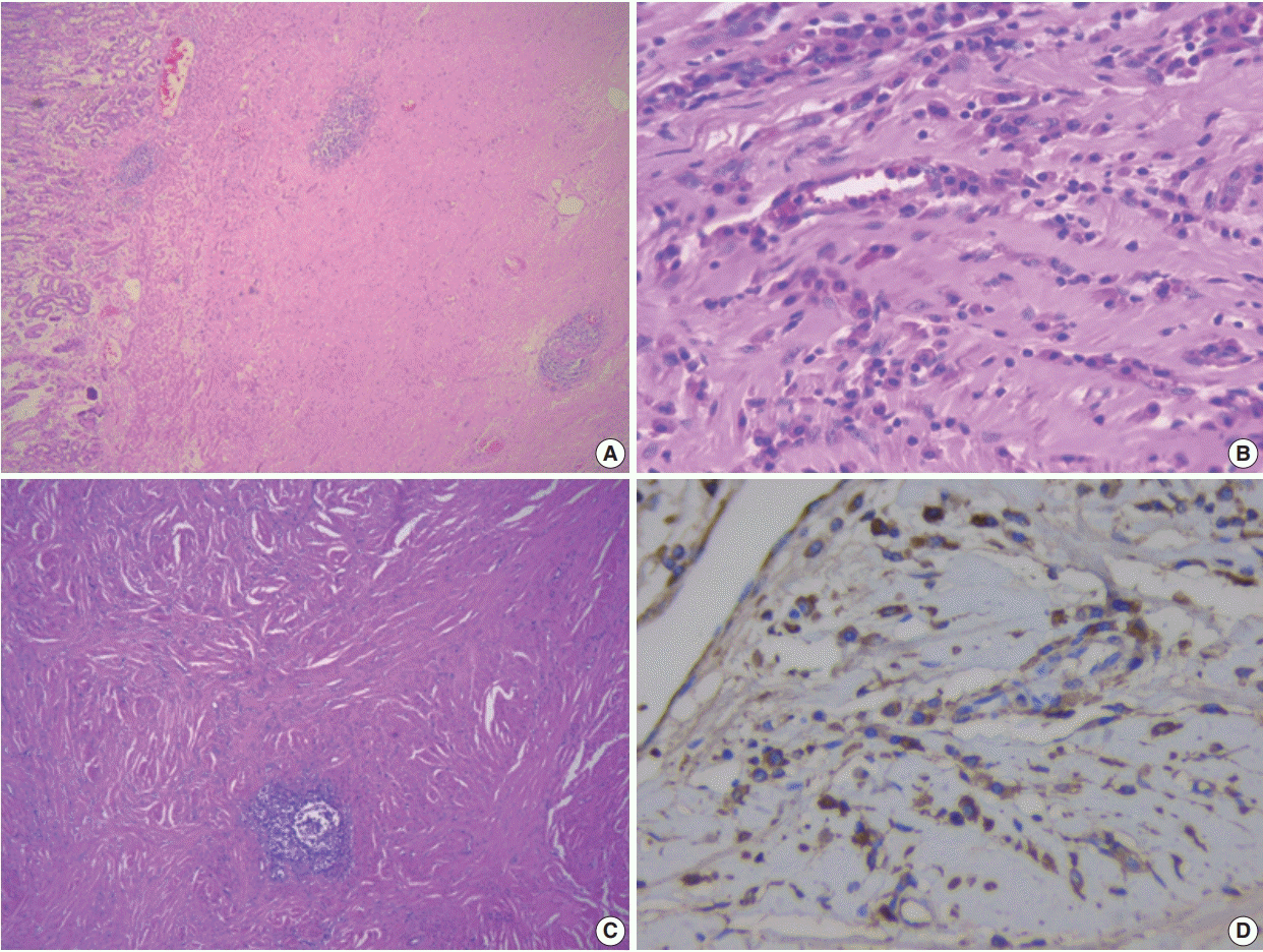
IgG4 immunohistochemistry (IHC) and serum IgG4 level were assessed. The IgG4 IHC showed 35–40 immunoreactive plasma cells per high power field (hpf), and serum IgG4 level was elevated (4.36 g/L). These tests confirmed the diagnosis of IgG4-RD. Subsequent serum levels after 3 and 4 months were 2.71g/L and 2.53 g/L, respectively. The patient was treated with steroids and azathioprine. He experienced postsurgery complications that required revision of the gastric anastomosis. During follow-up, his prognosis while receiving medical treatment has been good.
Case 2
The second patient was a 58-year-old male found to have erosive gastritis and a submucosal swelling in the body of the stomach in December 2017, while undergoing upper gastrointestinal endoscopy for investigation of dyspepsia. CT examination showed a well-defined, 3×2.9 cm, round, homogeneous, enhancing soft tissue lesion in the distal body of the stomach along the lesser curvature, which was suspected to be GIST or leiomyoma. He was advised to undergo follow-up and elective surgery. His gastric symptoms worsened over the next 10 months, and he underwent excision of the gastric submucosal lesion in October 2018. Histopathological examination revealed a wellcircumscribed globular mass measuring 4×3.3×3.5 cm, and whorling was seen on the cut surface. Microscopically, the lesion was composed of extensively fibrotic and sclerotic stroma with a storiform pattern of fibrosis in foci (Fig. 1C). Discrete, cords and clusters of plasma cells admixed with lymphocytes, a few eosinophils and a few scattered lymphoid aggregates and follicles were present. Perivascular aggregates of plasma cells were also present. There was no evidence of obliterative or nonobliterative phlebitis. Bundles of smooth muscle were identified at the periphery on one aspect. The possibility of IgG4-RD of the stomach was suggested, and IgG4 IHC and serum estimation were recommended. The IgG4 IHC revealed 20–30 immuno-reactive plasma cells/hpf (Fig. 1D). Serum IgG4 was elevated at 3.11g/L, well above the 1.35 g/L cut-off for diagnosis of IgG4-RD. The patient did not return for follow-up.
Ethics statement
The authors certify that they obtained patient consent for publication, and the study was approved by the Institutional Review Board of Sri Ramaswamy Memorial Institutes for Medical Science, Chennai, India (IEC NO: SIMS IEC/other/18/2019).
Go to : 
DISCUSSION
IgG4-RD is an immune-mediated fibroinflammatory lesion, first described in patients with sclerosing cholangitis associated with autoimmune pancreatitis type 1 [5]. Later, it was identified in other organs including the liver, bile ducts, salivary glands, retroperitoneum, lymph nodes, and lungs. It is characterized by diffuse or partial enlargement of the organ and histologically as dense lymphoplasmacytic infiltration with an increase in IgG4 plasma cells on immunostaining, a storiform pattern of fibrosis and obliterative or non-obliterative phlebitis, and an increase in serum IgG4 level.
IgG4-RD of the gastrointestinal tract is very rare and can present as diffuse wall thickening or as polyp or mass-like lesion [2,3]. Even though obliterative phlebitis was not present in these two cases, the presence of dense fibrosis, which was storiform in one case, and dense lymphoplasmacytic infiltration with lymphoid aggregates and follicles, presenting as a submucosal masslike lesion, suggested the possibility of IgG4-RD, which was confirmed by IHC and elevated serum IgG4 level. The presence of at least two histological features is required for confident diagnosis of IgG4-RD; in most cases, dense lymphoplasmacytic infiltrate and diffuse/storiform fibrosis are seen. Additional clinical, serological (serum level >135 mg/dL or 1.35 g/L), or radiological evidence is required to confirm IgG4-RD [1].
The cut-off point for the presence of IgG4 plasma cells in tissues varies and can range from >30 plasma cells/hpf to >50/hpf, which is highly specific [1,5]. In biopsy specimens, more than 10 IgG4 plasma cells/hpf were reported in one study [16]. However, the cut-off points vary depending on organ system. Some studies have suggested that IgG4+/IgG+ plasma cell ratio >0.4 is a marker of IgG4-RD in the presence of classic histopathological features and with a compatible clinical features [1,17].
IgG4-RD can involve multiple organs or any sites in the body synchronously or metachronously [18]. Patients can present with non-specific symptoms and swelling or mass-like lesion. Patients with biliary or pancreatic lesion can present with jaundice, weight loss, and vague abdominal pain. The disease can be an incidental finding during radiological examination and can be mistaken for malignancy, as there are no specific radiological features characteristic of this disease [18,19]. Most cases of gastric IgG4-RD have been reported in middle-aged patients, and both men and women are affected [3,6]. Both patients in this report were middle-aged men.
IgG4-RD of the stomach was first described in 2004 by Shinji et al. [20], presenting as a gastric ulcer. Because it is difficult to diagnose clinically, especially in isolated cases, most of the reported patients have undergone surgery. Because this disease involves a submucosal lesion in the stomach, these cases are often misdiagnosed as GIST [3,7,18,19] and are difficult to diagnose on endoscopic forceps biopsy, similar to our cases. Gastric lesions that have been mistaken for GIST or gastric cancer have been reported in the literature (Table 1) [3,6-15]. IgG4-RD of the stomach that involved the regional lymph nodes has also been reported [6].
Table 1.
Clinicopathological features of IgG4-related disease manifesting as gastric lesions
| Case No. | Age (yr) | Sex | Endoscopic finding/Clinical diagnosis | Location | Size (mm) | Histopathology/IHC | Serum IgG4 levels | Treatment | Study |
|---|---|---|---|---|---|---|---|---|---|
| 1 | 48 | F | Mass/GIST/NET | Mid body | 36 × 22 | SF, LP, OP, IgG4 + 210/hpf, IgG4/IgG ratio about 85% | NA | WR | Woo et al. [3] |
| 2 | 62 | F | Mass/gastic cancer | Antrum | 80 × 30 | SF, LP, OP, IgG4 + ve lymphoplasmacytes > 50/hpf | Elevated | DG | Bulanov et al. [6] |
| 3 | 59 | F | Mass/GIST | NA | 33 × 14 | Abundant LP, SF, lymphoid follicles, IgG4 > 50/hpf | Normal | WR | Kim et al. [7] |
| 4 | 56 | F | Mass/GIST | NA | 21 × 15 | Abundant LP, SF, calcification, IgG4 > 50/hpf | Normal | WR | Kim et al. [7] |
| 5 | 60 | F | Nodule/NA | Fundus | 10 × 15 | Fibrosis, dense LP, IgG4 > 80/hpf | Normal | WR | Chetty et al. [8] |
| 6 | 45 | M | Multiple nodules/NA | Antrum | Up to 22 | LP, many eosinophils, IgG4/IgG ratio 0.84 | NA | DG | Chetty et al. [8] |
| 7 | 56 | M | Nodule/NA | Body | 8 | SF, LP, IgG4-40-102/hpf IgG4/IgG ratio 80%–90% | NA | ESR | Na et al. [9] |
| 8 | 58 | M | Nodule/AIP | Fundus and body | 14 | Dense LP, extensive IgG and IgG4 + staining | Normal | Steroid | Baez et al. [10] |
| 9 | 55 | F | Nodule/GIST | Body | 20 | Dense hyalinization, LP, IgG4/IgG ratio 41% | Normal | ESR | Zhang et al. [11] |
| 10 | 75 | F | Polyp/GIST | Body | 56 × 50 | Fibrosis, LP, many eosinophils, IgG4–39/hpf | Normal | WR | Rollins et al. [12] |
| 11 | 44 | M | Mass/GIST | Body | 20 × 18 | Fibrosis, LP, IgG4 + ve lymphoplasmacytes | Normal | ESR | Otsuka et al. [13] |
| 12 | 27 | F | Mass/GIST/NET | Fundus | 40 | Dense fibrosis, LP, IgG4/IgG ratio 25.3% | Normal | WR | Cheong et al. [14] |
| 13 | 29 | F | Mass/GIST | Body | 20 × 15 | Fibrosis, LP, IgG4 + ve plasma cells 150/hpf | NA | WR | Skorus et al. [15] |
| 14 | 43 | M | Mass/GIST | Antrum | 70 × 50 | Dense LP, IgG4 + plasma cells 35–40/hpf | Elevated | WR + Steroids | Present case 1 |
| 15 | 58 | M | Mass/GIST | Distal body | 45 × 40 | LP, SP, IgG4 + ve plasma cells 20–30/hpf | Elevated | WR | Present case 2 |
IHC, immunohistochemistry; GIST, gastrointestinal stromal tumor; NET, neuroendocrine tumor; SF, storiform fibrosis; LP, lymphoplasmacytic infiltrate; OP, obliterative phlebitis; hpf, high power field; NA, not available; WR, wedge resection; DG, distal gastrectomy; ESR, endoscopic submucosal resection; AIP, autoimmune pancreatitis.
![]()
Although steroids are the first therapeutic option for treating IgG4-RD, it is difficult to diagnose gastric IgG4-RD without histopathological examination. Almost all cases have been reported after surgical resection. Therefore, this disease should be considered in the differential diagnosis of gastric submucosal mass lesion.
To conclude, we present two cases of IgG4-RD of the stomach that presented as a mass lesion and were clinically suspected to be GIST. The diagnosis was made only after histopathological examination of resection specimens. This highlights the importance of considering this disease in differential diagnosis to avoid surgical resection.
Go to :
 XML Download