

 PDF
PDF Citation
 Print
Print


Even before the term “primary age-related tauopathy (PART)” was proposed in 2014, pathologists had observed localized neurofibrillary degeneration in brains from aged people with relatively well-preserved cognitive function that was mostly restricted to medial temporal regions. These findings were somewhat informally described as “aging changes” because the features were considered insufficient for a diagnosis of Alzheimer disease (AD). The newly proposed consensus term (i.e., PART) includes features that range from the presence of isolated neurofibrillary tangles (NFTs) in cognitively normal aged brains to a subtype of frontotemporal lobe degeneration (FTLD) known as FTLD-tau, which is also referred to as tangle-only dementia, tangle-predominant senile dementia (TPSD), and preferential development of NFT without senile plaques [1,2]. However, these previous designations tended to accentuate unnecessarily the clinical aspects of cognitive impairment, leading to a biased understanding of the disease entity [1]. Although the word dementia is included in these terms, profound cognitive impairment that interferes with daily activities occurs in only a minority of affected individuals within this population [1].
Thus, the consensus term PART was suggested by researchers of neurodegenerative diseases as a more objective and quantitative description of pathological disease status separate from the clinical presentation. The term PART was inspired by the pathological classification system for AD of the National Institute on Aging-Alzheimer’s Association [1,3]. Since being introduced, the clinicopathological traits of PART have been clarified more precisely [4-8]. This review aims to increase recognition of this disease entity by Korean pathologists through a literature review and discussion of the clinicopathological implications of PART, and via a figurative presentation of a PART case recently diagnosed at Chonnam National University Hospital brain bank.
DEFINITION AND HISTOLOGICAL SPECTRUM OF PRIMARY AGE-RELATED TAUOPATHY
The diagnosis of PART is histological in nature and can be applied to patients exhibiting mild-to-moderate tau-positive NFTs but without, or with few, β-amyloid (Aβ) plaques [1,9]. The gross features of a brain with PART may include minimal atrophy that is primarily located in the medial temporal lobe; however, diffuse neocortical atrophy is also present in some cases. AD-type NFTs, including ghost tangles, are mainly distributed in the hippocampus and medial temporal lobe (Fig. 1A, in comparison with advanced AD, Fig. 1B), and correspond to Braak stages I–III in the majority of patients and to stage IV in rare cases [9]. The neuronal tauopathy of PART may also extend to granule cells of the dentate gyrus and neurons in the CA4 subregion of the hippocampus (Fig. 1C) [9]. Other than PART, the presence of ghost tangles or tau involvement in the dentate gyrus and CA4 are typically considered to be features of advanced AD (Fig. 1D) [9]. In addition to the hippocampus and medial temporal lobe, NFTs may also be observed in subcortical structures, such as the amygdala, nucleus basalis of Meynert, nucleus accumbens, hypothalamus, thalamus, and olfactory system (bulb and cortex), and in the brainstem, including the substantia nigra, locus coeruleus, dorsal raphe nucleus, and medulla oblongata, where NFTs develop at a younger age, sometimes even in teenagers [1,10,11]. Biochemical and immunohistochemical studies have revealed that the NFTs in PART contain mixed three-repeat (3R) and four-repeat (4R) isoforms of tau proteins, as seen in AD [1,9].
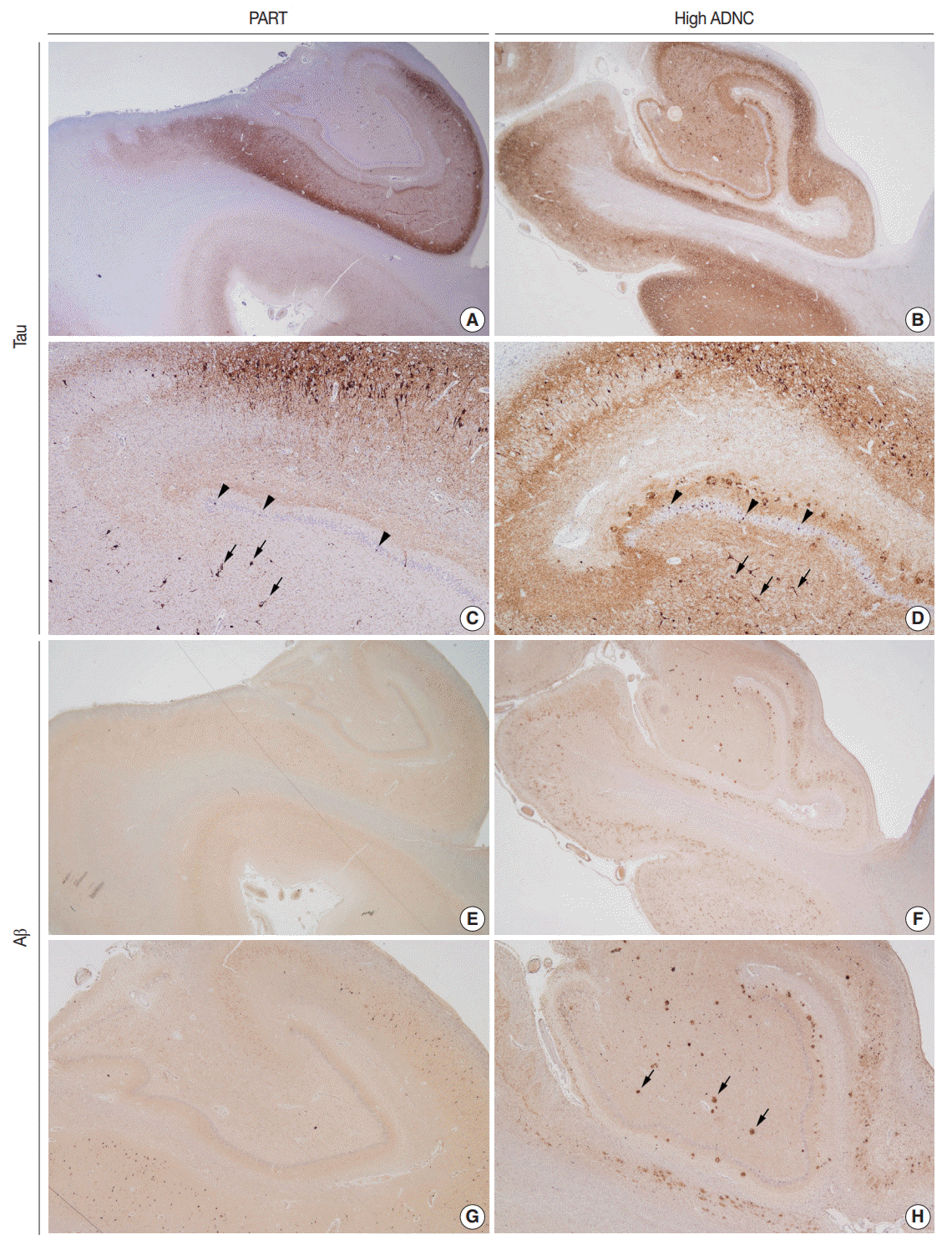 | Fig. 1.Histopathological findings of primary age-related tauopathy (PART) in a 92-year-old woman (A, C, E, G) compared with those of high– Alzheimer’s disease (AD) neuropathologic change (ADNC) in an 82-year-old man (B, D, F, H). Tau immunohistochemistry analyses revealed marked tauopathy centered in the hippocampus and subiculum of the PART brain (A) and the extension of tauopathy into the temporal neocortex of the high-ADNC brain (B) (AT8 immunohistochemistry). At a higher magnification, the granule cells of the dentate gyrus (arrowheads) and the neurons of the CA4 subregion (arrows) exhibited tau involvement in the PART brain (C), as well as in the brain of the advanced AD case (D) (AT8 immunohistochemistry). Although the hippocampus in the PART brain did not reveal any β-amyloid (Aβ)–positive plaques (E, G) (Aβ immunohistochemistry), the high ADNC brain showed Aβ deposition in the temporal neocortex through the CA4 subregion (arrows) that corresponded to Thal Aβ phase 4 (F, H) (Aβ immunohistochemistry).
|
The presence of NFTs in Braak stage IV or lower is a basic requirement for a histological diagnosis of PART; when combined with Thal Aβ phase 0, PART can be diagnosed definitively (Fig. 1E–G). If the required distribution of NFTs is observed together with Thal Aβ phase 1 or 2, then the pathology is categorized as possible PART [1]. The following is an example pathological diagnosis of PART: “Primary age-related tauopathy (PART), Definite, Braak stage III.” [1] Although Braak stage IV is considered a requirement for PART diagnosis, Braak stage IV pathology in the absence of Aβ plaques is rare and the possibility that cases such as these represent FTLD-tau needs to be considered [1].
Go to : 
GENETIC AND CLINICAL ASPECTS OF PART
PART brains are deficient in the apolipoprotein E (APOE) ε4 allele, which is highly associated with the risk of AD [2]. The frequency of APOE ε4 in PART is approximately 10% [1,6], whereas its prevalence in AD exceeds 45% [12,13]. A major genetic risk factor for PART is the microtubule-associated protein tau (MAPT) gene H1 haplotype, which is also an accepted risk factor of progressive supranuclear palsy (PSP), corticobasal degeneration (CBD), and argyrophilic grain disease (AGD) [2,14].
By definition, patients with the PART pathology present with a lack of, or minimal, cognitive impairment [2]. However, greater impairment has been noted as an aspect of TPSD, where the initial symptoms typically include memory disturbances [15]. During disease progression, deficits in cognitive function may extend to mild cognitive impairment (MCI) with a relatively well-preserved personality [15]. Mental derangements, such as disorientation (or rarely, delirium), depression and paranoid thinking have also been observed [15,16]. One feature of PART is that cases with higher NFT stages (Braak stage III or IV) are more likely to be associated with subjective memory impairments, which is a common complaint among the elderly population [17,18].
Go to :
DEBATE REGARDING WHETHER PART IS ON THE ALZHEIMER DISEASE CONTINUUM
Considering PART as being on the AD continuum, and especially comparing it with the limbic-predominant form of AD, has been discouraged for several reasons [6,9,16,19]. First, PART is associated with lower Braak NFT stages and fewer, or an absence of, Aβ plaques. Second, patients with PART have an older age of onset, shorter disease duration, and less severe cognitive impairment. Third, the frequency of APOE ε4 is much lower in PART than in the normal elderly population and the frequency of TDP-43 proteinopathy is higher in patients with limbic-predominant AD (67%) than in those with definite PART (29%) (Table 1) [20].
Table 1.
Comparison of PART with AD
| Variable | PART | AD |
|---|---|---|
| Primary pathology | ||
| Tau-positive NFTs | Medial temporal region-restricted | Diffuse cortical distribution |
| Braak stage ≤ IV (usually I–III) | Braak stage ≥ III (usually IV–VI) | |
| Aβ plaques | Thal phase 0–2 | Thal phase ≥ 3 |
| Definite PART: Thal phase 0 | ||
| Possible PART: Thal phase 1–2 | ||
| Tau isoforms | Mixed 3R and 4R tau | Mixed 3R and 4R tau |
| Genetic association | MAPT H1 haplotype | APOE ε4 |
| Clinical features | ||
| Cognition | Normal-to-mild impairment | Dementia |
| Duration (yr) | 5 | 9 [16] |
| Age at death (yr) | 86 | 79 [14,19] |
| Prevalence at ≥ 80 yr (%) | 20 | 80 [8,11] |
| Other co-existing pathology | ||
| TDP-43 proteinopathy (%) | 30 | 67 [14,19,20] |
| Hippocampal sclerosis (%) | 10 | 3 [16,20] |
| α-Synuclein positive Lewy bodies (%) | 10 | 30 [14,20] |
![]()
A discussion on the role that Aβ plays in tauopathy is inevitable when PART is compared with AD. In the absence of Aβ, as seen in definite PART, the severity of tau-positive NFTs tends to be greater with older age at death [6]. A proposed pathological step in late-onset AD is tauopathy, corresponding to PART, which is purported to occur at some point in the life cycle of almost every individual; amyloidosis may also occur as an independent event [21]. In this model, Aβ is not a catalyst for tau deposition in the brain, but rather serves to promote the spread of tauopathy. This hypothesis is supported by a study that used an established cell biosensor assay to show that the seeding activity of tauopathy is enhanced in the presence of Aβ-positive plaques [4]. However, another report found no clear distinction in tau seeding activity between PART and AD subjects [7]. The functional interaction between tau and Aβ, as well as the relationship between PART and AD, remain to be elucidated [2,22].
Go to :
COEXISTING PATHOLOGIES IN ELDERLY INDIVIDUALS WITH PRIMARY AGE-RELATED TAUOPATHY
Previous studies focused on brain pathology in the elderly have identified several pathological trends [8,11,18]. Roughly 20% of aged people in their 80s or older have PART [8,11,18,23], while the remaining 80% exhibit some degree of AD-type pathology characterized by both NFTs and neuritic plaques [11]. Moreover, there are few cases that are Aβ-positive only; most cases of Aβ deposition show some tau-positive NFTs [8,11]. These observations are consistent with prior reports showing that approximately 25% of elderly people with well-preserved cognitive function lack brain amyloidosis [1]. Even among a carefully selected group of centenarians, 20% were relatively free of Aβ deposition as detected by immunohistochemistry [11]. Thus, the relative proportions of PART and AD appear to be maintained in centenarian populations [8,11].
It has been proposed that a neuropathological diagnosis of TPSD or PART should be conservatively applied to cases where NFTs mainly affect the hippocampal/limbic area, and where there is a scarcity of Aβ deposits and no evidence of any other dementia characterized by NFTs [15]. The limits of the PART diagnosis explain the exclusion of PSP, CBD, and even Lewy body disease cases in previous studies on PART [6]. Despite the proposed limitations to its diagnostic criteria, PART has been shown to co-exist with other pathologies. For example, TDP-43 proteinopathy, including cerebral age-related TDP-43 with sclerosis, and AGD have been identified in approximately 30% of cases with definite PART [6]. Similarly, hippocampal sclerosis has been identified in approximately 10% of cases [16], while α-synuclein-positive Lewy bodies have been observed in fewer than 10% of PART cases [6].
Go to :
CONCLUSIONS
PART can be diagnosed in cases showing neurofibrillary degeneration restricted to the medial temporal region, an absence (or scarcity) of Aβ deposition, and a lack of cognitive impairment or MCI. However, several unanswered issues remain regarding the “gray zone” between PART and AD. The definition of clinicopathological PART requires refinement due to the presence of clinical PART in cases with higher Thal Aβ phases and moderate-to-frequent neuritic plaques [18]. The implications of the association of PART with diffuse amyloid and neuritic plaques also remain to be clarified, although a quantitative margin of Aβ deposition, up to Thal phase 2, has been proposed [1]. Neither an exact diagnostic threshold for Aβ deposition nor the methodology with which to detect its presence has been clearly defined. Furthermore, the term “age-related” is somewhat ambiguous. For example, the accumulation of abnormally phosphorylated tau proteins can begin before puberty and Braak stage I or II may be seen in individuals in their 20s [11]. Determining a common pathology in aged brains, with recognition of PART, will provide a firm foundation for a more profound understanding of age-related neurodegenerative changes. Additionally, accumulated neuropathology data reflecting the epidemiology of the Korean population will be a good resource for future neuroscientific research.
Go to :
 XML Download