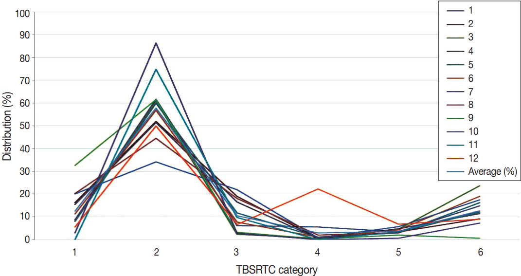
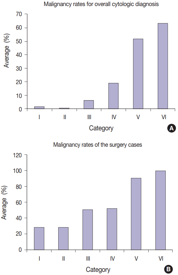
1. Kim M, Park HJ, Min HS, et al. The use of the Bethesda System for Reporting Thyroid Cytopathology in Korea: a nationwide multicenter survey by the Korean Society of Endocrine Pathologists. J Pathol Transl Med. 2017; 51:410–7.

2. Lipton RF, Abel MS. Aspiration biopsy and the thyroid in evaluation of thyroid dysfunction. Am J Med Sci. 1944; 208:736–42.
3. Crile G Jr, Hawk WA Jr. Aspiration biopsy of thyroid nodules. Surg Gynecol Obstet. 1973; 136:241–5.
4. Lee MH. Thyroid biopsy. Korean J Med. 1977; 20:731–6.
5. Park HS. Cytohistopathologic comparative study of aspiration biopsy cytology from various sites. Korean J Cytopathol. 1991; 2:8–19.
6. Lee K. The history of the Korean Society for Cytopathology. Seoul: The Korean Society for Cytopathology;2006. p. 15–9.
7. Kim WB, Kim TY, Kwon HS, et al. Management guidelines for patients with thyroid nodules and thyroid cancer. J Korean Endocr Soc. 2007; 22:157–87.

8. Yi KH, Lee EK, Kang HC, et al. 2016 Revised Korean Thyroid Association management guidelines for patients with thyroid nodules and thyroid cancer. Int J Thyroidol. 2016; 9:59–126.

9. Oh EJ, Jung CK, Kim DH, et al. Current cytology practices in Korea: a nationwide survey by the Korean Society for Cytopathology. J Pathol Transl Med. 2017; 51:579–87.

10. Cibas ES, Ali SZ; NCI Thyroid FNA State of the Science Conference. The Bethesda System For Reporting Thyroid Cytopathology. Am J Clin Pathol. 2009; 132:658–65.

11. Guidelines of the Papanicolaou Society of Cytopathology for the examination of fine-needle aspiration specimens from thyroid nodules. The Papanicolaou Society of Cytopathology Task Force on Standards of Practice. Diagn Cytopathol. 1996; 15:84–9.
12. Bongiovanni M, Spitale A, Faquin WC, Mazzucchelli L, Baloch ZW. The Bethesda System for Reporting Thyroid Cytopathology: a meta-analysis. Acta Cytol. 2012; 56:333–9.

13. Jung YY, Jung S, Lee HW, Oh YL. Significance of subcategory atypia of undetermined significance/follicular lesion of undetermined significance showing both cytologic and architectural atypia in thyroid aspiration cytology. Acta Cytol. 2015; 59:370–6.

14. Kwon KH, Jin SY, Lee DW. A study of usefulness of fine needle aspiration cytology of the thyroid lesions. Korean J Cytopathol. 1996; 7:111–21.
15. Park KM, Ko IH. Diagnostic accuracy of fine needle aspiration cytology in thyroid lesions: analysis of histologically confirmed 153 cases. Korean J Cytopathol. 1996; 7:122–33.
16. Park JH, Yoon SO, Son EJ, Kim HM, Nahm JH, Hong S. Incidence and malignancy rates of diagnoses in the bethesda system for reporting thyroid aspiration cytology: an institutional experience. Korean J Pathol. 2014; 48:133–9.

17. Lee YB, Cho YY, Jang JY, et al. Current status and diagnostic values of the Bethesda system for reporting thyroid cytopathology in a papillary thyroid carcinoma-prevalent area. Head Neck. 2017; 39:269–74.

18. Lee K, Jung CK, Lee KY, Bae JS, Lim DJ, Jung SL. Application of Bethesda system for reporting thyroid aspiration cytology. Korean J Pathol. 2010; 44:521–7.

19. The Korean Society of Pathologist. The History of the Korean Society of Pathologists 2006~2015. Seoul: The Korean Society of Pathologist;2016. p. 20–1.
20. Jung CK, Lee A, Jung ES, Choi YJ, Jung SL, Lee KY. Split sample comparison of a liquid-based method and conventional smears in thyroid fine needle aspiration. Acta Cytol. 2008; 52:313–9.

21. Kim ES, Lim DJ, Lee K, et al. Absence of galectin-3 immunostaining in fine-needle aspiration cytology specimens from papillary thyroid carcinoma is associated with favorable pathological indices. Thyroid. 2012; 22:1244–50.

22. Gweon HM, Kim JA, Youk JH, et al. Can galectin-3 be a useful marker for conventional papillary thyroid microcarcinoma? Diagn Cytopathol. 2016; 44:103–7.

23. Koo JS, Jung W, Hong SW. Cytologic characteristics and beta-catenin immunocytochemistry on smear slide of cribriform-morular variant of papillary thyroid carcinoma. Acta Cytol. 2011; 55:13–8.
24. Jung YY, Park IA, Kim MA, Min HS, Won JK, Ryu HS. Application of chemokine CXC motif ligand 12 as a novel diagnostic marker in preoperative fine-needle aspiration biopsy for papillary thyroid carcinoma. Acta Cytol. 2013; 57:447–54.

25. Lee SR, Yim H, Han JH, et al. VE1 antibody is not highly specific for the BRAFV600E mutation in thyroid cytology categories with the exception of malignant cases. Am J Clin Pathol. 2015; 143:437–44.
26. Kim YH, Yim H, Lee YH, et al. Evaluation of the VE1 antibody in thyroid cytology using ex vivo papillary thyroid carcinoma specimens. J Pathol Transl Med. 2016; 50:58–66.
27. Kwak JY, Kim EK, Kim JK, et al. Dual priming oligonucleotide-based multiplex PCR analysis for detection of BRAFV600E mutation in FNAB samples of thyroid nodules in BRAFV600E mutation-prevalent area. Head Neck. 2010; 32:490–8.
28. So YK, Son YI, Park JY, Baek CH, Jeong HS, Chung MK. Preoperative BRAF mutation has different predictive values for lymph node metastasis according to tumor size. Otolaryngol Head Neck Surg. 2011; 145:422–7.
29. Lee SE, Hwang TS, Choi YL, et al. Molecular profiling of papillary thyroid carcinoma in Korea with a high prevalence of BRAFV600E mutation. Thyroid. 2017; 27:802–10.
30. Kim Y, Choi KR, Chae MJ, et al. Stability of DNA, RNA, cytomorphology, and immunoantigenicity in residual ThinPrep specimens. APMIS. 2013; 121:1064–72.
31. Jung CK, Min HS, Park HJ, et al. Pathology reporting of thyroid core needle biopsy: a proposal of the Korean Endocrine Pathology Thyroid Core Needle Biopsy Study Group. J Pathol Transl Med. 2015; 49:288–99.


 PDF
PDF Citation
 Print
Print


 XML Download