Journal List > J Pathol Transl Med > v.50(3) > 1152176

Pyo, Sohn, and Kang: Core Needle Biopsy Is a More Conclusive Follow-up Method Than Repeat Fine Needle Aspiration for Thyroid Nodules with Initially Inconclusive Results: A Systematic Review and Meta-Analysis

Abstract

Background:

This study investigated the appropriate management of thyroid nodules with prior non-diagnostic or atypia of undetermined significance/follicular lesion of undetermined significance (AUS/FLUS) through a systematic review and meta-analysis.

Methods:

This study included 4,235 thyroid nodules from 26 eligible studies. We investigated the conclusive rate of follow-up core needle biopsy (CNB) or repeat fine needle aspiration (rFNA) after initial fine needle aspiration (FNA) with non-diagnostic or AUS/FLUS results. A diagnostic test accuracy (DTA) review was performed to determine the diagnostic role of the follow-up CNB and to calculate the area under the curve (AUC) on the summary receiver operating characteristic (SROC) curve.

Results:

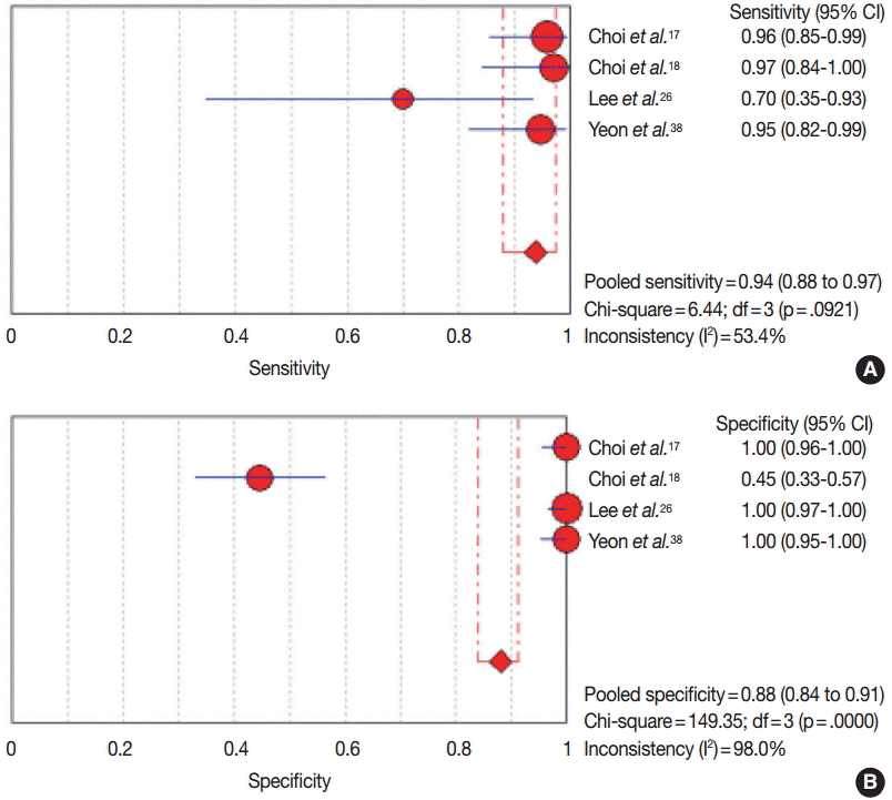
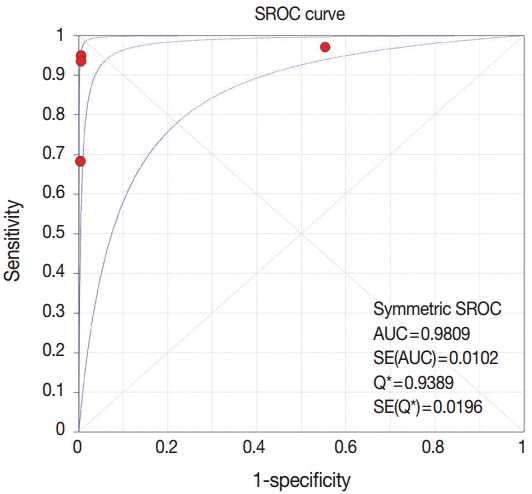
The conclusive rates of follow-up CNB and rFNA after initial FNA were 0.879 (95% confidence interval [CI], 0.801 to 0.929) and 0.684 (95% CI, 0.627 to 0.736), respectively. In comparison of the odds ratios of CNB and rFNA, CNB had more frequent conclusive results than rFNA (odds ratio, 5.707; 95% CI, 2.530 to 12.875). Upon subgroup analysis, follow-up CNB showed a higher conclusive rate than rFNA in both initial non-diagnostic and AUS/FLUS subgroups. In DTA review of followup CNB, the pooled sensitivity and specificity were 0.94 (95% CI, 0.88 to 0.97) and 0.88 (95% CI, 0.84 to 0.91), respectively. The AUC for the SROC curve was 0.981, nearing 1.

Conclusions:

Our results show that CNB has a higher conclusive rate than rFNA when the initial FNA produced inconclusive results. Further prospective studies with more detailed criteria are necessary before follow-up CNB can be applied in daily practice.

Papillary thyroid carcinoma, which has recently increased in incidence, is the most common malignant tumor in endocrine system [1]. The cause for the increased incidence of papillary thyroid carcinoma is not fully understood [1]. One possible cause is the improvement in ultrasonography and computed tomography [2,3]. In daily practice, the treatment and follow-up for thyroid nodules are based on the results of initial fine needle aspiration (FNA). According to current guidelines [4-6], FNA is recommended as the initially performed modality, and additional testing is suggested as indicated by the initial FNA results.
In daily practice, repeat FNA (rFNA) is recommended for thyroid nodules of non-diagnostic or atypia of undetermined significance/follicular lesion of undetermined significance (AUS/FLUS). In addition, for definite diagnosis of thyroid nodules, BRAFV600E mutation test or diagnostic surgery can also be performed. In previous studies, the rates of non-diagnostic and AUS/FLUS were 5%–17% and 3%–18%, respectively [7-9]. For thyroid nodule with non-diagnostic result, the possibility of an inconclusive reading might also be higher with rFNA. Although rFNA is recommended for non-diagnostic or AUS/FLUS thyroid nodules in the current guidelines, other modalities, such as core needle biopsy (CNB) or combination of FNA and CNB, have been introduced in recent reports [10,11]. However, the effectiveness or diagnostic role of these follow-up modalities has not been fully elucidated.
In this study, follow-up CNB was defined as CNB performed after initial non-diagnostic or AUS/FLUS findings. We investigated the conclusive rates of the follow-up procedures of CNB and rFNA in thyroid nodule with initial non-diagnostic or AUS/FLUS finding through a systematic review and meta-analysis. Indeed, the diagnostic test accuracy (DTA) review was performed to determine the diagnostic accuracy of follow-up CNB in thyroid nodules with initial inconclusive results.

MATERIALS AND METHODS

Literature search and selection criteria

Relevant articles were obtained from a search of PubMed and MEDLINE databases through December 31, 2015. The search was performed using ‘thyroid,’ ‘core needle biopsy,’ and ‘fine needle aspiration’ as search terms. The titles and abstracts of all returned articles were screened for exclusion. To find additional eligible studies, review articles were also screened. Search results were then reviewed and included if (1) initial FNA for a thyroid nodule was performed, and (2) there was information about CNB or FNA as a follow-up study for thyroid nodules with initial non-diagnostic or AUS/FLUS results. Articles were excluded if they were (1) non-original articles or case reports or (2) non-English language publications.

Data extraction

The following information was collected from the full texts of eligible studies and verified: name of first author, publication year, study location, number of patients analyzed, and method and results of initial and follow-up studies. We did not define a minimal number of patients to be included in a study. Any disagreements were resolved by consensus.

Statistical analysis

Data were analyzed using the Comprehensive Meta-Analysis software package (Biostat, Englewood, NJ, USA). We evaluated the conclusive rates of follow-up studies with follow-up CNB or rFNA after initial FNA with non-diagnostic or AUS/FLUS results. The conclusive results included benign, follicular neoplasm or suspicious for follicular neoplasm, suspicious for malignancy, and malignancy categories. The conclusive rates were measured by dividing the number of conclusive results into the total number of cases with a follow-up study. The heterogeneity between eligible studies was assessed using Q and I2 statistics and presented using p-values. A sensitivity analysis was performed to assess the impact of each study on the combined effect and the heterogeneity of eligible studies. To identify any publication bias, Egger’s test and Begg’s funnel plot were initially performed. When a significant publication bias was found, the fail-safe N and trimfill tests were additionally conducted to confirm the degree of bias. The results were considered statistically significant at p<0.05.
A DTA review was conducted using the Meta-Disc program ver. 1.4 [12]. The forest plots of pooled sensitivity and specificity and the summary receiver operating characteristic (SROC) curve were determined as described previously [13]. The diagnostic odds ratio (OR) and the value of the area under the curve (AUC) on SROC were investigated.

RESULTS

Selection and characteristics of studies

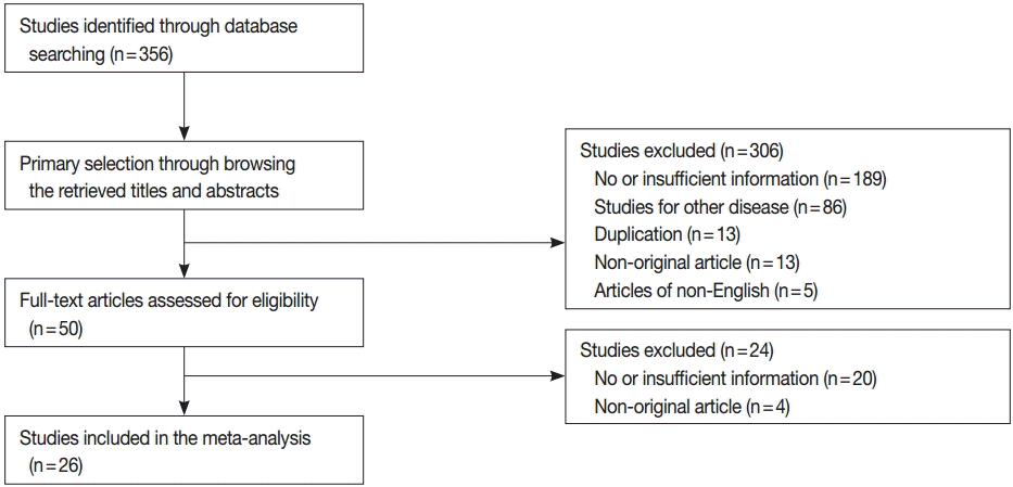
In the current study, 356 reports were identified in the database search. Among the search results, 209 reports were excluded due to insufficient information. In addition, 121 reports were excluded for the following reasons: focusing on other diseases (n=86), non-original articles (n=17), duplicate articles (n=13), and articles in a language other than English (n=5). Twenty-six eligible studies and 4,253 thyroid nodules were ultimately included in the current study (Table 1, Fig. 1) [14-39]. The characteristics of the included studies are shown in Table 1.

Higher conclusive rate in follow-up CNB than in rFNA

For initial FNA with non-diagnostic or AUS/FLUS, follow-up studies using CNB or rFNA showed an overall conclusive rate of 0.690 (95% confidence interval [CI], 0.675 to 0.704) and 0.748 (95% CI, 0.701 to 0.791) with the fixed and random effect models, respectively. For follow-up CNB, the conclusive rate was 0.775 (95% CI, 0.745 to 0.802) and 0.879 (95% CI, 0.801 to 0.929) with the fixed and random effect models, respectively. In rFNA, the conclusive rate was 0.670 (95% CI, 0.653 to 0.686) and 0.684 (95% CI, 0.627 to 0.736) with the fixed and random effect models, respectively. The ranges of conclusive rates were 0.631–0.981 and 0.286–0.942 for follow-up CNB and rFNA, respectively. Follow-up CNB showed a higher conclusive rate compared with rFNA (OR, 5.707; 95% CI, 2.530 to 12.875). The heterogeneity of eligible studies was significant in both follow-up CNB and rFNA groups (I2=90.4%, p<0.001 and I2=90.3%, p<0.001, respectively). In sensitivity analysis, no study had an effect on the concordance rates for either follow-up CNB (range, 0.864 to 0.896) or rFNA (range, 0.670 to 0.697). Subgroup analysis revealed a significant difference in conclusive rate between follow-up CNB and rFNA in both non-diagnostic (0.927; 95% CI, 0.848 to 0.966 vs 0.699; 95% CI, 0.596 to 0.785) and AUSL/FLUS cases (0.794; 95% CI, 0.675 to 0.877 vs 0.673; 95% CI, 0.608 to 0.732) with the random effect model (Table 2).
Egger’s test revealed that the follow-up CNB group showed a significant publication bias (p=.009). Additionally, fail-safe N and trim-fill tests were performed for confirmation of the degree of publication bias in the CNB group. The number of missing studies that would produce a p-value higher than alpha was 5,401 on the fail-safe N test. Because there were 11 observed studies, the publication bias was not large. In addition, the trim and fill test showed no significant difference between the observed and adjusted values. Therefore, we concluded that the publication bias in the follow-up CNB group was not significant through interpretation of Egger’s test, Begg’s funnel plot, the fail-safe N test, and the trim-fill test. In the rFNA group, there was no significant publication bias according to Egger’s test (p=.651) or Begg’s funnel plots.

DTA review of follow-up CNB as a follow-up study

For confirmation of the diagnostic accuracy of follow-up CNB, we conducted a DTA review. The pooled sensitivity and specificity values were 0.94 (95% CI, 0.88 to 0.97) and 0.88 (95% CI, 0.84 to 0.91), respectively (Fig. 2). The sensitivity and specificity of eligible studies ranged from 0.70 to 0.97 and from 0.45 to 1.00, respectively. On the SROC curve, the value of AUC was 0.981 (Fig. 3). In addition, the diagnostic OR was 448.73 (95% CI, 36.63 to 5,497.51).

DISCUSSION

The current guidelines recommend FNA as the initial test for thyroid nodules [4-6]. Although rFNA was applied to thyroid nodules with non-diagnostic and AUS/FLUS findings [4-6], the effectiveness of rFNA has not been fully elucidated. The present study is the first meta-analysis and DTA review of published studies on the diagnostic role of follow-up CNB compared with rFNA after initial FNA with non-diagnostic or AUS/FLUS results.
In the assessment of thyroid nodule, FNA is the most cost-effective initial test. Recently, FNA using ultrasonography has produced a higher quality of specimens for more precise diagnosis [10]. However, some cases were non-diagnostic or AUS/FLUS, and the rates of inconclusive results ranged from 10%–33.6% in previous studies [6,8,9,40]. Although the current guidelines recommend rFNA as a follow-up study for thyroid nodules with initial inconclusive results [4-6], rFNA does not ensure conclusive results. The inconclusive rates of rFNA in previous studies ranged from 9.9% to 72.0% [18,29,34,36]. In the current meta-analysis, the rate of inconclusive results for rFNA was 31.6%. If the cellularity of initial FNA is low, rFNA can also show low cellularity. In a previous study, rFNA was the most significant risk factor affecting repeat non-diagnostic results [18]. This situation might be caused by the nature of the thyroid nodule, including factors such as intratumoral calcification and cystic change [41-43]. In addition, the quality of the specimen might also be affected by the experience of the operator. Consequently, diagnostic surgery is recommended for thyroid nodule with repeat inconclusive results, and the incidence of diagnostic surgery has been reported as 22.2%–94.7% [24,44-46]. Therefore, to reduce the inconclusive rate, an alternative follow-up study, such as CNB, could be considered for thyroid nodules with inconclusive readings in initial FNA.
Considering the results of a previous meta-analysis, histologic examination using CNB as an initial test might be useful for obtaining conclusive results [47]. Other previous comparative studies of FNA and CNB as an initial test have reported that CNB has a suspected higher specificity, higher positive predictive value, and lower rate of inconclusive results [11,48]. However, definitive results for the sensitivity and specificity of CNB as a follow-up study have not yet been obtained [10,11,47]. CNB showed a lower sensitivity for thyroid glands than for other head and neck lesions [49]. However, the initial test might be more important for achieving higher sensitivity and patient safety. Furthermore, CNB has some limitations, including bleeding, tumor-cell displacement, and difficulty in approaching thyroid nodules in a posterior portion or close to critical structures, including the carotid artery or trachea; these factors limit its use as an initial test [48,49]. To obtain an adequate specimen for diagnosis, the experience of the operator may be more important for CNB than for FNA [48]. Whether CNB is appropriate as an initial test for thyroid nodules is not fully understood and could not be determined in the current systematic review. Despite a previous report that found follow-up CNB to have lower non-diagnostic and inconclusive rates than rFNA (7.2% vs 72.0%) [18], many previous studies have reported that CNB showed lower sensitivity than FNA. However, the diagnostic accuracy of follow-up CNB has not been fully elucidated. In the current DTA review, the pooled sensitivity and specificity of follow-up CNB were significantly high. For this reason, follow-up CNB after initial FNA might be useful for predicting malignancy and reducing the inconclusive rate in follow-up study.
There were some limitations in the current meta-analysis. First, most included studies were retrospective rather than prospective evaluations. Many thyroid nodules with initial inconclusive results only involved follow-up with ultrasonography. The conclusive and inconclusive rates of rFNA and follow-up CNB might have been affected by such cases. Therefore, cumulative prospective studies are needed to determine the effectiveness of follow-up procedures. Second, the current study included both non-diagnostic and AUS/FLUS subgroups, and subgroup analysis was performed. However, the DTA review could be not conducted for the rFNA subgroup due to insufficient information from eligible studies. Thus, a comparison of diagnostic accuracy between follow-up CNB and rFNA could not be performed. Third, because the current guidelines recommend initial FNA test for thyroid nodules [4-6], the current meta-analysis was performed only for studies with initial FNA. An investigation of the effectiveness of CNB as an initial test was therefore not conducted in the current study. Fourth, the current study was analyzed for initial non-diagnostic and AUS/FLUS categories. However, additional evaluations, including subgroup analysis for AUS and FLUS results, could not be performed due to lack of information on follow-up CNB from eligible studies [23]. Fifth, eligible studies used various criteria for nondiagnostic or indeterminate lesion. The rate of inconclusive results of CNB may have differed from the real value. Further studies are needed to establish guidelines of pathology reporting for thyroid CNB.
In conclusion, the current study showed that follow-up CNB had a higher conclusive rate than rFNA after initial FNA with inconclusive results. In addition, follow-up CNB had greater diagnostic accuracy for prediction of malignancy than rFNA. Additional prospective studies are required to determine standardized application criteria of follow-up CNB in daily practice.

Notes

Conflicts of Interest

No potential conflict of interest relevant to this article was reported.

REFERENCES

1. Sprague BL, Warren Andersen S, Trentham-Dietz A. Thyroid cancer incidence and socioeconomic indicators of health care access. Cancer Causes Control. 2008; 19:585–93.
crossref
2. Enewold L, Zhu K, Ron E, et al. Rising thyroid cancer incidence in the United States by demographic and tumor characteristics, 1980-2005. Cancer Epidemiol Biomarkers Prev. 2009; 18:784–91.
crossref
3. Mercante G, Frasoldati A, Pedroni C, et al. Prognostic factors affecting neck lymph node recurrence and distant metastasis in papillary microcarcinoma of the thyroid: results of a study in 445 patients. Thyroid. 2009; 19:707–16.
crossref
4. Cibas ES, Ali SZ. The Bethesda System for Reporting Thyroid Cytopathology. Thyroid. 2009; 19:1159–65.
crossref
5. American Thyroid Association (ATA) Guidelines Taskforce on Thyroid Nodules and Differentiated Thyroid Cancer, Cooper DS, Doherty GM, et al. Revised American Thyroid Association management guidelines for patients with thyroid nodules and differentiated thyroid cancer. Thyroid. 2009; 19:1167–214.
crossref
6. Gharib H, Papini E, Paschke R, et al. American Association of Clinical Endocrinologists, Associazione Medici Endocrinologi, and European Thyroid Association medical guidelines for clinical practice for the diagnosis and management of thyroid nodules: executive summary of recommendations. J Endocrinol Invest. 2010; 33(5 suppl 1):51–6.
crossref
7. Yang J, Schnadig V, Logrono R, Wasserman PG. Fine-needle aspiration of thyroid nodules: a study of 4703 patients with histologic and clinical correlations. Cancer. 2007; 111:306–15.
crossref
8. Yassa L, Cibas ES, Benson CB, et al. Long-term assessment of a multidisciplinary approach to thyroid nodule diagnostic evaluation. Cancer. 2007; 111:508–16.
crossref
9. Nayar R, Ivanovic M. The indeterminate thyroid fine-needle aspiration: experience from an academic center using terminology similar to that proposed in the 2007 National Cancer Institute Thyroid Fine Needle Aspiration State of the Science Conference. Cancer. 2009; 117:195–202.
10. Harvey JN, Parker D, De P, Shrimali RK, Otter M. Sonographically guided core biopsy in the assessment of thyroid nodules. J Clin Ultrasound. 2005; 33:57–62.
crossref
11. Renshaw AA, Pinnar N. Comparison of thyroid fine-needle aspiration and core needle biopsy. Am J Clin Pathol. 2007; 128:370–4.
crossref
12. Zamora J, Abraira V, Muriel A, Khan K, Coomarasamy A. Meta-DiSc: a software for meta-analysis of test accuracy data. BMC Med Res Methodol. 2006; 6:31.
crossref
13. Pyo JS, Sohn JH, Kang G. BRAF immunohistochemistry using clone VE1 is strongly soncordant with BRAF(V600E) mutation test in papillary thyroid carcinoma. Endocr Pathol. 2015; 26:211–7.
14. Anderson TJ, Atalay MK, Grand DJ, Baird GL, Cronan JJ, Beland MD. Management of nodules with initially nondiagnostic results of thyroid fine-needle aspiration: can we avoid repeat biopsy? Radiology. 2014; 272:777–84.
crossref
15. Baloch Z, LiVolsi VA, Jain P, et al. Role of repeat fine-needle aspiration biopsy (FNAB) in the management of thyroid nodules. Diagn Cytopathol. 2003; 29:203–6.
crossref
16. Chen JC, Pace SC, Chen BA, Khiyami A, McHenry CR. Yield of repeat fine-needle aspiration biopsy and rate of malignancy in patients with atypia or follicular lesion of undetermined significance: the impact of the Bethesda System for Reporting Thyroid Cytopathology. Surgery. 2012; 152:1037–44.
crossref
17. Choi YJ, Baek JH, Ha EJ, et al. Differences in risk of malignancy and management recommendations in subcategories of thyroid nodules with atypia of undetermined significance or follicular lesion of undetermined significance: the role of ultrasound-guided core-needle biopsy. Thyroid. 2014; 24:494–501.
crossref
18. Choi SH, Baek JH, Lee JH, et al. Thyroid nodules with initially nondiagnostic, fine-needle aspiration results: comparison of core-needle biopsy and repeated fine-needle aspiration. Eur Radiol. 2014; 24:2819–26.
crossref
19. Dincer N, Balci S, Yazgan A, et al. Follow-up of atypia and follicular lesions of undetermined significance in thyroid fine needle aspiration cytology. Cytopathology. 2013; 24:385–90.
crossref
20. Gocun PU, Karakus E, Bulutay P, Akturk M, Akin M, Poyraz A. What is the malignancy risk for atypia of undetermined significance? Three years’ experience at a university hospital in Turkey. Cancer Cytopathol. 2014; 122:604–10.
crossref
21. Gweon HM, Son EJ, Youk JH, Kim JA. Thyroid nodules with Bethesda system III cytology: can ultrasonography guide the next step? Ann Surg Oncol. 2013; 20:3083–8.
crossref
22. Ho AS, Sarti EE, Jain KS, et al. Malignancy rate in thyroid nodules classified as Bethesda category III (AUS/FLUS). Thyroid. 2014; 24:832–9.
crossref
23. Hyeon J, Ahn S, Shin JH, Oh YL. The prediction of malignant risk in the category “atypia of undetermined significance/follicular lesion of undetermined significance” of the Bethesda System for Reporting Thyroid Cytopathology using subcategorization and BRAF mutation results. Cancer Cytopathol. 2014; 122:368–76.
24. Jo VY, Vanderlaan PA, Marqusee E, Krane JF. Repeatedly nondiagnostic thyroid fine-needle aspirations do not modify malignancy risk. Acta Cytol. 2011; 55:539–43.
crossref
25. Lee SH, Oh HW, Fang Y, et al. Identification of plant compounds that disrupt the insect juvenile hormone receptor complex. Proc Natl Acad Sci USA. 2015; 112:1733–8.
crossref
26. Lee SH, Kim MH, Bae JS, Lim DJ, Jung SL, Jung CK. Clinical outcomes in patients with non-diagnostic thyroid fine needle aspiration cytology: usefulness of the thyroid core needle biopsy. Ann Surg Oncol. 2014; 21:1870–7.
crossref
27. Moon HJ, Kim EK, Yoon JH, Kwak JY. Malignancy risk stratification in thyroid nodules with nondiagnostic results at cytologic examination: combination of thyroid imaging reporting and data system and the Bethesda System. Radiology. 2015; 274:287–95.
crossref
28. Moslavac S, Matesa-Anić D, Matesa N, Kusić Z. When to repeat thyroid fine needle aspiration cytology? Acta Clin Croat. 2012; 51:549–54.
29. Na DG, Min HS, Lee H, Won JK, Seo HB, Kim JH. Role of core needle biopsy in the management of atypia/follicular lesion of undetermined significance thyroid nodules: comparison with repeat fine-needle aspiration in subcategory nodules. Eur Thyroid J. 2015; 4:189–96.
crossref
30. Na DG, Kim JH, Sung JY, et al. Core-needle biopsy is more useful than repeat fine-needle aspiration in thyroid nodules read as nondiagnostic or atypia of undetermined significance by the Bethesda system for reporting thyroid cytopathology. Thyroid. 2012; 22:468–75.
crossref
31. Nagarkatti SS, Faquin WC, Lubitz CC, et al. Management of thyroid nodules with atypical cytology on fine-needle aspiration biopsy. Ann Surg Oncol. 2013; 20:60–5.
crossref
32. Oertel YC, Miyahara-Felipe L, Mendoza MG, Yu K. Value of repeated fine needle aspirations of the thyroid: an analysis of over ten thousand FNAs. Thyroid. 2007; 17:1061–6.
crossref
33. Park KT, Ahn SH, Mo JH, et al. Role of core needle biopsy and ultrasonographic finding in management of indeterminate thyroid nodules. Head Neck. 2011; 33:160–5.
crossref
34. Park VY, Kim EK, Kwak JY, Yoon JH, Moon HJ. Malignancy risk and characteristics of thyroid nodules with two consecutive results of atypia of undetermined significance or follicular lesion of undetermined significance on cytology. Eur Radiol. 2015; 25:2601–7.
crossref
35. Samir AE, Vij A, Seale MK, et al. Ultrasound-guided percutaneous thyroid nodule core biopsy: clinical utility in patients with prior nondiagnostic fine-needle aspirate. Thyroid. 2012; 22:461–7.
crossref
36. Sullivan PS, Hirschowitz SL, Fung PC, Apple SK. The impact of atypia/follicular lesion of undetermined significance and repeat fine-needle aspiration: 5 years before and after implementation of the Bethesda System. Cancer Cytopathol. 2014; 122:866–72.
crossref
37. Trimboli P, Nasrollah N, Amendola S, et al. A cost analysis of thyroid core needle biopsy vs. diagnostic surgery. Gland Surg. 2015; 4:307–11.
38. Yeon JS, Baek JH, Lim HK, et al. Thyroid nodules with initially nondiagnostic cytologic results: the role of core-needle biopsy. Radiology. 2013; 268:274–80.
crossref
39. Yoon JH, Moon HJ, Kim EK, Kwak JY. Inadequate cytology in thyroid nodules: should we repeat aspiration or follow-up? Ann Surg Oncol. 2011; 18:1282–9.
crossref
40. Degirmenci B, Haktanir A, Albayrak R, et al. Sonographically guided fine-needle biopsy of thyroid nodules: the effects of nodule characteristics, sampling technique, and needle size on the adequacy of cytological material. Clin Radiol. 2007; 62:798–803.
crossref
41. Choi YS, Hong SW, Kwak JY, Moon HJ, Kim EK. Clinical and ultrasonographic findings affecting nondiagnostic results upon the second fine needle aspiration for thyroid nodules. Ann Surg Oncol. 2012; 19:2304–9.
crossref
42. Choi SH, Han KH, Yoon JH, et al. Factors affecting inadequate sampling of ultrasound-guided fine-needle aspiration biopsy of thyroid nodules. Clin Endocrinol (Oxf). 2011; 74:776–82.
crossref
43. Moon HJ, Kwak JY, Kim EK, Kim MJ. Ultrasonographic characteristics predictive of nondiagnostic results for fine-needle aspiration biopsies of thyroid nodules. Ultrasound Med Biol. 2011; 37:549–55.
crossref
44. Orija IB, Piñeyro M, Biscotti C, Reddy SS, Hamrahian AH. Value of repeating a nondiagnostic thyroid fine-needle aspiration biopsy. Endocr Pract. 2007; 13:735–42.
crossref
45. Hryhorczuk AL, Stephens T, Bude RO, et al. Prevalence of malignancy in thyroid nodules with an initial nondiagnostic result after ultrasound guided fine needle aspiration. Ultrasound Med Biol. 2012; 38:561–7.
crossref
46. Lubitz CC, Nagarkatti SS, Faquin WC, et al. Diagnostic yield of nondiagnostic thyroid nodules is not altered by timing of repeat biopsy. Thyroid. 2012; 22:590–4.
crossref
47. Li L, Chen BD, Zhu HF, et al. Comparison of pre-operation diagnosis of thyroid cancer with fine needle aspiration and core-needle biopsy: a meta-analysis. Asian Pac J Cancer Prev. 2014; 15:7187–93.
crossref
48. Yoon JH, Kim EK, Kwak JY, Moon HJ. Effectiveness and limitations of core needle biopsy in the diagnosis of thyroid nodules: review of current literature. J Pathol Transl Med. 2015; 49:230–5.
crossref
49. Novoa E, Gürtler N, Arnoux A, Kraft M. Role of ultrasound-guided core-needle biopsy in the assessment of head and neck lesions: a meta-analysis and systematic review of the literature. Head Neck. 2012; 34:1497–503.
crossref

Fig. 1.
Flow chart of study search and selection methods.
jptm-2016-02-15f1.tif
Fig. 2.
The sensitivity (A) and specificity (B) of follow-up core needle biopsy for prediction of papillary thyroid carcinoma after prior fine needle aspiration with non-diagnostic or atypia/follicular lesion of undetermined significance in thyroid nodules.
jptm-2016-02-15f2.tif
Fig. 3.
The summary receiver operating characteristics (SROC) curve of follow-up core needle biopsy for prediction of papillary thyroid carcinoma after prior fine needle aspiration with non-diagnostic or atypia/follicular lesion of undetermined significance in thyroid nodules. AUC, area under the curve.
jptm-2016-02-15f3.tif
Table 1.
Main characteristics of eligible studies
Study Location Diagnosis of initial FNA Follow-up study No. Results of follow-up study
Conclusive Inconclusive
Anderson et al. [14] (2014) USA Non-diagnostic FNA 336 263 73
Baloch et al. [15] (2003) USA Non-diagnostic FNA 123 92 31
Chen et al. [16] (2012) USA AUS/FLUS FNA 26 17 9
Choi et al. [17] (2014) Korea Non-diagnostic CNB 128 120 8
FNA 140 40 100
Choi et al. [18] (2014) Korea AUS/FLUS CNB 107 103 4
Dincer et al. [19] (2013) Turkey AUS/FLUS FNA 74 51 23
Gocun et al. [20] (2014) Turkey AUS/FLUS FNA 118 73 45
Gweon et al. [21] (2013) Korea AUS/FLUS FNA 86 81 5
Ho et al. [22] (2014) USA AUS/FLUS FNA 116 74 42
Hyeon et al. [23] (2014) Korea AUS/FLUS FNA 274 214 60
Jo et al. [24] (2011) USA Non-diagnostic FNA 363 267 96
Lee et al. [25] (2015) Korea AUS/FLUS CNB 34 28 6
FNA 118 74 44
Lee et al. [26] (2014) Korea Non-diagnostic CNB 121 114 7
FNA 357 239 118
Moon et al. [27] (2015) Korea AUS/FLUS FNA 246 185 61
Moslavac et al. [28] (2012) Croatia Non-diagnostic FNA 38 31 7
Na et al. [29] (2015) Korea AUS/FLUS CNB 158 110 48
FNA 158 75 83
Na et al. [30] (2012) Korea Non-diagnostic CNB 45 43 2
FNA 45 32 13
AUS/FLUS CNB 104 82 22
FNA 104 75 29
Nagarkatti et al. [31] (2013) USA AUS/FLUS FNA 51 28 23
Oertel et al. [32] (2007) USA Non-diagnostic FNA 27 23 4
Park et al. [33] (2011) Korea Non-diagnostic CNB 54 53 1
FNA 142 73 69
Park et al. [34] (2015) Korea AUS/FLUS FNA 236 155 81
Samir et al. [35] (2012) USA Non-diagnostic CNB 69 51 18
FNA 69 36 33
Sullivan et al. [36] (2014) USA AUS/FLUS FNA 86 48 38
Trimboli et al. [37] (2015) Italy Indeterminate CNB 198 125 73
Yeon et al. [38] (2013) Korea Non-diagnostic CNB 116 108 8
Yoon et al. [39] (2011) Korea Non-diagnostic FNA 99 91 8

FNA, fine needle aspiration; CNB, core needle biopsy; AUS/FLUS, atypia/follicular lesion of undetermined significance.

Table 2.
Conclusive rates of second CNB and repeat FNA after prior FNA with non-diagnostic or AUS/FLUS significance in thyroid nodules
No. of subsets No. of patients Fixed effect model (95% CI) Heterogeneity (p-value) Random effect model (95% CI) Egger’s test
Overall 35 4,566 0.690 (0.675–0.704) < .001 0.748 (0.701–0.791) .009
Second CNB 11 1,134 0.775 (0.745–0.802) < .001 0.879 (0.801–0.929) < .001
 Non-diagnostic 6 533 0.897 (0.864–0.923) < .001 0.927 (0.847–0.966) .122
 AUS/FLUS 5 601 0.712 (0.671–0.749) < .001 0.794 (0.675–0.877) .030
Repeat FNA 24 3,432 0.670 (0.653–0.686) < .001 0.684 (0.627–0.736) .651
 Non-diagnostic 11 1,739 0.674 (0.650–0.697) < .001 0.699 (0.596–0.785) .773
 AUS/FLUS 13 1,693 0.666 (0.642–0.689) < .001 0.673 (0.608–0.732) .719

CNB, core needle biopsy; FNA, fine needle aspiration; AUS/FLUS, atypia of undetermined significance/follicular lesion of undetermined significance; CI, confidence interval.

TOOLS
Similar articles