Abstract
In this July issue of Clinical Endoscopy, state-of-the-art articles selected from the lectures delivered during the 52nd Seminar of the Korean Society of Gastrointestinal Endoscopy (KSGE) on March 29, 2015 are covered, focusing on highlighted educational contents relevant to either diagnostic or therapeutic gastrointestinal (GI) endoscopy. Our society, the KSGE, has continued to host this opportunity for annual seminars twice a year over the last 26 years and it has become a large-scale prestigious seminar accommodating over 4,000 participants. Definitely, the KSGE seminar is considered as one of the premier state-of-the-art seminars dealing with GI endoscopy, appealing to both the beginner and advanced experts. Lectures, live demonstrations, hands-on courses, as well as an editor school, which was an important consensus meeting on how to upgrade our society journal, Clinical Endoscopy, to a Science Citation Index (Expanded) designation were included in this seminar. The 52nd KSGE seminar consisted of more than 20 sessions, including special lectures, concurrent sessions for GI endoscopy nurses, and sessions exploring new technologies. This is a very special omnibus article to highlight the core contents divided into four sessions: upper GI tract, lower GI tract, pancreatobiliary system, and other specialized sessions.
The aim of a seminar in encyclopedia format is that of academic instruction offered by either a commercial or a professional organization, which has the core function of bringing together small groups of experts for recurring meetings, focusing each time on a particular subject. Therefore, it is essentially a place where assigned literature is discussed, questions can be raised, and debates conducted, quite comparable to the lecture system of academic institutions. In a modern Social Network Service society, gathering for a seminar seems to be foolish in a sense due to the inconvenience of access as well as being cumbersome to attend. However, the merits include specific expertise, state-of-art lectures, as well as participation in advanced technology and exhibitions.
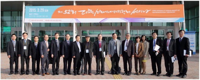
In this July issue 2015 of Clinical Endoscopy, journal editors have selected and invited core review articles among the state-of-the-art lectures delivered during the 52nd Seminar of Korean Society of Gastrointestinal Endoscopy (KSGE) (Fig. 1), with the aim of helping readers to scan and grasp the core contents at a glance.
Upper endoscopy (esophagogastroduodenoscopy, EGD) is used widely for the diagnosis and treatment of esophageal, gastric, and duodenal diseases and is a powerful screening tool for neoplastic and preneoplastic lesions. It is safely performed by a well-trained endoscopist, and the procedures should allow a complete evaluation of the upper gastrointestinal (GI) tract. During the procedure, photo documentation is mandatory for clinically significant abnormal lesions as well as for capturing at least eight normal parts of the tract in high-quality images.1 Standard measures for EGD should be followed strictly in any situation and should be properly documented.2
Information obtained during endoscopy needs to be described in detail so that it can be transferred as a medical record. The standardized report includes patient identification, indication(s) of procedure, patient demographics, medication history or comorbidities, pre-procedural medications, and procedural personnel. A detailed description of the observation with special emphasis on abnormal findings, diagnoses, endoscopic interventions, and details of the pathology specimens should also be included. All description needs to be recorded using standardized terminology rather than difficult to understand abbreviations or acronyms. Abnormal findings must be described according to standard classifications provided for erosive esophagitis or for gastroesophageal varices, according to the Paris classification for GI neoplastic lesions or to the Forrest classification of ulcer bleeding. Appropriate documentation of endoscopic procedures is a critical component for patient care.
Endoscopists should pay attention to the technique and devices used for obtaining adequate biopsy specimens, because pathological evaluation is essential for proper diagnosis of many upper GI lesions. The location and the number of biopsy specimens obtained should be recorded properly and the information regarding the lesion should be clearly provided to pathologists. Biopsy specimens need be taken from the portion, where the highest diagnostic yield is expected. Diagnostic impression of the target lesion and expected treatment should be considered in determining the number of biopsy specimens to be taken. Obtaining too many biopsy specimens results in artificial ulcer and subsequent fibrosis, which could render endoscopic resections for neoplastic lesions difficult.
Sedation during endoscopy is a drug-induced depression in the level of consciousness. The purpose of sedation is to lessen the patient anxiety and discomfort before and during the procedure, and to induce short-term amnesia to relieve the uncomfortable memory of the procedure, which will enhance future compliance for the procedure. Most endoscopic procedures can be performed under "conscious sedation" with the patient under moderate sedation. Informed consent should be obtained, and baseline evaluation of the patient's condition is necessary. Intraprocedural monitoring includes the level of consciousness, respiration with blood oxygen saturation, and blood pressure and pulse rates. Sedation must be provided under the thorough understanding of medications, and an individual should be dedicated to observe and monitor the patient without being involved in any procedure-related activities in case of deep sedation. Before leaving, the endoscopy room after the procedure, evaluation, and documentation of the patient's recovery state is equally important.3
Early esophageal cancer (EEC) is defined as a cancer confined to the mucosal layer without any lymph node metastasis. In contrast to the early gastric cancer (EGC) definition, esophageal cancer involving the mucosal or submucosal layer regardless of lymph node metastasis is considered superficial esophageal cancer. EEC in white light endoscopy shows a fine erythematous change reflecting irregular dilation of the intraepithelial papillary capillary loops (IPCLs). Chromoendoscopy using Lugol's solution or image-enhanced endoscopy such as narrow-band imaging (NBI) with magnification endoscopy (ME) is a useful tool to detect EEC. EEC appears as a brownish, well-demarcated lesion in the NBI mode. Identification of abnormal findings in the IPCLs such as dilation, tortuosity, caliber change, and various shape changes enable early detection of esophageal cancer using NBI with ME.
Although the incidence of Barrett's adenocarcinoma has increased over the past few decades in Western countries, such an epidemiologic change is not evident in Korea. The incidence of esophageal adenocarcinoma arising from Barrett's esophagus is about 0.5%. The standard treatment for Barrett's adenocarcinoma is surgical treatment; however, this is associated with significant morbidity. Thus, endoscopic treatment including argon plasma coagulation, photodynamic therapy, or endoscopic resection is recommended for early treatment of Barrett's adenocarcinoma. Sentinel polyp is an inflammatory lesion commonly associated with gastroesophageal reflux diseases (GERDs). Because the lesion is often confused with gastric cardia cancer, it is prudent to take a biopsy for pathological confirmation. Current standard treatment is medical treatment for GERD because of the benign nature of the disease without progression. However, follow-up endoscopy is recommended after proton pump inhibitor (PPI) treatment to rule out malignancy
Although the most common cause of esophageal inflammation is reflux esophagitis, there are numerous other forms of esophagitis. Infectious esophagitis may be caused by fungal (Candida, Aspergillus, Cryptococcus), viral (herpes simplex, Varicella zoster, cytomegalovirus), bacterial pathogens including mycobacteria and Treponema pallidum, or parasitic agents. Infectious esophagitis is usually associated with an immune compromised state, but it may occur in an immune-competent host. Systemic diseases that may be associated with esophageal inflammation include Behcet's disease, epidermolysis bullosa, pemphigus vulgaris, inflammatory bowel diseases (Crohn's disease), collagenous diseases, and eosinophilic esophagitis. Drugs, chemotherapy, radiation therapy, and corrosive agents are also important causes of esophagitis. Rapid diagnosis and proper treatment is challenging for many of these conditions, but is essential for patient care.
The gold standard tests for esophageal motility disorders are esophageal manometry and barium esophagography for bolus transit evaluation. The major role of endoscopy is to exclude other underlying diseases such as malignancy and to apply therapeutic measures. Usually there is no significant abnormality in endoscopic examinations at early stages of the disease, but many signs are suggestive of motility diseases such as achalasia, scleroderma, or spastic disorders of esophagus as the disease stage progresses.
Recently, the EGC detection rate has been increasing owing to improvements in screening endoscopy in Korea. To detect gastric cancer in early stages, the stomachs of high-risk patients having underlying atrophy and/or intestinal metaplasia should be observed carefully.4 Endoscopists should pay attention not to leave blind spots at the cardia and corpus. Systemic observation needs be verified by photo records. Knowledge of subtle changes suggesting malignancy is essential in order not to miss small EGCs. Chromoendoscopy using indigo carmine spray is the most commonly used method to contrast EGC lesions. NBI can be useful in characterizing the detected lesion and in delineating the tumor margins despite the limitations of initial detection of EGC. Accurate target biopsy is important for final pathological diagnosis to obtain an adequate specimen especially from very small lesions and ulcerative EGCs.
The prevalence of GERD is increasing and it imposes health burden as obesity and the popularity of a westernized diet increases in Korea. GERD can be classified according to the Montreal classification as symptomatic syndrome including typical reflux syndrome or reflux chest pain syndrome, syndromes involving esophageal injury including reflux esophagitis, reflux stricture, Barrett's esophagus, or adenocarcinoma, as well as extraesophageal syndromes including cough, laryngitis, asthma, or dental erosions.5 Endoscopic evaluation for GERD provides information about the presence of mucosal defects and complications such as stricture or Barrett's esophagus, and information to rule out other non-GERD underlying diseases. Prevalence of asymptomatic silent reflux esophagitis is known to be 11.6% to 45.3% in patients with EGD-proven erosive esophagitis from Asian studies,6 and the clinical significance of this condition is still controversial; long-term follow up evaluation is warranted.
The definition of gastritis has not been clearly elucidated and the disease has a diverse etiology. The Sydney classification for gastritis was established to provide a universal classification to include both endoscopic and pathological findings as well as the etiology of the disease.7 According to the guidelines, gastritis can be classified as erythematous/exudative gastritis, flat erosive gastritis, raised erosive gastritis, atrophic gastritis, hemorrhagic gastritis, rugal hyperplastic gastritis, and enterogastric reflux gastritis. Atrophic gastritis is a well-known risk factor for gastric cancer development and the definition has changed from "loss of glands" to "loss of appropriate glands," which encompasses the replacement of normal glands with metaplastic glands. Endoscopic classification of the severity of atrophy is commonly used because no additional biopsy specimen is required. Intestinal metaplasia is the common endpoint of severe Helicobacter pylori-related gastritis and subtypes suggest different levels of gastric cancer risk. Interobserver agreement for diagnosis of various types of gastritis is not satisfactory showing, only 40% to 60% concordance.
The endoscopic ultrasonography (EUS) session for hollow viscus consisted of four lectures: (1) normal anatomy: mediastinum, gut wall and perirectal organs; (2) cancer staging: TNM staging of hollow viscus cancer with EUS; (3) subepithelial lesion: characteristic EUS findings; and (4) EUS-guided fine needle aspiration for subepithelial and mediastinal lesions. A review article regarding the role of EUS in staging of GI cancer is published elsewhere in this issue of Clinical Endoscopy.8
The prevalence of gastric subepithelial tumor in Korea is approximately 1% in asymptomatic individuals and is usually found incidentally during screening evaluations. Characteristics of the lesion including shape, mobility, consistency, color, size, and surface mucosal change should be defined. EUS can provide information for diagnosis and subsequent treatment plans. Benign lesions include lipoma, leiomyoma, varix, heterotopic pancreas, duplication cyst, and inflammatory fibroid polyp. Potential malignant or malignant lesions are represented by GI stromal tumor, carcinoid, glomus tumor, lymphoma, and metastatic cancer. Algorithms for further plans/interventions should be tailored according to the tumor size and EUS characteristics suggesting potential malignancy.
Neuroendocrine tumor (NET) of the stomach is a heterogeneous group of tumors and shows different clinicopathological features and behavior according to the type identified. Its incidence is increasing, especially in Asian countries, as asymptomatic lesions are usually detected during screening endoscopy. Gastric NETs are classified according to the World Health Organization 2010 classification of digestive neuroendocrine neoplasm as grade 1 NETs (G1-NETs), G2-NETs, G3 neuroendocrine carcinomas, mixed adenoneuroendocrine carcinomas, and hyperplastic and preneoplastic lesions. Curative therapy for gastric NETs requires complete resection using an endoscopic technique for small (≥1 cm) NETs, or local surgical resection for larger early stage tumors. Radical surgery with lymph node dissection is a standard treatment for advanced NETs. It is necessary to obtain an adequate assessment of the clinical and pathological characteristics of the tumor to apply optimal therapeutic approach in patients with gastric NETs.
Peptic ulcer bleeding (PUB) accounts for 30% to 60% of upper GI bleeding. The main role of endoscopy is to identify high-risk stigmata lesions, and to provide endoscopic hemostasis. Candidate selection and timing of endoscopy should be carefully evaluated using clinical information including careful history taking, physical examination, and laboratory findings.
Endoscopic therapy is better indicated for patients having ulcers with high-risk stigmata, such as active bleeding or visible vessels. Endoscopic treatments including injection, ablative, mechanical or combination therapies can be applied based on the characteristics of the bleeding lesion. Combined therapy has been demonstrated to be superior to injection treatment alone, but single treatment with mechanical or thermal methods is safe and effective for PUB. Endoscopic hemostatic techniques clearly decrease rebleeding risk and subsequent risk of PUB-related mortality. Ulcer-covering hemospray or endoscopic suturing devices are new promising tools.
Peptic ulcer disease (PUD) has long been established a chronic disabling disease showing recurrent exacerbation and remission, but the management of PUD has dramatically changed over the past decades. The recognition of H. pylori infection as the main causative agent and effective eradication regimens for the organism such as antisecretory agents, including PPIs, have made this disease a curable one using medical treatment. However, refractory PUD often occurs and it is necessary to determine possible etiologies and to provide an adequate management for the condition to prevent severe PUD-related complications.9
Possible conditions to be considered first in refractory ulcer include persistent H. pylori infection due to antimicrobial resistance and a false negative H. pylori test. Nonsteroidal anti-inflammatory drugs, tobacco or alcohol use, and hypersecretory conditions such as Zollinger-Ellison syndrome should be sought. Systemic diseases including vasculitis or Crohn's disease, and other rare infections such as syphilis, tuberculosis, or viral diseases should be considered. Gastric cancer should be ruled out carefully as a cause of intractable ulcer. Comprehensive history taking and physical examination are the main corner stones for identification of the etiology and subsequent proper management. Intractable PUDs are mainly controlled by medical treatment but rare severe complications still require surgical intervention.
Screening endoscopy can detect most gastric cancers as an EGC, which is sometimes difficult to diagnose even by an experienced endoscopist. Elevated type EGC often resembles adenoma or submucosal tumor. Flat type EGCs might resemble gastritis, and shows ill-defined margins especially in EGCs with undifferentiated type histology. Ulcerative type EGCs are common but many are confused with benign gastric ulcers or erosions, especially small ones. Obtaining an adequate biopsy specimen and follow-up endoscopy after treatment are crucial for proper diagnosis and management of the unusual type of EGCs.
Informed consent must be patient-centered so that patients can get enough information about the colonoscopic procedure, make a decision about whether to undergo the procedure or not on the basis of fully disclosed information, and make individual judgments based on the opportunity to inquire about the procedure. The images and findings from colonoscopy are very important because they are useful to decide on the strategy for future treatment, provide explanations to the patients, and allow medical staff members to improve communication with each other, as well as to analyze and report the lesions.
Lectures on the basic techniques of insertion of the colonoscope—bowel shortening, bending and rotation using a knob, torque, right turn shortening, hooking the fold, jiggling, shaking, a slalom technique, and a sliding technique—and on its supplementary techniques—position changes, abdominal wall compression, breath-holding, and liquid infusion—were conducted. Colonoscopy may be conducted with relative ease in the rectum and in the descending and ascending colons, which may barely stretch because they are fixed to the retroperitoneal wall and are less likely to vary among patients. However, it is important to note that the sigmoid and transverse colons and those regions connected to them are difficult to access.
What is most important for both the operator and the patient in conducting colonoscopy is not to miss the lesion but, instead, to diagnose it accurately. It is necessary to practice good bowel preparation and inserting a colonoscope for the purpose of thorough and accurate observation. The mucosa should be observed even while inserting the colonoscope. It is important to reduce blind spots through good control of insufflation and aspiration and to identify any blind spot and observe the region of the spot through repetitive insertions and withdrawals. The proximal side of colonic folds should be checked and observed several times. Any lesion having subtle changes, such as color change, loss of the vascular pattern, deformity of the bowel wall, hemorrhage, and white spots should never be missed.
It is essential to adjust the position of the monitor and the patient's bed in order to maintain good posture for the colonoscopist. The monitor needs to be located directly in front of the operator's eyes; the distance between the monitor and his/her eyes must be ≥50 cm; his/her eyes need to look 15° to 25° downward; and it is necessary that the monitor's height be adjustable as a function of the operator's height and the operation posture. The height of the bed needs to be adjusted so that the position of the insertion site is 10 cm lower than the operator's elbow while inserting the colonoscope. It is desirable to have a suitable rest intraoperatively since the left thumb and wrist may suffer from accumulative tension and fatigue during the procedure at the colonoscopy site, particularly in cases of prolonged colonoscopy. It is important to adopt beneficial working habits such as regular stretching and have adequate working conditions; once symptoms occur, it is essential to examine the condition correctly and receive proper treatment.
Bowel preparation for colonoscopy is a complex process that involves both pre-colonoscopic diet modification and selection and administration of preparation medicines. Good bowel preparation is essential for a more efficient colonoscopy because it is closely associated with the cecal intubation rate and the adenoma detection rate, both of which are indexes of quality control in the colonoscopic procedure. As the importance of the bowel preparation has steadily been emphasized in recent years, the European Society of Gastrointestinal Endoscopy and the US Multi-Society Task for Colorectal Cancer have presented bowel preparation guidelines in 2013 and 2014, respectively.1011 It is important to instruct the operator to choose a suitable means of administering the preparation medicine and to administer it properly, taking into consideration of the patient's characteristics, such as his/her age and disease status, and the properties of the preparation.
The 20% to 25% of patients may experience poor bowel preparation for various reasons even if they undergo diet restriction or take a preparation medicine correctly as instructed. Patients who are aged, male, obese, or diabetic, as well as those who are inpatients, take at least eight medicines concomitantly, have neurological diseases (e.g., stroke, spinal injury, and Parkinson's disease), or have undergone GI operations, patients who take antidepressants or narcotic analgesics frequently, and those experiencing severe constipation will require special attention because they are highly likely to experience poor bowel preparation.12 For patients who are expected to have poor bowel preparation, it is desirable to recommend split dose of a preparation medicine and conduct colonoscopy 3 to 5 hours after they have taken the final dose of the medicine; in addition, an intensive bowel cleansing strategy can be selected preemptively. It is also necessary to learn how to manage situations presenting with poor bowel preparation and encourage good bowel preparations in at least 90% of the patients.
Every year, 10% of patients taking antithrombotics, such as antiplatelet agents and anticoagulants, undergo an operation or invasive procedure requiring temporary discontinuation of the medication.13 It is necessary to take into consideration the urgency of the endoscopy, the potential risk of hemorrhage as a consequence of antithrombotic therapy, the risk of hemorrhage due to endoscopy during concomitant antithrombotic treatment, and the risk of thromboembolism due to discontinuation of antithrombotic treatment prior to the endoscopy procedure in these patients. It is also necessary to learn other techniques that may replace endoscopy, or antithrombotics that can be used as an alternative medicine, and the tests that can be used to monitor antithrombotic treatment status.13 While guidelines have been suggested for the use and discontinuation of treatment with the more traditional antiplatelet agents and anticoagulants based on specific evidence and expert opinions, it is not easy to decide how to manage those patients taking antiplatelet agents and anticoagulants that have be more recently introduced to clinical practice.
For pediatric colonoscopy, small caliber colonoscopy, or upper endoscopy can be helpful when it is difficult to pass through the sigmoid colon. It has been reported that such basic insertion techniques as torquing, jigging, hooking the fold, pull back, sliding, and aspiration of an excessive amount of air may enable successful insertion into the cecum without the necessity of special tools or changing endoscopes in three-fourths of patients with a redundant colon.14 A variable stiffness of the scope and balloon enteroscopy can also be of help. A transparent hood, CO2 gas insufflation, a water immersion method, and a sigmoid overtube may be used as supplementary tools. Computed tomography colonography, barium enema, and colon capsule endoscopy can be used as alternative techniques to examine the colon.
When a colonic ulcer or inflammation is detected, clinical, endoscopic, laboratory, image, histologic, and operation findings are generally assembled to formulate a diagnosis. However, it is difficult to endoscopically distinguish Crohn's disease, intestinal tuberculosis, or intestinal Behcet's disease, each of which presents with lesions located in the ileocecal region. In particular, it is not easy to differentiate these three diseases by relying solely on colonoscopic findings as highlighted in reported cases of an ileocecal ulcer found accidentally by colonoscopy among asymptomatic patients.1516
In addition to idiopathic inflammatory bowel diseases, there are many other diseases that may cause colonic inflammation: intestinal tuberculosis, infectious colitis, parasitic infection, ischemic colitis, drug related colitis, radiation colitis, eosinophilic colitis, and diversion colitis.
Colonoscopy, which has recently been used in clinical practice, may not only provide visual findings based on white light but also the through the use of image enhancement findings including optical techniques such as NBI, and is very helpful in making a correct diagnosis in combination with more conventional techniques, including chromoendoscopy. Kudo's pit pattern classification is a typical approach used to classify the surface mucosal structures of polypoid epithelial lesions. The more recently available high-resolution endoscopes may make it possible to guess the pit pattern without assistance from chromoendoscopy or magnifying endoscopy. The techniques that may provide a better understanding of the surface microstructure or vascular pattern using optical technique include NBI, flexible spectral imaging color enhancement, and iScan. Of these, NBI may enable a better observation of the surface microvascular pattern, while the NBI international colorectal endoscopic classification, which can postulate a histologic diagnosis, is recommended as a helpful technique to determine the pit pattern.17 Endoscopic diagnosis of polypoid epithelial lesions is important because whether or not the lesion requires endoscopic resection may be determined on the basis of the endoscopic diagnosis. It is necessary to become accustomed to formulating an endoscopic diagnosis with the aim of avoiding unnecessary endoscopic resections or any unnecessary surgical procedures for polypoid epithelial lesions and therefore constructing a good treatment plan.
A review article about colorectal subepithelial lesion is available separately in this issue of Clinical Endoscopy.18
As the most definitive approach of preventing colorectal cancer, polypectomy is expected to decrease the overall prevalence and mortality of colorectal cancer. By polypectomy, polyps ≤5 mm can be removed using biopsy forceps; in contrast, polyps >5 mm are mostly removed using a snare. A doctor initiating a polypectomy procedure needs to have a good understanding of the clips available for preventing and treating hemorrhage and perforation, both of which are common complications of the technique, as well as the of biopsy forceps, the snare, and electrosurgical units.
Polypectomy is an established method of preventing colorectal cancer and reduces its incidence by ≥40%.19 Colonic polyps may be divided into minute (≤5 mm), small (6 to 9 mm), large (≥1 cm), and huge polyps (≥3 cm) based on size; since 90% of colonic polyps are clinically known to be <1 cm size, learning how to use basic polypectomy techniques may enable the majority of polyps to be easily removed endoscopically in an outpatient clinic setting. However, since advanced lesions are found in nearly 9% of the minute polyps, it is important to improve procedural integrity even for tiny polyps through repetitive training in basic techniques for the purpose of safe polypectomy without likelihood of recurrence.
Most pathologists complain that endoscopists do not provide phone consultations, do not provide records, and do not present any clinical reports, whereas the latter tend to believe that the former do not require any additional information since they can make a full diagnosis based only on microscopic findings. Given the consequences of a diagnosis based solely on inappropriate histologic tests and its costs, good communication with the pathologist is the most essential and fundamental requirement for the patient's sake. It is therefore necessary to take the habit of recording as much information as possible about the patient, and in addition to requesting the pathological testing, the endoscopist should make an effort to consult regularly and establish mutual communication with the pathologist.
Approximately 27.3% of patients undergoing polypectomy may reportedly have a colorectal cancer occurrence due to incomplete resection20 and are likely to have complications, such as procedure-related perforation or hemorrhage. Thus, polypectomy needs to be performed in the optimally in pursuit of a safe and complete resection, taking into account the properties of polyps. First, it is necessary to differentiate lesions requiring resection from those not requiring resection and then to select the proper polypectomy technique based on the polyp's properties, such as its location, size, shape, and count. An additional surgical procedure is recommended when the endoscopic resection of a malignant polyp is deemed incomplete, when cancer invasion into vessels or lymphatics is found on histopathological tests, when a cancer invasion into the submucosal layer ≥1,000 µm is detected, when poor cell differentiation is observed, or when cancer invasion is observed ≤2 mm from the resection margin. If any positive non-lifting sign is identified, it is recommended to stop the endoscopic resection and provide surgical treatment.
Sedation for endoscopy strives to seek a balance between patient comfort and drug-related side effects. Optimal sedation allows the patient the greatest degree of comfort while preserving the greatest degree of safety. This session introduced sedation drugs and methods based on age, sex, and comorbidity according to the sedation guidebook recently published by KSGE.
Bleeding is one of main complications observed during endoscopic retrograde cholangiopancreatography (ERCP); meta-analysis has shown that the incidence was 1.3%. However, this incidence can increase in patients on antithrombotic therapy. Antithrombotic therapy is used to reduce the risk of thromboembolic events in patients with certain cardiovascular conditions, deep venous thrombosis, hypercoagulable states, and endoprostheses. This session included the risk factors for bleeding complications, risk stratification upon discontinuation of anticoagulant therapy, preparation methods before ERCP procedures for reducing bleeding complications, and management of bleeding after ERCP.
This session included evaluation of radiation risk and injuries for the endoscopist during ERCP procedures, and practical guidelines for radiation safety and protection. This topic introduced practical information regarding radiation biology, doses to endoscopists during ERCP, factors that limit radiation exposure, protection principles, and practical methods for reducing radiation exposure while performing ERCP.
This session dealt with the formulation of an endoscopic record and the minimal standard terminology to be adopted for GI endoscopy. This topic introduced the initial version of a computerized endoscopic reporting program in Korea that was prepared by the Korean Pancreatobiliary Association in 2014.
Selective cannulation is a basic technique for therapeutic ERCP. This topic included basic techniques for selective bile duct cannulation. For selective cannulation, a specific understanding of the anatomy surrounding the ampulla of Vater is necessary. This session included the insertion method for the duodenoscope, the standard technique for bile duct cannulation, bile duct cannulation with a sphincterotome, cannulation using wires, and bile duct cannulation after pancreatic duct stent insertion.
It is sometimes rather difficult to reach into the desired duct using standard techniques with a cannula or pull type sphincterotome. Thus, precut and needle knife fistulotomies are sometimes necessary for selective cannulation. This topic included the complications and risk factors resulting from failed selective cannulation, timing of precut sphincterotomies, and precut access using a needle knife and transpancreatic septostomy.
The success rate of selective cannulation is known to be 70% to 90% in patients with a surgically altered anatomy. This topic introduced the accessories, endoscopic insertion methods, and tips for selective bile duct cannulation for surgically altered anatomy.
Usually, pancreatic duct cannulation is easier than selective bile duct cannulation. However, pancreatic duct cannulation is sometimes difficult and a repeated trail can induce post-ERCP pancreatitis. This topic introduced indications for minor papilla cannulation, the basic technique for major and minor pancreatic duct cannulation, and pancreatic sphincterotomy.
The endoscopic treatment of pancreatic massed and cysts is not yet standardized, and has been an experimental treatment modality until now. This topic introduced the EUS-guided ethanol ablation in pancreatic cysts and radiofrequency ablation in pancreatic solid masses.21
Percutaneous transhepatic gallbladder drainage (PTGBD) is the most widely established salvage therapy for gallbladder drainage (GBD) in patients unresponsive to medical treatments or who are at high risk for cholecystectomy. The perceived advantages of EUS-guided GBD (EUS-GBD) include the avoidance of external drainage, unlike PTGBD, and the potential for no risk of post-ERCP pancreatitis or cholangitis, unlike transpapillary drainage. EUS-GBD can be used as a bridge to surgery in patients unsuitable for emergent cholecystectomy. This topic introduced the indications, methods, efficacies, complications, and long-term results of EUS-GBD.
Endoscopic drainage can be achieved internally by cystogastrostomy or cystenterostomy through direct puncture using ERCP or through EUS guidance. Prior studies have shown comparable success rates using either approach when pseudocysts with visible luminal compression were targeted. However, EUS-guided drainage of pseudocysts is technically more successful and likely safer, particularly in the setting of portal hypertension and nonbulging pseuodocysts. This session introduced the indications for pseudocyst drainage and techniques for EUS-guided drainage.
ERCP for biliary access and drainage is successful in 90% to 95% of cases. In cases of failure of ERCP to achieve biliary drainage due to difficult cannulation or abnormal anatomy, the alternative has been percutaneous transhepatic biliary drainage. EUS-guided ERCP can be performed in a number of ways depending on the route of access or the route of drainage. If the papilla is accessible, a rendezvous technique can be adopted wherein EUS is used to puncture the bile duct and a wire is negotiated through the papilla, further therapy is carried out through ERCP. If the papilla is not accessible, EUS is used to access the bile duct and create a fistula for placement of a stent, the so-called the transmural technique. This session introduced basic techniques, safety, and efficacy of EUS-guided biliary drainage.
Obesity is an enormous health-related issue nowadays. Definition, indication for treatment, and pharmacological therapy for obesity was introduced along with endoscopic and surgical treatment methods in this session. Restrictive methods including intragastric balloon therapy, transoral gastroplasty with sleeve stapler and endoluminal vertical gastroplasty were introduced. The EndoBarrier GI liner (EndoBarrier, Hertogenbosch, The Netherlands), which is an endoscopic GI bypass device, was also presented. However, all these methods require further investigation and validation.
Adequate sedation for any endoscopic procedure is very important, and recently, KSGE published guidelines for conscious sedation. In this session, results of a survey regarding the status of sedated endoscopy in Korea were handed out. Advisory lectures for safe sedation in a private clinic setting and a practical how-to use guidebook were also included. The medicolegal review of deaths related to propofol administration was very informative.
Live demonstrations of upper and lower GI diagnostic and therapeutic cases were popular with attendees. Hands-on courses for colonoscopy and scope disinfection processes were also one of the most popular sessions of the seminar as expected.
Intense reviews were prepared to upgrade our society journal, Clinical Endoscopy, to qualify as a Science Citation Index (SCI) journal under the session name of Editor School. In this session, we discussed how to foster efforts to improve the Clinical Endoscopy journal in order to be included among the SCI Expanded SCI(E) journals. Separate from the current highlight article, two special articles have been included in this special issue of Clinical Endoscopy regarding this aim and scope, and the specific strategy for Clinical Endoscopy
The KSGE has held seminars twice a year for the last 26 years and has always attempted to cover topics for the endoscopist that would appeal to the very beginner to advanced experts in both diagnostic and therapeutic fields. The 52nd Seminar of the KSGE, which was held on March 29, 2015, consisted of 26 sessions filled with very informative lectures, discussions, live demonstrations, and hands-on courses with over 4,100 attendees including physicians, nurses, and technicians was also very successful. Endoscopy continues to grow with innovative new techniques, allowing it to evolve from a basic diagnostic modality to a more advanced diagnostic and therapeutic armamentarium. The lectures presented in the seminars covered basic to advanced topics in the field of gastroenterology and attendees participated in every session actively and willingly. The varied content of the meeting could offer beneficial information for doctors and for patients in their future practice.
References
1. Park KS. Introduction to starting upper gastrointestinal endoscopy: proper insertion, complete observation, and appropriate photographing. Clin Endosc. 2015; 48:279–284.
2. Park WG, Shaheen NJ, Cohen J, et al. Quality indicators for EGD. Gastrointest Endosc. 2015; 81:17–30. PMID: 25480101.
3. Standards of Practice Committee of the American Society for Gastrointestinal
Endoscopy. Lichtenstein DR, Jagannath S, et al. Sedation and anesthesia in GI endoscopy. Gastrointest Endosc. 2008; 68:815–826. PMID: 18984096.
4. Moon HS. Improving the endoscopic detection rate in patients with early gastric cancer. Clin Endosc. 2015; 48:291–296.
5. Vakil N, van Zanten SV, Kahrilas P, Dent J, Jones R. Global Consensus Group. The Montreal definition and classification of gastroesophageal reflux disease: a global evidence-based consensus. Am J Gastroenterol. 2006; 101:1900–1920. PMID: 16928254.
6. Lu CL. Silent gastroesophageal reflux disease. J Neurogastroenterol Motil. 2012; 18:236–238. PMID: 22837870.
7. Dixon MF, Genta RM, Yardley JH, Correa P. Classification and grading of gastritis. The updated Sydney System. International Workshop on the Histopathology of Gastritis, Houston 1994. Am J Surg Pathol. 1996; 20:1161–1181. PMID: 8827022.
8. Cho JW. The role of endosonography in the staging of gastrointestinal cancers. Clin Endosc. 2015; 48:297–301.
9. Kim HU. Diagnostic and treatment approaches for refractory peptic ulcers. Clin Endosc. 2015; 48:285–290.
10. Hassan C, Bretthauer M, Kaminski MF, et al. Bowel preparation for colonoscopy: European Society of Gastrointestinal Endoscopy (ESGE) guideline. Endoscopy. 2013; 45:142–150. PMID: 23335011.
11. Johnson DA, Barkun AN, Cohen LB, et al. Optimizing adequacy of bowel cleansing for colonoscopy: recommendations from the US multi-society task force on colorectal cancer. Gastroenterology. 2014; 147:903–924. PMID: 25239068.
12. Hassan C, Fuccio L, Bruno M, et al. A predictive model identifies patients most likely to have inadequate bowel preparation for colonoscopy. Clin Gastroenterol Hepatol. 2012; 10:501–506. PMID: 22239959.
13. Desai J, Granger CB, Weitz JI, Aisenberg J. Novel oral anticoagulants in gastroenterology practice. Gastrointest Endosc. 2013; 78:227–239. PMID: 23725876.
14. Rex DK, Goodwine BW. Method of colonoscopy in 42 consecutive patients presenting after prior incomplete colonoscopy. Am J Gastroenterol. 2002; 97:1148–1151. PMID: 12014719.
15. Almadi MA, Ghosh S, Aljebreen AM. Differentiating intestinal tuberculosis from Crohn's disease: a diagnostic challenge. Am J Gastroenterol. 2009; 104:1003–1012. PMID: 19240705.
16. Epstein D, Watermeyer G, Kirsch R. Review article: the diagnosis and management of Crohn's disease in populations with high-risk rates for tuberculosis. Aliment Pharmacol Ther. 2007; 25:1373–1388. PMID: 17539977.
17. Oba S, Tanaka S, Sano Y, Oka S, Chayama K. Current status of narrow-band imaging magnifying colonoscopy for colorectal neoplasia in Japan. Digestion. 2011; 83:167–172. PMID: 21266811.
19. Nishihara R, Wu K, Lochhead P, et al. Long-term colorectal-cancer incidence and mortality after lower endoscopy. N Engl J Med. 2013; 369:1095–1105. PMID: 24047059.
20. Pabby A, Schoen RE, Weissfeld JL, et al. Analysis of colorectal cancer occurrence during surveillance colonoscopy in the dietary Polyp Prevention Trial. Gastrointest Endosc. 2005; 61:385–391. PMID: 15758908.