Abstract
Tumor bleeding is not a rare complication in patients with inoperable gastric cancer. Endoscopy has important roles in the diagnosis and primary treatment of tumor bleeding, similar to its roles in other non-variceal upper gastrointestinal bleeding cases. Although limited studies have been performed, endoscopic therapy has been highly successful in achieving initial hemostasis. One or a combination of endoscopic therapy modalities, such as injection therapy, mechanical therapy, or ablative therapy, can be used for hemostasis in patients with endoscopic stigmata of recent hemorrhage. However, rebleeding after successful hemostasis with endoscopic therapy frequently occurs. Endoscopic therapy may be a treatment option for successfully controlling this rebleeding. Transarterial embolization or palliative surgery should be considered when endoscopic therapy fails. For primary and secondary prevention of tumor bleeding, proton pump inhibitors can be prescribed, although their effectiveness to prevent bleeding remains to be investigated.
Go to : 
Tumor bleeding accounts for up to 5% of upper gastrointestinal bleeding (UGIB) cases.1,2,3,4,5 Of these, primary gastric cancer is the most common cause of tumor bleeding, accounting for between 36% and 58% of bleeding cases resulting from upper gastrointestinal malignancies.1,3,4 However, little information is available regarding the current management of gastric cancer bleeding. Of the information available, 8% of patients undergo urgent endoscopy due to hematemesis,5 and bleeding is implicated in 37% of gastric cancer patients in the emergent setting and 10% in the outpatient setting.6 In addition, 79% of patients presenting with tumor bleeding are unaware of their cancer, and 75% of those have cancer metastasis.4 For these patients, curative surgical or endoscopic resection of gastric cancer may be the definite treatment for cancer bleeding. However, tumor bleeding from inoperable gastric cancer is still problematic because of the difficulty in treating bleeding and frequent recurrent bleeding events after successful hemostasis.
Endoscopy is important for the diagnosis and primary treatment of UGIB, and guidelines recommended endoscopy within 24 hours of presentation.7,8,9,10 Endoscopy is also important for the management of tumor bleeding due to inoperable gastric cancer, but limited studies have investigated the effects and roles of endoscopy in the diagnosis and treatment of these cases. Furthermore, guidelines regarding the management of tumor bleeding are not well established. In this review, we will discuss the current data and considerations for managing tumor bleeding due to inoperable gastric cancer.
Go to : 
Several possible reasons may explain why patients with inoperable gastric cancer are vulnerable to UGIB, other than bleed-ing directly from the gastric cancer. First, nausea and vomiting due to the toxicities of chemotherapy or gastric outlet obstruction may be associated with Mallory-Weiss syndrome.11,12 Second, thrombocytopenia due to bone marrow suppression after chemotherapy may cause coagulopathy,13 and severe thrombocytopenia (platelet count <50,000/mm3) may increase the risk of spontaneous bleeding, including UGIB.14 Third, patients with multiple bone metastases may have coagulopathy because of direct bone marrow suppression.15 Moreover, UGIB in patients with gastric cancer can result from benign causes, including peptic ulcers, esophageal and gastric varices, hemorrhagic gastritis, and angiodysplasia.12,13 Therefore, clinicians must pay attention to these special considerations when managing tumor bleeding in patients with inoperable gastric cancer.
Go to : 
When inoperable gastric cancer patients present with UGIB, initial assessments should be performed according to the general guidelines for the management of UGIB.7,8,9,10,16 The initial assessments include observation of the symptoms and signs of UGIB, hemodynamic status (blood pressure and pulse rate), and laboratory findings such as complete blood counts, including hematocrit measurement. In addition to the initial assessments, initial resuscitation including intravenous fluids and transfusion may be required for some patients. Furthermore, thrombocytopenia should be corrected in patients with platelet counts below 40,000/mm3 before endoscopy if possible.17
Recent studies have reported that several scoring systems such as the Glasgow-Blatchford bleeding score (GBS)18 and the Rockall score,19 which utilize several clinical criteria, are useful for stratifying risk and predicting interventions for the management of UGIB. Among scoring systems, GBS shows better performance than the Rockall score in discriminating low-risk patients who could be treated as outpatients.20 Thus, guidelines recommend the use of these scoring systems to stratify the risk in patients with UGIB.8,9,10 However, studies investigating the efficacy or role of these scoring systems in patients with tumor bleeding are limited. In a recent study, Kim et al.21 evaluated the bleeding score systems for their ability to predict the need for interventions and the clinical outcomes in patients with tumor bleeding due to inoperable gastric cancer. The post-endoscopic Rockall score was useful for predicting the need for procedures, including endoscopic therapy (ET), transarterial embolization (TAE), and surgery (area under the receiver operating characteristics curve, 0.77; p<0.001). GBS was superior in predicting the need for all interventions, including transfusion and other procedures (area under the receiver operating characteristics curve, 0.81; p<0.001). These results indicate that bleeding scoring systems are helpful in managing tumor bleeding due to inoperable gastric cancer, although further studies are needed.
The UGIB management guidelines define the roles and uses of prokinetic agents and proton pump inhibitors (PPIs). A recent meta-analysis reported that utilizing intravenous prokinetics such as erythromycin or metoclopramide immediately before endoscopy reduces the need for a repeat endoscopy (odds ratio [OR], 0.55; 95% confidence interval [CI], 0.32 to 0.94), but does not improve the number of blood transfusions, hospital stays, or the need for surgery.22 Furthermore, a Cochrane meta-analysis of six randomized controlled trials found that utilizing PPIs before endoscopy significantly reduces the proportion of patients with high-risk endoscopic stigmata of recent hemorrhage (SRH) according to the Forrest classification (OR, 0.67; 95% CI, 0.54 to 0.84).23 However, pre-endoscopic PPI therapy does not decrease mortality, rebleeding, or the need for surgery.
Current guidelines do not recommend the routine use of pre-endoscopic prokinetic agents, but these agents could be used for a better diagnostic yield during endoscopy for UGIB patients with suspected fresh blood or clots in the stomach.7,9 However, pre-endoscopic PPI therapy is recommended for all patients with acute UGIB because of several beneficial effects.7,8,9,16 Similar guidelines may be beneficial if applied for the management of tumor bleeding due to inoperable gastric cancer. However, the effects and roles of prokinetic agents and PPIs have not been well studied in these patients.
Go to : 
For the treatment of UGIB, ET is generally recommended as the first-line treatment.7,8,9,16 ET reduces further bleeding (OR, 0.38; 95% CI, 0.32 to 0.45), the need for surgery (OR, 0.36; 95% CI, 0.28 to 0.45), and mortality (OR, 0.55; 95% CI, 0.40 to 0.76).24 However, the efficacy of ET has not been well studied, and limited studies have reported the success rates and instances of rebleeding after ET in patients with tumor bleeding due to gastric cancer.2,3,4,25,26,27,28
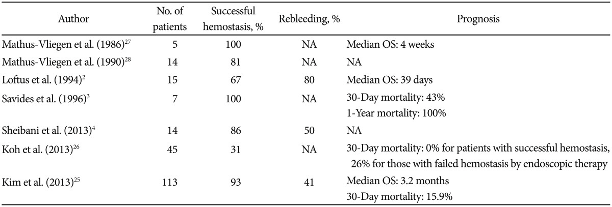
Table 1 summarizes the results of those studies. The rate of successful hemostasis by ET in patients with tumor bleeding due to gastric cancer was between 67% and 100%, except in one study which showed a low success rate of 31%.26 These high rates were similar to those observed following ET in patients with peptic ulcer bleeding (definite hemostasis in 75% to 89% of patients).29 However, rebleeding rates in patients that underwent ET for gastric cancer bleeding were higher (ranging from 41% to 80%) than those for patients with peptic ulcer bleeding (ranging from 8% to 24%).10,30 In a previous study, 43 of 105 patients (41%) that achieved successful hemostasis following ET developed rebleeding, and 18 of 43 (42%) underwent repeat ET for treatment of rebleeding with a success rate of 89%.25 Taken together, these data indicate that ET may be an effective treatment for achieving initial hemostasis as well as for the management of rebleeding in patients with tumor bleeding from gastric cancer.
The Forrest classification of endoscopic SRH31 has been commonly used for predicting the need for ET, the risk of rebleeding after initial hemostasis, and mortality.32,33,34 The Forrest classification is also useful for planning treatment strategies for tumor bleeding due to inoperable gastric cancer. Sheibani et al.4 reported that the distribution of patients with tumor bleeding among the Forrest classification groups was 1% with spurting hemorrhage (Forrest Ia), 29% with oozing hemorrhage (Forrest Ib), 2% with a non-bleeding visible vessel (Forrest IIa), and 6% with adherent clots (Forrest IIb). Kim et al.25 showed that the distribution of endoscopic SRH in patients undergoing ET for tumor bleeding due to inoperable gastric cancer was 15% Forrest Ia, 52% Forrest Ib, 20% Forrest IIa, and 12% Forrest IIb. However, no studies have investigated the association between the Forrest classification of endoscopic SRH and rebleeding or mortality after ET for tumor bleeding.
The ET modalities that can be used for hemostasis are injection therapy, mechanical therapy, ablative therapy, and a combination of several modalities.10,16,35 Injection therapy includes epinephrine, ethanol, ethanolamine, and polidocanol. Hemostasis by these agents results from the volume effect, direct tissue injury, and thrombosis.10,36 Of these, injection therapy with epinephrine has been widely used, and higher volumes of epinephrine injection (>13 mL of 1:10,000 solution) are particularly effective for hemostasis.37 However, meta-analyses of the risk of persistent bleeding or rebleeding after initial hemostasis revealed that the need for emergency surgery is significantly higher following epinephrine injection alone than following combination treatment with epinephrine injection and hemoclips or thermal methods for peptic ulcer bleeding.29,38 In addition, injection therapy using normal saline, thrombin, fibrin sealant, and cyanoacrylate glue is available, but those agents have limited roles and are not currently used in clinical practice.35
Mechanical therapy includes hemoclip placement, balloon tamponade, and band ligation devices.10,35 Among these methods, hemoclips are the most widely used and can achieve immediate hemostasis by obstructing the vessel, and have the advantage of not causing additional tissue damage.39,40 Hemoclip usage for treating non-variceal UGIB is superior to injection alone,29 but is comparable to thermal methods in achieving definite hemostasis.29,41
Thermocoagulation using a heater probe, electrocoagulation using monopolar or bipolar electrocautery, and argon plasma coagulation (APC) are among the ablative methods of ET.35 Ablative therapies promote hemostasis by coagulation of tissue proteins, activation of thrombocoagulation, and tissue destruction.42 These methods are as effective as hemoclips for achi-eving hemostasis,29,41 and in some cases, are easier to use than hemoclips.41
All ET modalities can be used for hemostasis in patients with tumor bleeding due to inoperable gastric cancer. Although no specific guidelines have been established, the Forrest classification of endoscopic SRH may be useful for choosing a method among several ET methods.
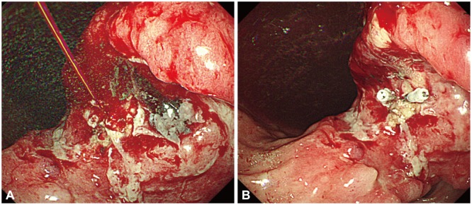
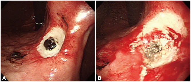
In spurting hemorrhages (Forrest Ia) and non-bleeding visible vessels (Forrest IIa), hemoclips or ablative therapy using thermocoagulation or electrocoagulation alone or with combination injection therapy can be selected (Figs. 1, 2). In a previous study, the success rate of initial hemostasis was 88% in patients undergoing electrocoagulation and 70% in those receiving hemoclips.25 In this study, electrocoagulation using hemostatic forceps (Radial Jaw 3, Boston Scientific, Heredia, Costa Rica) with an ERBE VIO 300D generator (ERBE Elektrome-dizin GmbH, Tübingen, Germany; effect 8, 80 W) was most commonly used for controlling spurting hemorrhages and non-bleeding visible vessels and effectively controlled the bleeding. Meanwhile, mechanical therapy using hemoclips was performed in a small number of patients, and the rate of initial hemostasis was low, resulting from difficult hemoclip placement because of tumor bed characteristics, such as marked fibrosis and hardness of the adjacent mucosa.43 In addition, hemoclip placement is difficult for treating a bleed resulting from a peptic ulcer with fibrotic and hard mucosa.41 Thus, electrocoagulation may be a better option than hemoclips in cases with a spurting hemorrhage and non-bleeding visible vessels on endoscopy.
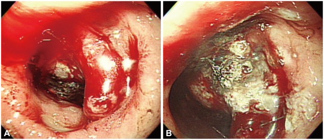
In patients with active tumor bleeding due to gastric cancer, the most common form of endoscopic SRH is oozing hemorrhage (Forrest Ib), occurring in 52% to 97% of patients.4,25 In this type of hemorrhage, ablative therapy with or without other therapeutic methods can be chosen. Ablative therapy seems to be effective for controlling active bleeding because the bleeding focus tends to be diffuse in this endoscopic SRH. Among ablative therapies, electrocoagulation using APC with an APC2 generator (ERBE Elektromedizin GmbH; effect 2, flow 2 L/min, 40 W) was the most common method used for this form of SRH in a previous study (Fig. 3),25 because APC is effective for the treatment of superficially extensive, diffuse lesions such as gastric antral vascular ectasia44,45 and radiation proctitis.46
The proportion of endoscopic SRH classified as adherent clots (Forrest IIb) in patients with tumor bleeding due to gastric cancer was between 6% and 12%.4,25 In the management of non-variceal UGIB, aggressive endoscopic clot removal is favored by most endoscopists because of the possibility of concealed underlying lesions, including Dieulafoy lesions and non-bleeding visible vessels, and the high risk of rebleeding or continued bleeding after medical therapy alone.35,47,48,49 In patients with tumor bleeding due to inoperable gastric cancer and adherent clots, endoscopic clot removal and proper management with ET may also be beneficial.
Go to : 
Despite comparable success rates of initial hemostasis by ET, patients undergoing ET for tumor bleeding due to inoperable gastric cancer have more frequent rebleeding events (41% to 80%)2,4,25 than those with UGIB due to other benign causes (8% to 24%).10,30 Rebleeding is associated with poor overall survival in patients who achieve successful hemostasis by ET for tumor bleeding due to gastric cancer, especially in patients with early rebleeding (≤3 days after initial hemostasis); median overall survival was 4.3 months in those without rebleeding, 3.1 months in those with late rebleeding, and 1.0 months in those with early rebleeding (p=0.004).25
A systematic review showed that hemodynamic instability, comorbid illness, active bleeding (spurting hemorrhage and oozing hemorrhage on endoscopy), large ulcer size (>2 cm), and ulcers in the lessor curvature are independent risk factors associated with rebleeding in patients with peptic ulcer bleeding.30 However, the factors associated with rebleeding after successful hemostasis in patients with tumor bleeding due to gastric cancer are not well studied. Kim et al.25 analyzed the factors associated with early rebleeding after successful hemostasis by ET. Transfusion with greater than five units is the only independent factor (adjusted OR, 4.75; 95% CI, 1.45 to 15.57; p=0.010).25 Sheibani et al.4 also reported that age ≤60 years (adjusted OR, 2.49; 95% CI, 1.06 to 5.81; p=0.04) and unstable hemodynamic status (adjusted OR, 2.42; 95% CI, 1.08 to 5.46; p=0.03) are independent factors associated with delayed rebleeding.
In general, rebleeding can be effectively controlled by repeat ET after initial hemostasis by ET in patients with peptic ulcer bleeding.7,9,10,50,51 Similarly, ET may be a primary treatment option for rebleeding after initial hemostasis by ET in UGIB due to inoperable gastric cancer with a demonstrated success rate of 89%.25 When rebleeding occurs after initial hemostasis by ET or ET fails to control the bleeding, TAE or surgery may be the other treatment options in patients with non-variceal UGIB.8,9,16,52 A review of a large case series of 819 patients with rebleeding after ET or ET failure reported that TAE has high technical (69% to 100%) and clinical success rates (63% to 97%) for controlling non-variceal UGIB.53 For salvage treatment after failure of ET, TAE is an effective treatment in patients with tumor bleeding from inoperable gastric cancer.26 Meanwhile, palliative surgery is rarely used for controlling bleeding in patients with tumor bleeding due to inoperable gastric cancer, even though surgery can achieve definitive hemostasis; only a small number of patients underwent palliative surgery in our study.25
Go to : 
Prescription of an acid suppressive agent such as H2-receptor antagonists or PPIs is an important strategy for primary prevention of UGIB, especially for patients who used aspirin, nonsteroidal anti-inflammatory drugs (NSAIDs), or antithrombotic drugs. Among acid suppressive agents, PPIs are preferred because these drugs provide more powerful mucosal protection against UGIB resulting from the use of aspirin, NSAIDs, or antithrombotic drugs than H2-receptor antagonists.54,55 Lin et al.54 showed that patients who currently used PPIs for more than 1 month had significantly reduced risks for UGIB due to NSAIDs or antithrombotic drugs. Targownik et al.55 reported that concurrent use of PPIs and celecoxib, which is a cyclooxygenase-2 (COX-2) inhibitor-a NSAID with less gastrointestinal mucosal toxicity-resulted in the greatest risk reduction for NSAID-related UGI complications. Although PPIs can be prescribed to prevent tumor bleeding due to gastric cancer, no studies have been performed that investigated the preventive roles of PPIs in patients with gastric cancer. Currently, a randomized controlled trial investigating the effect of oral PPI therapy on the prevention of tumor bleeding due to advanced gastric cancer (ClinicalTrials.gov ID, NCT02150447) is ongoing.
Guidelines recommend several strategies to reduce rebleeding events after successful hemostasis in non-variceal UGIB.8,9,52 First, in patients with Helicobacter pylori-associated bleeding ulcers, H. pylori eradication is recommended. Second, in patients with NSAID- or aspirin-induced ulcer bleeding, reassessment of the need for those drugs should be considered. If those drugs are resumed, concurrent use of PPIs or switch to COX-2 inhibitors is the strategy for secondary prevention of bleeding in non-variceal UGIB. However, no recommendations or studies exist for secondary prevention of tumor bleeding due to gastric cancer.
Go to : 
Tumor bleeding is not a rare complication in patients with inoperable gastric cancer. Endoscopy is important for the diagnosis of SRH and provides a relevant treatment for tumor bleeding in these patients. The success rate of initial hemostasis by ET in tumor bleeding is high and is similar to the success rate for ET in bleeding due to benign causes. Ablative therapy using electrocoagulation methods such as hemostatic forceps or APC are among several methods of ET that can effectively control tumor bleeding. ET may also be a treatment option for controlling rebleeding with a high success rate. However, TAE or palliative surgery should be considered when ET fails to control rebleeding.
Go to : 
References
1. Allum WH, Brearley S, Wheatley KE, Dykes PW, Keighley MR. Acute haemorrhage from gastric malignancy. Br J Surg. 1990; 77:19–20. PMID: 2302505.

2. Loftus EV, Alexander GL, Ahlquist DA, Balm RK. Endoscopic treatment of major bleeding from advanced gastroduodenal malignant lesions. Mayo Clin Proc. 1994; 69:736–740. PMID: 8035627.

3. Savides TJ, Jensen DM, Cohen J, et al. Severe upper gastrointestinal tumor bleeding: endoscopic findings, treatment, and outcome. Endoscopy. 1996; 28:244–248. PMID: 8739741.

4. Sheibani S, Kim JJ, Chen B, et al. Natural history of acute upper GI bleeding due to tumours: short-term success and long-term recurrence with or without endoscopic therapy. Aliment Pharmacol Ther. 2013; 38:144–150. PMID: 23710797.

5. Moreno-Otero R, Rodriguez S, Carbó J, Mearin F, Pajares JM. Acute upper gastrointestinal bleeding as primary symptom of gastric carcinoma. J Surg Oncol. 1987; 36:130–133. PMID: 3498860.

6. Blackshaw GR, Stephens MR, Lewis WG, et al. Prognostic significance of acute presentation with emergency complications of gastric cancer. Gastric Cancer. 2004; 7:91–96. PMID: 15224195.

7. Barkun AN, Bardou M, Kuipers EJ, et al. International consensus recommendations on the management of patients with nonvariceal upper gastrointestinal bleeding. Ann Intern Med. 2010; 152:101–113. PMID: 20083829.

8. Sung JJ, Chan FK, Chen M, et al. Asia-Pacific Working Group consensus on non-variceal upper gastrointestinal bleeding. Gut. 2011; 60:1170–1177. PMID: 21471571.

9. Laine L, Jensen DM. Management of patients with ulcer bleeding. Am J Gastroenterol. 2012; 107:345–360. PMID: 22310222.

10. Hwang JH, Fisher DA, Ben-Menachem T, et al. The role of endoscopy in the management of acute non-variceal upper GI bleeding. Gastrointest Endosc. 2012; 75:1132–1138. PMID: 22624808.

11. Imbesi JJ, Kurtz RC. A multidisciplinary approach to gastrointestinal bleeding in cancer patients. J Support Oncol. 2005; 3:101–110. PMID: 15796441.
12. Yarris JP, Warden CR. Gastrointestinal bleeding in the cancer patient. Emerg Med Clin North Am. 2009; 27:363–379. PMID: 19646642.
13. Heller SJ, Tokar JL, Nguyen MT, Haluszka O, Weinberg DS. Management of bleeding GI tumors. Gastrointest Endosc. 2010; 72:817–824. PMID: 20883861.

14. Maltz GS, Siegel JE, Carson JL. Hematologic management of gastrointestinal bleeding. Gastroenterol Clin North Am. 2000; 29:169–187. PMID: 10752021.

15. Kwon JY, Yun J, Kim HJ, et al. Clinical outcome of gastric cancer patients with bone marrow metastases. Cancer Res Treat. 2011; 43:244–249. PMID: 22247710.

16. Kim KB, Yoon SM, Youn SJ. Endoscopy for nonvariceal upper gastrointestinal bleeding. Clin Endosc. 2014; 47:315–319. PMID: 25133117.

17. Schiffer CA, Anderson KC, Bennett CL, et al. Platelet transfusion for patients with cancer: clinical practice guidelines of the American Society of Clinical Oncology. J Clin Oncol. 2001; 19:1519–1538. PMID: 11230498.

18. Blatchford O, Davidson LA, Murray WR, Blatchford M, Pell J. Acute upper gastrointestinal haemorrhage in west of Scotland: case ascertainment study. BMJ. 1997; 315:510–514. PMID: 9329304.

19. Rockall TA, Logan RF, Devlin HB, Northfield TC. Risk assessment after acute upper gastrointestinal haemorrhage. Gut. 1996; 38:316–321. PMID: 8675081.

20. Stanley AJ, Ashley D, Dalton HR, et al. Outpatient management of patients with low-risk upper-gastrointestinal haemorrhage: multicentre validation and prospective evaluation. Lancet. 2009; 373:42–47. PMID: 19091393.

21. Kim YI, Choi IJ, Cho SJ, et al. Risk scoring systems in predicting intervention and clinical outcomes of bleeding in patients with unresectable gastric cancer. Gastrointest Endosc. 2014; 79(5 Suppl):AB298.
22. Barkun AN, Bardou M, Martel M, Gralnek IM, Sung JJ. Prokinetics in acute upper GI bleeding: a meta-analysis. Gastrointest Endosc. 2010; 72:1138–1145. PMID: 20970794.

23. Sreedharan A, Martin J, Leontiadis GI, et al. Proton pump inhibitor treatment initiated prior to endoscopic diagnosis in upper gastrointestinal bleeding. Cochrane Database Syst Rev. 2010; 7:CD005415. PMID: 20614440.

24. Cook DJ, Guyatt GH, Salena BJ, Laine LA. Endoscopic therapy for acute nonvariceal upper gastrointestinal hemorrhage: a meta-analysis. Gastroenterology. 1992; 102:139–148. PMID: 1530782.

25. Kim YI, Choi IJ, Cho SJ, et al. Outcome of endoscopic therapy for cancer bleeding in patients with unresectable gastric cancer. J Gastroenterol Hepatol. 2013; 28:1489–1495. PMID: 23662891.

26. Koh KH, Kim K, Kwon DH, et al. The successful endoscopic hemostasis factors in bleeding from advanced gastric cancer. Gastric Cancer. 2013; 16:397–403. PMID: 23053826.

27. Mathus-Vliegen EM, Tytgat GN. Laser photocoagulation in the palliative treatment of upper digestive tract tumors. Cancer. 1986; 57:396–399. PMID: 2417676.

28. Mathus-Vliegen EM, Tytgat GN. Analysis of failures and complications of neodymium: YAG laser photocoagulation in gastrointestinal tract tumors. A retrospective survey of 18 years' experience. Endoscopy. 1990; 22:17–23. PMID: 1689658.
29. Sung JJ, Tsoi KK, Lai LH, Wu JC, Lau JY. Endoscopic clipping versus injection and thermo-coagulation in the treatment of non-variceal upper gastrointestinal bleeding: a meta-analysis. Gut. 2007; 56:1364–1373. PMID: 17566018.

30. Elmunzer BJ, Young SD, Inadomi JM, Schoenfeld P, Laine L. Systematic review of the predictors of recurrent hemorrhage after endoscopic hemostatic therapy for bleeding peptic ulcers. Am J Gastroenterol. 2008; 103:2625–2632. PMID: 18684171.

31. Forrest JA, Finlayson ND, Shearman DJ. Endoscopy in gastrointestinal bleeding. Lancet. 1974; 2:394–397. PMID: 4136718.

32. Jensen DM. Spots and clots: leave them or treat them? Why and how to treat. Can J Gastroenterol. 1999; 13:413–415. PMID: 10377473.
34. Enestvedt BK, Gralnek IM, Mattek N, Lieberman DA, Eisen G. An evaluation of endoscopic indications and findings related to nonvariceal upper-GI hemorrhage in a large multicenter consortium. Gastrointest Endosc. 2008; 67:422–429. PMID: 18206878.
35. Cappell MS. Therapeutic endoscopy for acute upper gastrointestinal bleeding. Nat Rev Gastroenterol Hepatol. 2010; 7:214–229. PMID: 20212504.

36. Asge Technology Committee. Conway JD, Adler DG, et al. Endoscopic hemostatic devices. Gastrointest Endosc. 2009; 69:987–996. PMID: 19410037.

37. Lin HJ, Hsieh YH, Tseng GY, Perng CL, Chang FY, Lee SD. A prospective, randomized trial of large- versus small-volume endoscopic injection of epinephrine for peptic ulcer bleeding. Gastrointest Endosc. 2002; 55:615–619. PMID: 11979239.

38. Vergara M, Bennett C, Calvet X, Gisbert JP. Epinephrine injection versus epinephrine injection and a second endoscopic method in high-risk bleeding ulcers. Cochrane Database Syst Rev. 2014; 10:CD005584. PMID: 25308912.

39. Ohta S, Yukioka T, Ohta S, Miyagatani Y, Matsuda H, Shimazaki S. Hemostasis with endoscopic hemoclipping for severe gastrointestinal bleeding in critically ill patients. Am J Gastroenterol. 1996; 91:701–704. PMID: 8677932.
40. Binmoeller KF, Thonke F, Soehendra N. Endoscopic hemoclip treatment for gastrointestinal bleeding. Endoscopy. 1993; 25:167–170. PMID: 8491134.

41. Leung Ki EL, Lau JY. New endoscopic hemostasis methods. Clin Endosc. 2012; 45:224–229. PMID: 22977807.

42. Jensen DM, Machicado GA. Endoscopic hemostasis of ulcer hemorrhage with injection, thermal, and combination methods. Tech Gastrointest Endosc. 2005; 7:124–131.

44. Lecleire S, Ben-Soussan E, Antonietti M, et al. Bleeding gastric vascular ectasia treated by argon plasma coagulation: a comparison between patients with and without cirrhosis. Gastrointest Endosc. 2008; 67:219–225. PMID: 18226684.

45. Chiu YC, Lu LS, Wu KL, et al. Comparison of argon plasma coagulation in management of upper gastrointestinal angiodysplasia and gastric antral vascular ectasia hemorrhage. BMC Gastroenterol. 2012; 12:67. PMID: 22681987.

46. Karamanolis G, Triantafyllou K, Tsiamoulos Z, et al. Argon plasma coagulation has a long-lasting therapeutic effect in patients with chronic radiation proctitis. Endoscopy. 2009; 41:529–531. PMID: 19440956.

47. Kahi CJ, Jensen DM, Sung JJ, et al. Endoscopic therapy versus medical therapy for bleeding peptic ulcer with adherent clot: a meta-analysis. Gastroenterology. 2005; 129:855–862. PMID: 16143125.

48. Jensen DM, Kovacs TO, Jutabha R, et al. Randomized trial of medical or endoscopic therapy to prevent recurrent ulcer hemorrhage in patients with adherent clots. Gastroenterology. 2002; 123:407–413. PMID: 12145792.

49. Bleau BL, Gostout CJ, Sherman KE, et al. Recurrent bleeding from peptic ulcer associated with adherent clot: a randomized study comparing endoscopic treatment with medical therapy. Gastrointest Endosc. 2002; 56:1–6. PMID: 12085028.

50. Saeed ZA, Cole RA, Ramirez FC, Schneider FE, Hepps KS, Graham DY. Endoscopic retreatment after successful initial hemostasis prevents ulcer rebleeding: a prospective randomized trial. Endoscopy. 1996; 28:288–294. PMID: 8781792.

51. Lau JY, Sung JJ, Lam YH, et al. Endoscopic retreatment compared with surgery in patients with recurrent bleeding after initial endoscopic control of bleeding ulcers. N Engl J Med. 1999; 340:751–756. PMID: 10072409.

52. Wong SH, Sung JJ. Management of GI emergencies: peptic ulcer acute bleeding. Best Pract Res Clin Gastroenterol. 2013; 27:639–647. PMID: 24160924.

53. Loffroy R, Rao P, Ota S, De Lin M, Kwak BK, Geschwind JF. Embolization of acute nonvariceal upper gastrointestinal hemorrhage resistant to endoscopic treatment: results and predictors of recurrent bleeding. Cardiovasc Intervent Radiol. 2010; 33:1088–1100. PMID: 20232200.

54. Lin KJ, Hernández-Díaz S, García Rodríguez LA. Acid suppressants reduce risk of gastrointestinal bleeding in patients on antithrombotic or anti-inflammatory therapy. Gastroenterology. 2011; 141:71–79. PMID: 21458456.

55. Targownik LE, Metge CJ, Leung S, Chateau DG. The relative efficacies of gastroprotective strategies in chronic users of nonsteroidal anti-inflammatory drugs. Gastroenterology. 2008; 134:937–944. PMID: 18294634.

Go to : 


 PDF
PDF ePub
ePub Citation
 Print
Print


 XML Download