Abstract
Jejunal polypoid arteriovenous malformations (AVMs) and jejunojejunal intussusceptions are both rare. Here, we present the case of a 61-year-old woman who suffered intermittent episodes of abdominal pain over the course of 13 years. A computed tomography scan of her abdomen and pelvis revealed a distal jejunojejunal intussusception. A suspected low density mass was observed at the tip of the intussusception. Treatment comprised laparoscopic small bowel resection with end-to-end jejunostomy. The final diagnosis was a polypoid AVM measuring 5×3.5×3 cm. We suggest that polypoid AVM should be considered as a differential diagnosis in patients presenting with small intestinal neoplasms.
Vascular malformations of the gastrointestinal tract can occur
at any age, and patients may present with a variety of symptoms. Most malformations are asymptomatic, though some can present with acute hemorrhage or chronic anemia, and others can result in obstruction or intussusception when the vascular anomaly is tumorous.1
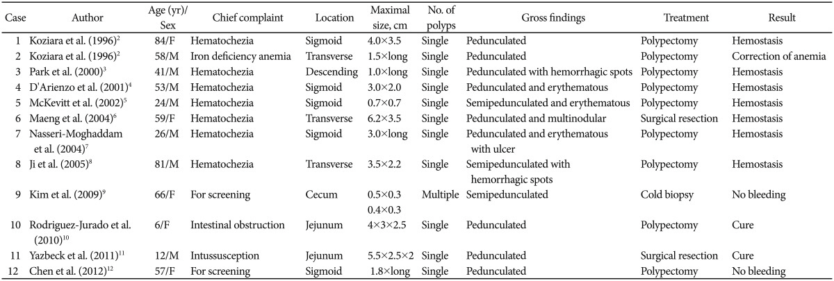
Polypoid arteriovenous malformations (AVMs) in the bowel are very rare and, to the best of our knowledge, only 12 cases have been reported in the medical literature thus far: 10 in adults and two in children.2,3,4,5,6,7,8,9,10,11,12 However, there are no reported adult cases of polypoid AVM presenting with intussusception, although one case was reported in a child.11
Here, we report an adult case of jejunal polypoid AVM, which manifested as a jejunojejunal intussusception, and review
the relevant literature.
A 61-year-old woman was admitted to our department with a 13-year history of intermittent cramping abdominal pain in the left lower quadrant, which radiated throughout the entire abdomen. There was no accompanying nausea, vomiting, melena, hematochezia, changes in bowel habits, or weight loss. She underwent esophagogastroduodenoscopy and colonoscopy 2 years ago and there were no significant findings. She refused further diagnostic workup because herabdominal pain resolved spontaneously. She had undergone a hysterectomy and left oophorectomy 20 years ago, and was diagnosed with hypertension 3 years ago. She took amlodipine (5 mg) and atrovastatin (10 mg) once per day.
Her vital signs were stable. She reported tenderness in left lower quadrant. There was no rebound tenderness or any palpable mass. Bowel sounds were normal. Laboratory results did not identify any significant abnormalities. Routine blood chemistry results were as follows: white blood cell count, 4,400/mm3; hemoglobin, 12.1 g/dL; hematocrit, 36.8%; and platelet count, 238,000/mm3. Tumor markers, including carcinoembryonic antigen, CA 19-9 and CA 72-4 were normal.
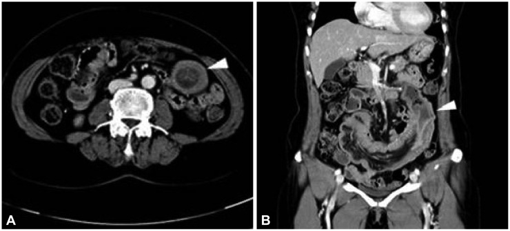
There were no significant findings on chest or abdominal radiography. Esophagogastroduodenoscopy (GIF-H260; Olympus, Tokyo, Japan) revealed gastric ulcer scars and duodenitis, and findings upon colonoscopy (CF-H260AL; Olympus) were unremarkable. However, intravenous contrast enhanced abdominal-pelvic computed tomography showed a small bowel intussusception in the mid-to-distal jejunum, with a suspected low density mass at its tip (Fig. 1).
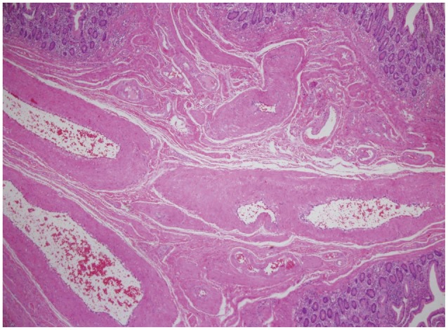
The patient underwent laparoscopic small bowel resection and primary anastomosis. A polypoid mass (5×3.5×3 cm) was found within the intussusceptum (Fig. 2). Histological examination revealed a normal mucosa, but abnormal dilated arteries and veins of variable size were observed in the submucosa, which is consistent with AVM (Fig. 3). Four days after surgery, the patient was discharged without any complications. At 3 months postdischarge, she was free of symptoms with no evidence of recurrence.
Here, we report the very unusual case of a 61-year-old woman presenting with a 13-year history of intermittent cramping abdominal pain. She eventually presented with a small bowel intussusception due to a polypoid AVM, which acted as a pathologic lead point. Intussusception is defined as the prolapse of a proximal bowel segment into a distal segment (rather like a telescope). It is rare in adults, representing only 1% to 5% of bowel obstruction cases. In contrast to those in children, approximately 70% to 90% of intussusceptions in adults occur secondary to a pathological organic lesion, and the nature of the lead point varies greatly. Apart from benign lesions, including lipomas, adenomas, leiomyomas, hamartomas, and inflammatory polyps, approximately half of intussuscepting lesions are malignant (e.g., adenocarcinomas, metastatic melanomas, or metastatic lymphomas). It is recommended that patients with small bowel lesions undergo surgical resection because of the high incidence of associated malignancy and to prevent recurrent intussusceptions.13 In the present case, the intussusception was caused by a polypoid AVM and was treated surgically.
Intestinal polypoid AVMs are rare in all age groups and to date there are only 12 reported cases in the literature (Table 1).2,3,4,5,6,7,8,9,10,11,12 Patients are usually over 40 years, and are equally likely to be male or female. The main initial symptom in adults is hematochezia, whereas children present with intestinal obstructions or intussusceptions. All reported adult cases have involved the colon, whereas the two reported pediatric cases involved the jejunum. All cases were pedunculated or semi-pedunculated, and nine were treated by polypectomy. One adult case was treated by cold biopsy. To the best of our knowledge, the adult case of polypoid AVM reported in the present study is the first in which intussusception was identified as the cause.
In conclusion, we report the unusual case of an adult presenting with chronic abdominal pain caused by an unusual benign small bowel polypoid AVM manifesting as an intussusception. The case was successfully treated by surgical resection. This case suggests that polypoid AVM should be considered as a differential diagnosis in patients (including adults) presenting with small intestinal neoplasms.
References
1. Gordon FH, Watkinson A, Hodgson H. Vascular malformations of the gastrointestinal tract. Best Pract Res Clin Gastroenterol. 2001; 15:41–58. PMID: 11355900.

2. Koziara FJ, Brodmerkel GJ, Boylan JJ, Ciambotti GF, Agrawal RM. Bleeding from polypoid colonic arteriovenous malformations. Am J Gastroenterol. 1996; 91:584–586. PMID: 8633515.
3. Park ER, Yang SK, Jung SA, et al. A case of pedunculated arteriovenous malformation presenting with massive hematochezia. Gastrointest Endosc. 2000; 51:96–97. PMID: 10625812.

4. D'Arienzo A, Manguso F, D'Armiento FP, et al. Colonoscopic removal of a polypoid arteriovenous malformation. Dig Liver Dis. 2001; 33:435–437. PMID: 11529657.
5. McKevitt EC, Attwell AJ, Davis JE, Yoshida EM. Diminutive but dangerous: a case of a polypoid rectal arteriovenous malformation. Endoscopy. 2002; 34:429. PMID: 11972282.

6. Maeng L, Choi KY, Lee A, Kang CS, Kim KM. Polypoid arteriovenous malformation of colon mimicking inflammatory fibroid polyp. J Gastroenterol. 2004; 39:575–578. PMID: 15235876.

7. Nasseri-Moghaddam S, Mohamadnejad M, Malekzadeh R, Tavangar SM. Images of interest. Gastrointestinal: polypoid arteriovenous malformation of the colon. J Gastroenterol Hepatol. 2004; 19:1419. PMID: 15610319.
8. Ji JS, Choi KY, Lee BI, et al. A large polypoid arteriovenous malformation of the colon treated with a detachable snare: case report and review of literature. Gastrointest Endosc. 2005; 62:172–175. PMID: 15990846.

9. Kim BK, Han HS, Lee SY, Kim CH, Jin CJ. Cecal polypoid arteriovenous malformations removed by endoscopic biopsy. J Korean Med Sci. 2009; 24:342–345. PMID: 19399283.

10. Rodriguez-Jurado R, Morales SS. Polypoid arteriovenous malformation in the jejunum of a child that mimics intussusception. J Pediatr Surg. 2010; 45:E9–E12. PMID: 20105573.

11. Yazbeck N, Mahfouz I, Majdalani M, Tawil A, Farra C, Akel S. Intestinal polypoid arteriovenous malformation: unusual presentation in a child and review of the literature. Acta Paediatr. 2011; 100:e141–e144. PMID: 21299613.

12. Chen CH, Yan SL. Colonic arteriovenous malformation presenting as a pedunculated polyp. Dig Endosc. 2012; 24:485. PMID: 23078454.

Fig. 1
(A, B) An abdominopelvic computed tomography scan showing a long segmental small bowel intussusception with a suspected low density mass at the tip (arrowheads).

Fig. 2
(A, B) Macroscopic appearance of the specimen after segmental resection. A well-defined, polypoid mass can be seen in the small intestinal mucosa.


 PDF
PDF ePub
ePub Citation
 Print
Print


 XML Download