

 PDF
PDF Citation
 Print
Print


INTRODUCTION
Human inflammatory bowel disease (IBD), consisting of Crohn disease (CD) and ulcerative colitis (UC), is a chronic condition characterized by acute exacerbations followed by remissions. Despite many years of extensive research, its pathogenesis is still poorly understood [1]. Inflammatory process frequently involves the more distal parts of the colon; in fact, distal colitis (DC) comprises 60%–85% of all UC cases upon admission. The upper border of the DC can be reached through topical treatment [2]. Currently, aminosalicylates, steroids, immunosuppressants, and biologic agents are used for treatment of UC. Despite various side effects of these treatments, many studies in recent years have suggested the importance of medical plants and their bioactive substances in management of this condition [3].
A large number of experimental models related to chemically-UC have been studied during the last decades. Acetic acid (AA) induced colitis model is one of the most widely used models preferred to induce inflammation and ulceration in the distal colon of rodents [34]. Mesalazine (5-amino salicylic acid; 5-ASA) is one of the principal medicines for the treatment of IBD and it has a significant anti-inflammation effect [56]. Ganoderma lucidum polysaccharides have anti-inflammatory and immunomodulatory effects. It has been previously reported that G. lucidum has a therapeutic potential to alleviate dextransulfate sodium-induced colitis [78].
In the light of this knowledge, our study aimed to compare the healing effects of mesalazine and G. lucidum in separate and combined treatments against AA-induced DC.
METHODS
Our experimental study was approved by Kırıkkale University Animal Experiments Local Ethics Committee on December 19, 2019 (No. 2019-13/65). A total of 24 Wistar albino male rats weighing 200–280 g were randomly divided into 4 groups; colitis, mesalazine treatment, G. lucidum treatment, and mesalazine-G. lucidum combined treatment groups. All animals were kept in collective cages under a controlled temperature (24℃) in daylight and dark conditions with water and rat chow provided ad libitum. G. lucidum group animals and combined treatment group animals were administered G. lucidum orally (dose of 100 mg/body weight [kg], dissolved in 0.2 mL water) for 14 days before the experimental colitis procedure. Twelve hours before the experimental procedure, feeding of animals was stopped and they were only allowed to drink water. All animals were anesthetized by using 8 mg/body weight (kg) of ketamine, intramuscularly, for the experimental colitis procedure. All animals were positioned in the Trendelenburg position and the anal canals of the animals were first washed with 1-mL saline. Then, 2 mL of 4% AA was administered to anal canal mucosa to induce experimental colitis. Colitis group animals had no additional procedure. Mesalazine treatment group animals were given a 200 mg/kg mesalazine dose rectally for a 5-day period after the colitis procedure. G. lucidum treatment group animals were administered 100 mg/kg dose of G. lucidum orally for a 5-day period. Combined treatment group animals were administered 100 mg/kg dose of G. lucidum orally and a 200 mg/kg dose of mesalazine rectally for a 5-day period. The animals were kept in the Trendelenburg position for a further 2 minutes so that the administered drugs did not come out of the rectum. On the 6th day of medical treatments, all animals were sacrificed. Laparotomy was done with a 2-cm midline incision. Distal colon tissue samples were removed and washed with isotonic NaCl. Distal colon tissue samples were divided into 2 pieces for histopathologic and biochemical examinations.
Tissue homogenization and total protein assay
DC specimens were taken for this study. Tissue samples were homogenized with phosphate buffer saline of pH 7.4 by using a homogenizer (FastPrep-24, MP Biomedicals, Santa Ana, CA, USA). The total amount of protein was measured by the Bradford method (Pierce BCA, Thermo Fisher Scientific, Waltham, MA, USA) in all tissue samples by spectrophotometer (Multiskan FC, Thermo Fisher Scientific).
Evaluation of interleukin and CRP levels
Samples were thawed and TNF-α ELISA kit (catalog No. E-EL-R0019, Elabscience, Houston, TX, USA), IL 1-β ELISA kit (catalog No. E-EL-R0012, Elabscience), IL-6 ELISA kit (catalog No. E-EL-R0015, Elabscience) and CRP ELISA kit (catalog No. E-EL-R0022, Elabscience) were used for the quantitative measurement of TNF-α, IL-1β, IL-6, and CRP in tissue homogenates. Samples and standards were added to appropriate wells which are precoated with the anti-human monoclonal antibody before incubation. Biotin was added to all wells and combined with streptavidin and horseradish peroxidase conjugate to form an immunocomplex; then incubation was carried out again, then uncombined enzymes were identified. Then chromogen solution A, B were added to observe whether the color of the liquid would change into blue. At the effect of the acid, the color finally became yellow. Optical density was read on a standard automated plate reader at 450 nm (Microplate Reader, Thermo Fisher Scientific). The detection range of kits were 78.13–5,000 pg/mL for TNF-α, 31.25–2,000 pg/mL for IL-1β, 12.5–800 pg/mL for IL-6, and 0.31–20 ng/mL for CRP.
Macroscopic examination of colitis
Distal colon tissue samples were prepared and placed in a 10% neutral buffered formalin solution then sliced at 0.4-cm intervals. Sliced tissue samples were left during one night for histopathologic examination.
Histopathologic examination of colitis
After the tissues were embedded in paraffin blocks, they were cut 4-µm thick and stained with hematoxylin and eosin. Histopathological findings were evaluated under light microscope by the pathologist who was blind to the treatment groups.
Histopathological scoring was determined according to the highest area for the disease, and mucosal damage and inflammation scoring was performed. By conducting semi-quantitatively, 3 categories (0, absent; 1, mild; 2, severe) were determined and the parameters were scored accordingly. We used the parameters “fibrosis, crypt distortion, crypt loss, cryptitis, crypt abscess, and mucin depletion” in the mucosal damage scoring and “lymphoplasmacytic inflammatory cell infiltration (ICI), neutrophil infiltration, and granuloma” parameters in the scoring of the inflammation.
We created our own algorithm based on the histopathological examinations mentioned in the study by Koçak et al. [9]. In this way, we were able to document it in a simpler and more understandable way. The classification of mucosal damage according to defined scores was reported as follows: grade 1, 0–3 (mild mucosal damage); grade 2, 4–6 (moderate mucosal damage); and grade 3, 7–10 (severe mucosal damage). We rated the inflammation grading as grade 1 of 0–1 (mild inflammation), grade 2 of 2–3 (medium intensity inflammation), and grade 3 of 4–5 (intense inflammation). In addition, the ulcer depth (0, none; 1, <2 mm; 2, ≥2 mm) and the depth of the disease (0, mucosa; 1, submucosal layer; 2, muscular layer; and 3, serosal layer) were scored.
Statistical analyses
All results are reported as mean ± standard error of the mean. The statistical analyses were performed using SPSS ver. 16.0 for Windows (SPSS Inc., Chicago, IL, USA). Due to the limited number of rats in each group, nonparametric methods were used for statistical analysis. Kruskal-Wallis variance analysis, which is used to compare the means of 3 or more groups, was used to determine whether there was a statistical difference between the groups. The Mann-Whitney U-test, which is used to compare the means of 2 groups, was used to determine from which group the significant difference originated. A P-value of <0.05 was considered significant.
RESULTS
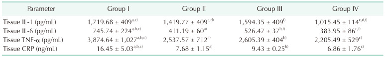
For the evaluation of oxidation, biochemical evaluation was performed as Özden et al. [10]. The evaluation of tissue biochemical results were detailed in Table 1. The highest tissue IL-1, IL-6, TNF-α, and CRP levels were observed in colitis group animals. The lowest tissue IL-1, IL-6, TNF-α, and CRP levels were noted in the combined treatment group. Compared to the colitis group, the mean tissue IL-1 levels were found to be significantly lower in combined treatment group animals (P < 0.05). There was also a significant difference in mean tissue IL-1 between colitis and mesalazine/mesalazine and combined treatment groups (P < 0.05). In tissue IL-6 evaluation, there was a significant difference between the colitis group and the treatment groups (P < 0.05). There was also a significant difference between the G. lucidum treatment group and the combined treatment group (P < 0.05). For TNF-α, a significant difference was observed only between the colitis group and the treatment groups (P < 0.05). Similar results were obtained with the CRP level. A significant difference was observed only between the colitis group and the treatment groups (P < 0.05). There was no significant difference between the mesalazine and G. lucidum treatment groups animals in terms of mean tissue IL-1, IL-6, TNF-α, and CRP levels (P = 0.131, P = 0.131, P = 0.586, and P = 0.939, respectively).
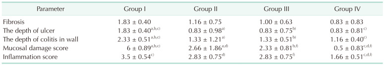
The evaluation of histopathologic results were detailed in Table 2. Histopathologic examinations of tissue samples were done by using parameters including the presence of fibrosis, the depth of the ulcer, and the depth of the colitis in the intestinal wall. There was no significant difference between any groups in terms of fibrosis (P = 0.066, P = 0.131, P = 0.090, P = 0.818, P = 0.488, and P = 0.818, respectively). There was a significant difference between the colitis group and the treatment groups in terms of the depth of the ulcer (P < 0.05). There was also a significant difference between the colitis and the treatment groups in terms of the degree of intestinal wall involvement (the depth of colitis in the intestinal wall) (P < 0.05) (Fig. 1). There were also significant differences between the colitis and the treatment groups in terms of mucosal damage (P < 0.05). At the same time, there were significant differences between the mesalazine and the combined treatment groups/the G. lucidum treatment group and the combined treatment groups in terms of mucosal damage (P < 0.05). There were significant results between the colitis, mesalazine, and G. lucidum treatment groups and the combined treatment group in terms of inflammation scores (P < 0.05).
DISCUSSION
About 3/2 of the patients suffering from UC have colitis limited to the left side of the colon. Physiological factors influencing vulnerability differ between the left and right sides of the colon and this difference is explained by metabolic differences between the 2 sides. DC is a complex, challenging problem and resistant to standard therapy [9].
AA-induced UC is a commonly preferred model for the induction of UC and has similar characteristics in human intestinal inflammations in terms of the development of an ulcer, inflammatory mediators, free radicals, and neutrophil infiltration [11]. DC was evoked by intrarectal administration of 2-mL 4% AA in our study. In experimental studies for IBDs, colon samples are terminated on the 5th to 7th days, taking into account the periods when the collagen structure is highest [12]. Tissue healing is considered to be highest during this period. Following the study of Mitchell et al. [13], the experiment was terminated on the 6th day.
Although the etiology and the pathophysiology of UC are not clearly elucidated, it is widely accepted that factors such as genetic, environmental, and gastrointestinal immunity play an important role in the pathophysiology of UC [341415]. AA-induced colitis is a model of UC that bears similar characteristics to human UC in terms of pathogenesis, histopathological features, and an inflammatory mediator profile. Intrarectal administration of a dilute solution of AA activates the intestinal immune system and leads to increased neutrophil infiltration into the intestinal tissue and vascular dilatation. The infiltration of neutrophils results in oxidative stress and contributes to progression of tissue necrosis and mucosal dysfunction [1114]. Oxidative stress is one of the important pathogenic factors associated with UC. Excessive and sustained production of reactive oxygen substrates can damage cellular proteins, membrane lipids, and DNA, leading to cell death [16]. Inflammatory responses in UC are characterized by an increased production of proinflammatory cytokines, such as TNF-α, IL-1, IL-6, prostaglandins, and leukotrienes. Previous clinical and experimental studies showed that there is a positive correlation between the increased proinflammatory cytokines and severity of the colitis in patients with IBD [7111517]. Blood and tissue IL-1, IL-6, and TNF-α levels were used in the comparison of the study groups in terms of severity of disease and the healing effects of therapeutic agents.
TNF-α is one of the principal inflammatory cytokines and plays an important role in the pathogenesis of IBD [18]. It is believed to be produced primarily by macrophages and has multiple biological activities involved in colitis. Thus, its inhibition and regulation is important goal in the treatment of UC. TNF-α agents have been commonly used in the management of patients with UC [111617]. The results of our study showed that induction of DC caused a significant elevation in levels of tissue TNF-α, particularly in colitis group animals.
IL-1 is also released from macrophages and plays a role in regulatory and proinflammatory functions in the pathogenesis of UC. IL-1β leads to an increase in intestinal permeability and stimulates the accumulation of neutrophils to the inflamed colonic tissue [1619]. In our study, the highest tissue IL-1 levels were observed in animals with untreated DC.
IL-6 is a potent pleiotropic cytokine and plays a central role in adaptive immune response in IBD. IL-6 also regulates the differentiation of macrophages and T cells. Blocking of IL-6 signaling pathway precludes T cell-mediated colitis. IL-6 also induces the expression of acute phase proteins during acute inflammation. It has been previously reported that the levels of IL-1 and IL-6 colon tissue correlate with the severity of intestinal inflammation and disease activity [16202122]. Our findings were consistent with these findings. The highest colon tissue IL-1 and IL-6 levels were noted in the colitis group of animals.
Mortensen et al. [23] showed that CRP levels were significantly elevated in patients with active disease for both UC and CD. Beigel et al. [24] reported that low CRP seems to be a reliable and easy-to-use predictor for mucosal healing in patients with IBD. Tissue CRP levels were used as a parameter to evaluate the healing effects of mesalazine and G. lucidum in our study. Our results showed that tissue CRP levels were found to be consistent with the histopathologic findings.
The extent of the inflammatory response in UC can be assessed by histopathologic changes in intestinal tissue. A scoring system based on the loss of crypt architecture, ICI, muscle thickening, goblet cell depletion, and crypt abscess is used for assessment of colonic histological damage. Quantitatively, tissue levels of IL-1, IL-6, and TNF-α can be determined by conventional ELISA methods [15]. Histopathologic changes due to AA-induced colitis are defined as extensive epithelial loss, destruction and/or loss of crypts, and severe ICI with marked goblet cell depletion [25]. The histopathologic changes in colonic tissue samples of our study groups were similar to these findings. Histopathologic changes were scored as a mucosal damage score and an inflammation score. Additionally, findings such as the presence of fibrosis, the depth of the ulcer, and intramural depth of colitis were also compared in our study. Mucosal damage and inflammatory response were considerably alleviated in colonic tissue of mesalazine and G. lucidum treatment groups animals in our study.
Current treatment options for UC are aminosalicylates, corticosteroids, immunomodulators, biologics, and a new category of small molecule drugs. Mesalazine (5-ASA) is accepted as the first-line therapy. It seems to be highly effective in clinical, endoscopic, and histologic remission [262728]. Mesalazine has a significant anti-inflammation effect and affects a variety of mediators and signaling pathways associated with leukocyte chemotaxis and function and epithelial barrier [3]. In our study, statistical comparisons of tissue IL-1, IL-6, TNF-α, and CRP levels showed that mesalazine has significant healing effects on AA-induced colitis. The healing effects of mesalazine were explained by its anti-inflammatory potential as mentioned in the literature [3]. Mucosal damage scores and inflammatory scores of mesalazine treated group animals have supported this opinion.
G. lucidum comprises about 400 different bioactive compounds including polysaccharides, triterpenoids, proteins, and enzymes, and has been proved to have several therapeutic potentials to control various diseases. G. lucidum exerts a wide spectrum of pharmacologic actions such as anti-inflammatory, immunomodulation, antioxidant, anticancer, and anti-ulcer effects [29]. Liu et al. [30] showed that G. lucidum inhibited the production of proinflammatory cytokines by peripheral blood mononuclear cells and inflamed colonic mucosa due to the blcockage of nuclear factor kappa B (NF-κB) activation. Tissue proinflammatory cytokines levels of G. lucidum treatment group animals were found to be lower compared to those of colitis group animals. Mucosal damage and inflammatory scores of G. lucidum treatment group animals were in concordance with these findings in our study.
In the literature, it has been proven that treatment with the combination of an antioxidant or anti-inflammatory agent with an anticolitis drug may be more effective than monotherapy in their separate use [29]. Our study examined the possible healing and tissue protective effects of mesalazine and G.
lucidum in separate and in combined treatment in AA-induced DC.
As it is known, UC or CD are intestinal inflammatory diseases that go with inflammation. The search for effective solutions in the treatment of these diseases continues. For this, single or combined treatment protocols are tried. In our study, we searched for an effective treatment for IBDs. In our study, we used mesalazine, which is known to have an effect and is in use, and G. lucidum, an easily obtainable substance that is consumed in many ways in daily life, has protective effects on the immune system. We thought that comfortable use of G. lucidum would be effective in maintaining the well-being of the patients. In the active phase of the disease, the use of mesalazine will thus be more effective. According to the results of our study, we saw that G. lucidum was effective when the G. lucidum treatment group results were compared with the colitis group results. The mesalazine group was more effective than the G. lucidum treatment group. However, the combined treatment gave much more effective results than the 2 groups. These results showed us that G. lucidum may be involved in the treatment of UC and CD.
In conclusion, the results of our study showed that the intrarectal administration of AA causes severe mucosal damage in the colon and activates severe intestinal inflammatory response characterized by elevated proinflammatory cytokine levels. Separate and combined treatments with mesalazine and G. lucidum exerted colon tissue protective effects and decreased the severity of intestinal inflammatory response. Combined treatment with mesalazine and G. lucidum was found to be significantly more effective than those of separate use in the treatment of DC.
This study has a limitation in that the experimental group is insufficient. If we had used more experimental animals in the study groups, we could have obtained statistically more valuable results. More extensive studies are needed.

 XML Download