Abstract
Confocal laser endomicroscopy (CLE) is a novel in vivo imaging technique that can provide real-time optical biopsies in the evaluation of pancreaticobiliary strictures and pancreatic cystic lesions (PCLs), both of which are plagued by low sensitivities of routine evaluation techniques. Compared to pathology alone, CLE is associated with a higher sensitivity and accuracy for the evaluation of indeterminate pancreaticobiliary strictures. CLE has the ability to determine the malignant potential of PCLs. As such, CLE can increase the diagnostic yield of endoscopic retrograde cholangiopancreatography and endoscopic ultrasound, reducing the need for repeat procedures. It has been shown to be safe, with an adverse event rate of ≤1%. Published literature regarding its cost-effectiveness is needed.
Confocal laser endomicroscopy (CLE) is an endoscopic imaging technique that can provide real-time optical biopsies during endoscopic retrograde cholangiopancreatography (ERCP) and endoscopic ultrasound (EUS) in the evaluation of pancreaticobiliary strictures (PSs) and pancreatic cystic lesions (PCLs). The basis of CLE is the use of a low-power laser beam focused onto tissue through an objective and the subsequent detection of fluorescent light gathered by the same objective through a small aperture [1]. As the aperture is placed in the same conjugate focal plane as the tissue specimen, only the light emitted from a desired focal spot is allowed to pass through [1].
CLE is performed via one of two miniprobes: the CholangioFlex or AQ-Flex miniprobe (Cellvizio; Mauna Kea Technologies, Paris, France). Measuring up to 0.94 mm in outer diameter, the miniprobe can be introduced through the working channel of a standard duodenoscope or echoendoscope via a biliary catheter or a 19 gauge fine needle aspiration (FNA) needle, respectively, and subsequently placed in contact with the tissue of interest. The miniprobe is attached to a laser scanning unit and confocal processor, providing real-time image processing. CLE performed via a biliary catheter with the CholangioFlex miniprobe is termed probed-based CLE (pCLE), whereas CLE performed via the AQ-Flex miniprobe through an FNA needle is termed needle-based CLE (nCLE).
The objective of this review is to summarize the role of CLE in pancreaticobiliary disease. As the sensitivity of cytological brushing or biopsy in the evaluation of PS and PCLs remains poor, CLE has the ability to increase the diagnostic yield of ERCP and EUS.
pCLE at the time of ERCP is indicated for the evaluation of indeterminate pancreaticobiliary strictures (IPSs). The prevalence of malignancy within IPSs is 63%, the majority due to cholangiocarcinoma or pancreatic cancer [2,3]. Confirming malignancy is essential to initiate treatment; however, ERCP is plagued by the poor sensitivity of routine sampling. In a meta-analysis, the pooled sensitivity of endoscopic brush cytology (EBC) and intraductal biopsy (IB) was 45% and 48.1%, respectively [2]. Even when you combine EBC with IB, the sensitivity remains low at 59.4% [2]. The result is delayed diagnoses, the need for repeat procedures, which is associated with increased cost and complications, and unfortunately for some patients, the progression of disease to advanced stages not amenable to curative resection.
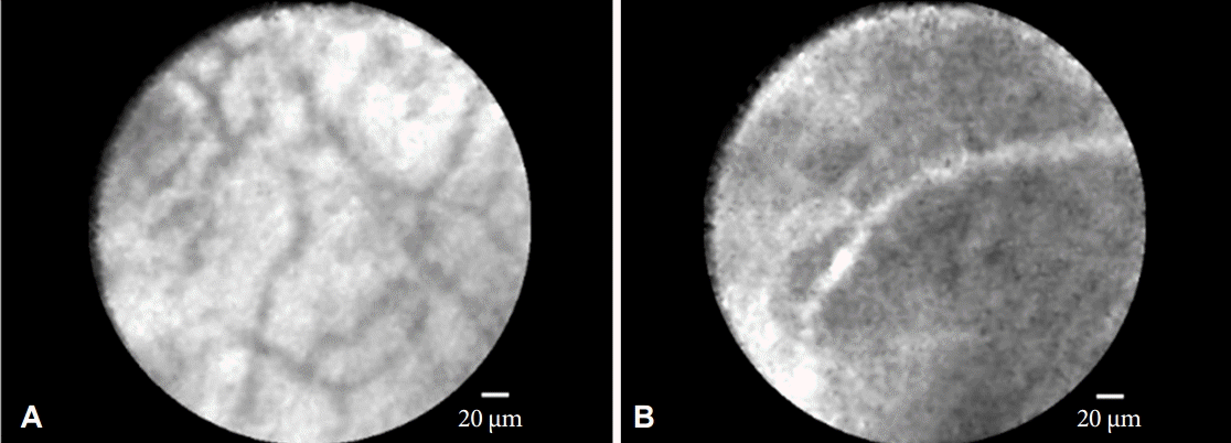
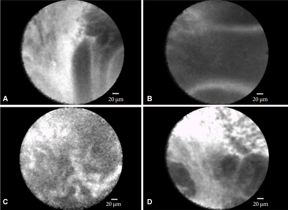
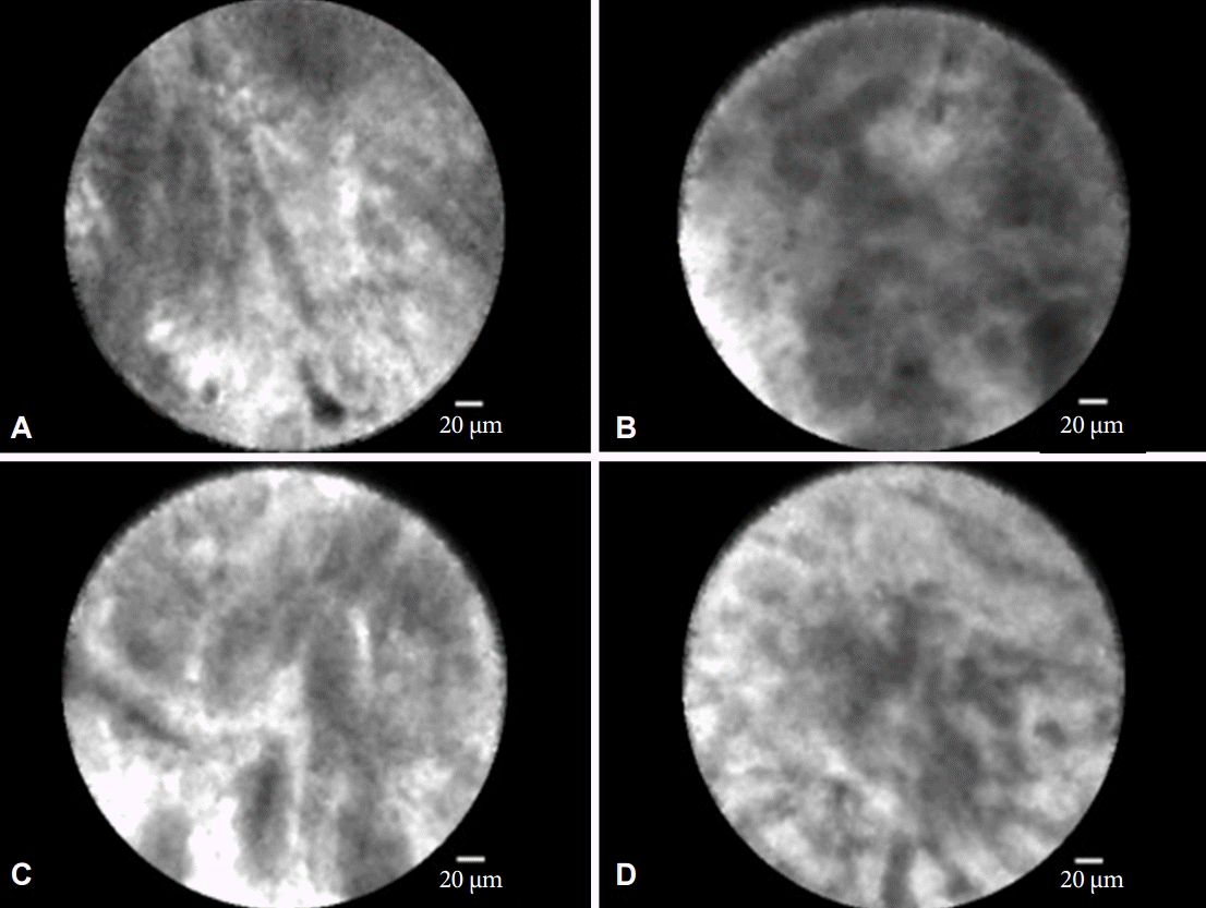
pCLE has been shown in multiple studies to improve the diagnostic sensitivity of ERCP. After pilot studies showed differential findings in the healthy bile duct (Fig. 1) compared to malignant and inflammatory PSs, the Miami classification (MC) for malignant PS was initially created, consisting of four criteria: (1) epithelial structures, (2) thick dark bands (>40 µm), (3) thick white bands (>20 µm), and (4) dark clumps (Fig. 2). Initial studies evaluating the MC found that the presence of three criteria was highly suggestive of malignancy, with sensitivities ranging from 96% to 97%; however, due to the false positives in the setting of inflammation, the specificity was low at 33% to 67% [4,5]. Highlighting the need for refined imaging criteria, the Paris classification (PC) for inflammatory strictures was created, which also consisted of four criteria: (1) vascular congestion, (2) granular pattern with scales, (3) increased inter-glandular space, and (4) thickened reticular structures (Fig. 3). The simultaneous evaluation of an IPS with both the MC and PC increases the specificity to 83%, with an overall accuracy of 82% (compared to 75% for index pathology) [5].
pCLE for the evaluation of IPS was validated in a multi-center, international, prospective study of 112 patients. Similar to prior studies, the sensitivity and accuracy of pCLE (89% and 82%, respectively) were superior to those of index pathology alone (56% and 72%, respectively) [3]. The combination of pCLE and pathology was found to have a sensitivity, specificity, and accuracy of 89%, 88%, and 88%, respectively [3]. Lastly, in a recently published meta-analysis, the pooled sensitivity for pCLE in IPS was 90% [6].
Just as the diagnosis of IPS can prove difficult, so is the accurate classification of PCLs. With the widespread use of cross-sectional imaging resulting in increased detection of PCLs, determining the malignant potential of PCLs is a frequent challenge. EUS-FNA with cyst fluid analysis for cytology, chemistry, and tumor markers is limited by sampling error and non-diagnostic samples, again leading to missed diagnoses and subjecting patients to repeat procedures or unnecessary surgical resection [7,8].
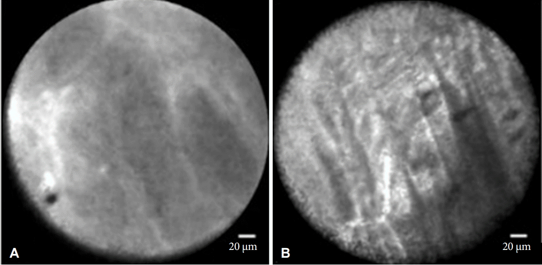
nCLE has the potential to provide valuable information at the time of EUS-FNA. However, due to the heterogeneity of PCLs, an nCLE classification system that classifies PCLs as benign, premalignant, or malignant does not exist. Nonetheless, initial nCLE studies proposed histologic correlates for a variety of findings in the evaluation of PCLs, including finger-like papillary projections in villous structures, dark clumps of cells concerning for neoplasia, white bands as blood vessels, dark lobular structures as acinar cells, and bright white particles consistent with debris or calcifications [9]. Two subsequent studies revealed two nCLE imaging findings as having 100% specificity for certain types of PCLs—villous structures in mucinous PCLs and superficial vascular networks in serous cystadenomas (Fig. 4) [9,10]. Furthermore, the absence of villous structures in combination with negative cytology and low carcinoembryonic antigen has a 100% sensitivity for mucinous PCLs [9]. Lastly, a recently published meta-analysis found the pooled specificity of nCLE for PCLs to be 90% [6].
nCLE is limited by the heterogeneity of PCLs. In addition to their malignant potential, PCLs can also vary in their size, location, and locularity, which can render their thorough evaluation via nCLE difficult. Furthermore, multiple nCLE findings can be present in PCLs of differing etiologies [9]. As a result, the pooled sensitivity for nCLE is low at 68%, the accuracy for classifying PCLs based on their malignant potential is low at 46%, and the interobserver agreement (IOA) for identification of nCLE findings is slight (κ=0.13) [6,11].
No current literature exists reviewing the safety of CLE for pancreaticobiliary indications. In theory, complications from CLE can arise from one of two methods: (1) the administration of intravenous fluorescein prior to performing CLE, and (2) the introduction and evaluation of tissue with the miniprobe.
CLE requires the use of a fluorophore, which is a dye that fluoresces when stimulated by the laser beam, allowing for contrast enhancement. Fluorescein (Ak-Fluor; Akorn Inc., Lake Forest, IL, USA) is the fluorophore used in pancreaticobiliary CLE. It is also used routinely in diagnostic angiography of the retina due to its low rate of adverse drug reaction (~1%), with the majority being mild nausea and vomiting [12].
Our group performed an extensive literature search, identifying only one adverse event out of >500 CLE procedures (<1% adverse event rate), an episode of pancreatitis [13]. This occurred in a patient undergoing ERCP with biliary pCLE using a GastroFlexUHD miniprobe (Mauna Kea Technologies), which has a larger diameter (2.6 mm vs. 0.94 mm) compared to the CholangioFlex miniprobe that is typically used.
When considering the costs of performing pancreaticobiliary CLE, it is important to keep in mind the need for repeat procedures due to the low sensitivity of sampling in both IPSs and PCLs. On average, patients with IPS have 2.5 ERCPs prior to being referred for CLE [3]. As the addition of CLE to ERCP has been shown to have higher sensitivity and accuracy than pathology alone, CLE has the ability to reduce the need for repeat procedures.
Performing both pCLE and nCLE requires the purchase of the CellVizio CLE system and their respective miniprobes. Initial costs of the CLE system ranged from $100,000 to $150,000, while miniprobes cost approximately $4,000 (reusable up to 10 times) [13]. We recommend you contact Mauna Kea Technologies (www.maunakeatech.com) directly for updated pricing information.
In the current literature, there is little data regarding the cost-effectiveness of pancreaticobiliary CLE. In a single-center, retrospective study of 67 patients undergoing CLE, 14 (20.1%) went on to have surgical procedures, generating on average $109,667 gross revenue per patient [14]. In this study, the net profit per patient was $38,231 and the overall profit margin was 34%. Furthermore, our group recently presented an abstract at Digestive Disease Week 2016 evaluating the cost-effectiveness of pCLE for IPS, in which we found pCLE to be more cost-effective than routine ERCP without pCLE at willingness-to-pay threshold of $50,000 [15]. At this time, a manuscript detailing our results is in progress.
CLE has been shown in multiple studies to be safe and effective at providing useful diagnostic information at the time of ERCP and EUS. pCLE in particular has been shown to have higher performance characteristics in the evaluation of IPS compared to EBC and IB, possibly reducing the need for repeat procedures and decreasing cost. nCLE, though not as extensively studied as pCLE, has shown promise in detecting imaging criteria that can differentiate pre-malignant mucinous lesions from non-mucinous lesions. However, further nCLE studies and refinement of imaging classifications of PCLs are needed to improve its accuracy and IOA.
REFERENCES
1. Nwaneshiudu A, Kuschal C, Sakamoto FH, Anderson RR, Schwarzenberger K, Young RC. Introduction to confocal microscopy. J Invest Dermatol. 2012; 132:e3.

2. Navaneethan U, Njei B, Lourdusamy V, Konjeti R, Vargo JJ, Parsi MA. Comparative effectiveness of biliary brush cytology and intraductal biopsy for detection of malignant biliary strictures: a systematic review and meta-analysis. Gastrointest Endosc. 2015; 81:168–176.

3. Slivka A, Gan I, Jamidar P, et al. Validation of the diagnostic accuracy of probe-based confocal laser endomicroscopy for the characterization of indeterminate biliary strictures: results of a prospective multicenter international study. Gastrointest Endosc. 2015; 81:282–290.

4. Meining A, Shah RJ, Slivka A, et al. Classification of probe-based confocal laser endomicroscopy findings in pancreaticobiliary strictures. Endoscopy. 2012; 44:251–257.

5. Caillol F, Filoche B, Gaidhane M, Kahaleh M. Refined probe-based confocal laser endomicroscopy classification for biliary strictures: the Paris Classification. Dig Dis Sci. 2013; 58:1784–1789.

6. Fugazza A, Gaiani F, Carra MC, et al. Confocal laser endomicroscopy in gastrointestinal and pancreatobiliary diseases: a systematic review and meta-analysis. Biomed Res Int. 2016; 2016:4638683.

7. Afify AM, al-Khafaji BM, Kim B, Scheiman JM. Endoscopic ultrasound-guided fine needle aspiration of the pancreas. Diagnostic utility and accuracy. Acta Cytol. 2003; 47:341–348.
8. Eloubeidi MA, Jhala D, Chhieng DC, et al. Yield of endoscopic ultrasound-guided fine-needle aspiration biopsy in patients with suspected pancreatic carcinoma. Cancer. 2003; 99:285–292.

9. Konda VJ, Meining A, Jamil LH, et al. A pilot study of in vivo identification of pancreatic cystic neoplasms with needle-based confocal laser endomicroscopy under endosonographic guidance. Endoscopy. 2013; 45:1006–1013.

10. Napoléon B, Lemaistre AI, Pujol B, et al. A novel approach to the diagnosis of pancreatic serous cystadenoma: needle-based confocal laser endomicroscopy. Endoscopy. 2015; 47:26–32.

11. Karia K, Waxman I, Konda VJ, et al. Needle-based confocal endomicroscopy for pancreatic cysts: the current agreement in interpretation. Gastrointest Endosc. 2016; 83:924–927.

12. Kwan AS, Barry C, McAllister IL, Constable I. Fluorescein angiography and adverse drug reactions revisited: the Lions Eye experience. Clin Experiment Ophthalmol. 2006; 34:33–38.

13. Karia K, Saul A, Tyberg A, Gaidhane M, Kahaleh M. Cholangioscopy and biliary confocal laser endoscopy. In : Konda VJ, Waxman I, editors. Endoscopic Imaging Techniques and Tools. New York: Springer;2016.
14. Cellvizio . Downstream revenue generated by probe-based confocal laser endomicroscopy (pCLE) in undetermined pancreaticobiliary lesions [Internet]. Paris: Mauna Kea Technologies;c2015 [cited 2016 Aug 9]. Available from: http://www.cellvizio.net/sites/default/files/issu_presentation/ICCU2014%20-%20Sharaiha.pdf.
15. Karia K, Gaidhane M, Kahaleh M, Sharaiha RZ. Probe-based confocal laser endomicroscopy for the evaluation of indeterminate biliary strictures: is it cost effective? Gastrointest Endosc. 2016; 83(5 Suppl):AB191.
Fig. 1.
Probed-based confocal laser endomicroscopy findings in the healthy bile duct. (A) Reticular network of thin dark branching bands (<20 µm) with light gray background. (B) Vessels <20 µm.

Fig. 2.
The Miami classification for malignant pancreaticobiliary strictures. (A) Epithelial structures. (B) Thick dark bands (>40 µm). (C) Thick white bands (>20 µm). (D) Dark clumps.


 PDF
PDF Citation
 Print
Print


 XML Download