Abstract
Colorectal polypectomy is an effective method for prevention of colorectal cancer. Many endoscopic instruments have been used for colorectal polypectomy, such as snares, forceps, endoscopic clips, a Coagrasper, retrieval net, injector, and electrosurgery generator unit (ESU). Understanding the characteristics of endoscopic instruments and their proper use according to morphology and size of the colorectal polyp will enable endoscopists to perform effective polypectomy. I reviewed the characteristics of endoscopic instruments for colorectal polypectomy and their appropriate use, as well as the basic principles and settings of the ESU.
The polypectomy method and instruments used depend on the colorectal polyp size and shape, or the preference of the endoscopist. Proper removal of colorectal polyps requires understanding of the characteristics and correct use of endoscopic instruments to reduce the occurrence of incomplete polypectomy, which is a major cause of interval colon cancer.
Thus, this review discusses various polypectomy methods, endoscopic instruments used for polypectomy (forceps, snares, and others), and the electrosurgery generator unit (ESU).
Polyps are classified as diminutive (<5 mm), small (5 to 9 mm), and large (≥10 mm). Generally, all polyps, including diminutive polyps, should be removed because of their malignant potential [1,2]. The choice of instruments often depends on the size and morphology of the polyp.
Techniques of polypectomy are classified according to removal instruments, such as forceps and snares, and according to whether submucosal injection and an electrical unit are used [3]. “Hot” refers to techniques using the ESU and “cold” means without use of the ESU.
Advantages of polypectomy using forceps are ease of use, high retrieval rate, and low complication rate. A recent prospective study of removal methods for diminutive lesions reported that the complete resection rate of 1 to 3 mm polyps by forceps was 92%, and 76% for 4 to 5 mm polyps [5].
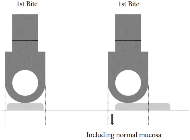
Forceps size refers to the cup. Cup length ranges from 5 to 8 mm and various types of forceps are available, including forceps with jaws, with spikes, and with a central cup hole. The diameter of the forceps cup is about 3 mm (Fig. 1). Because one-bite forceps polypectomy is not adequate, two bites with a forceps or use of a jumbo forceps can be applied for removal of 4 to 5 mm polyps. During two-bite cold forceps polypectomy, the first bite should include a normal mucosal margin to reduce remnant tissue (Fig. 2).
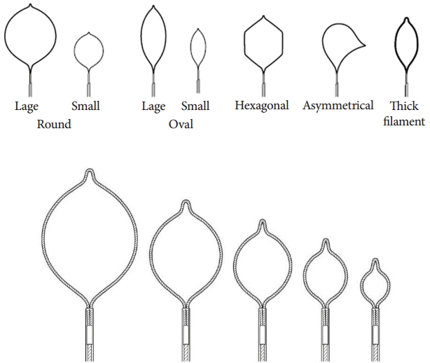
Snare polypectomy has been widely used for small colorectal polyps. Available snares vary in shapes and sizes. Snare size refers to the maximal width of the snare, and ranges from 10 to 30 mm. Generally, oval and hexagonal snares, and especially mini-oval snares (diameter 10 to 15 mm) are most widely used, because 90% of colorectal polyps are small or diminutive [8]. A crescent snare is often used in EMR, and is the proper instrument for resection of a polyp behind the colonic folds (Fig. 3).
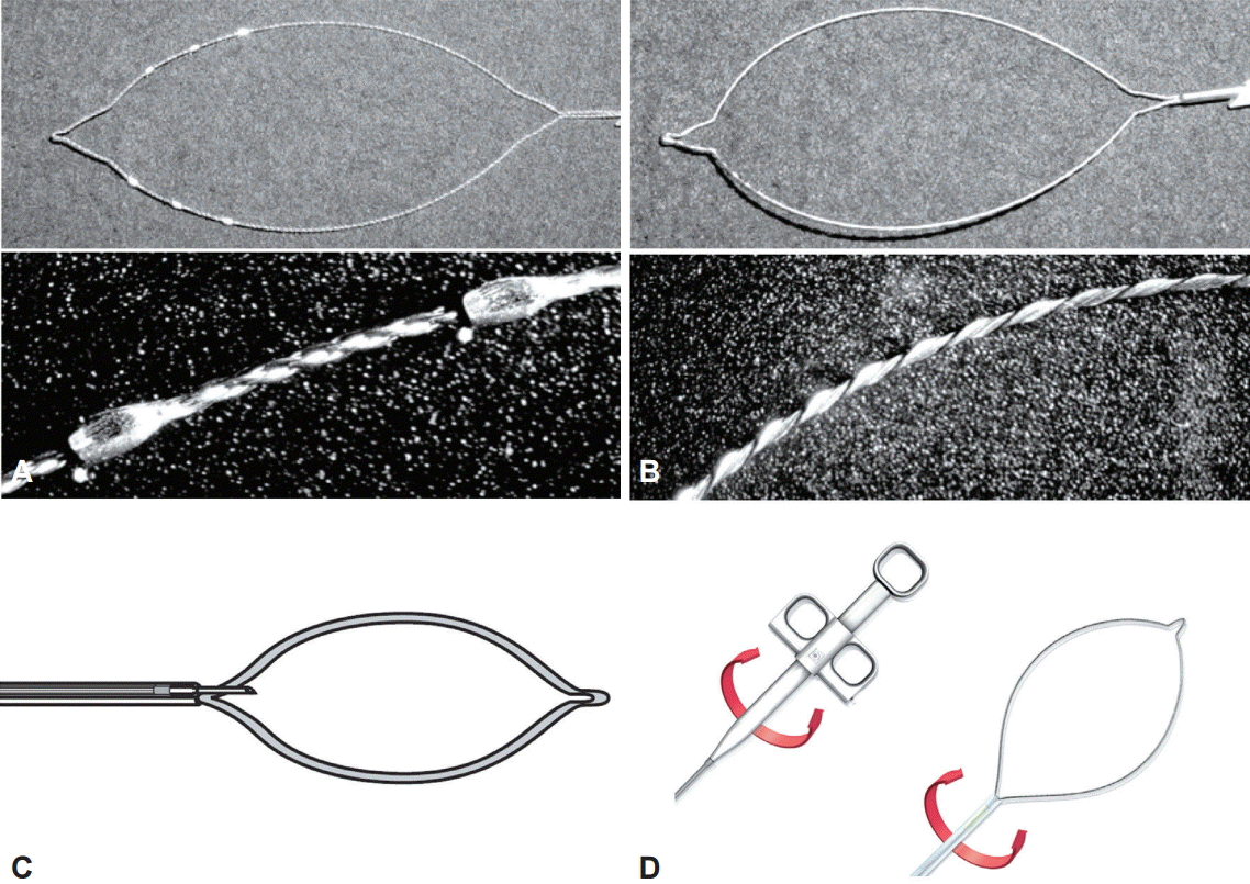
Specific types of snares are available. Barbed snares (oval snares with spikes or spiral snares) can be used for polyps with flat or sessile morphology, to ensure a firm grasp or prevent slippage [9]. A rotatable snare may be useful when the approach is not optimal, such that rotation of the snare is necessary. A combination snare-injection needle (snare-inflator) enables rapid snaring of the polyp just after submucosal injection, so that EMR can be performed effectively with a full submucosal cushion (Fig. 4).
Endoscopic clips are used for management or prevention of complications such as bleeding or perforation during a procedure. Three types of endoscopic clips are available according to length: short (5 mm), standard (7 mm), and long (9 mm). Generally, an acute-angle (90o) clip is used for perforation, and a right-angle clip (120o) is used for bleeding. A tip for use of a clip is to apply a small amount of air suction when closing the clip, which can help avoid clip slippage. Resolution clips which can be reopened are available.
A needle is used in EMR or ESD for submucosal injection. Generally, the needle is 4 mm long with 24-gauge thickness. The angle of the needle to the tissue plane is less than 30o and a volume of submucosal injection of 2 to 3 mL is optimal for EMR polypectomy. A tip for submucosal injection is to perform slow injection with slow withdrawal after slightly deep advancement of the injection needle to avoid creating a mucosal blister.
A detachable snare (Endoloop; Ethicon, Somerville, NJ, USA) can be used for prevention of postpolypectomy bleeding, especially in large pedunculated polyps containing a large vessel within the stalk. Tightening around the low portion of the stalk or base of the polyp should be performed before the polypectomy [10].
The Coagrasper is a coagulation forceps with a tapered cup that can produce focal coagulation with less electrical injury than a coagulation probe. Postpolyectomy bleeding from a relatively large vessel can be an indication for use of this instrument. A tip for bleeding control with the Coagrasper is to grasp the exact bleeding point, which can partly stop the bleeding before the use of electrical coagulation current.
An endoscopic net can be used for retrieval of a resected large polyp or tissue that will not pass through the suction channel, especially in the right colon [11].
Various types of ESU have been developed, with various electrical current modes. The selection of devices and modes is at the endoscopist’s discretion, which requires that the endoscopist understand the basic concept of the ESU.
The basic principle of the ESU is that heat can be produced without a neuromuscular response when an alternating current with high frequency between 300 KHz and 3 MHz (radiofrequency) passes through tissue [12]. ESU is a commonly used endoscopic tool for cutting or coagulating tissue. A strong electrical current can heat intracellular contents rapidly, resulting in bursting of cell membranes, and can be used for electrosurgical cutting. In contrast, electrosurgical coagulation is achieved by a less intense electrical current, which can desiccate and shrink cells without bursting the cell membrane [13].
Polypectomy with a cutting current has a risk of immediate postpolypectomy bleeding because the tissues are cut before coagulation of vessels, whereas polypectomy with a coagulation current may be associated with delayed bleeding and can obscure the tissue diagnosis [14].
Heated cells can be cooled by alternately applying and stopping current, so that cellular desiccation occurs without bursting of cell membranes. Modulating the frequency of the electrical current (duty cycle) and adjusting the peak voltage can produce a blended current, which exerts a cutting effect on the tissue with a coagulating effect at the resected margin [15].
There are no uniform and standard guidelines for ESU settings during polypectomy. Although a previous survey showed that US endoscopists have preferences for various electrical currents for polypectomy, such as pure coagulation current, blended current, and pure cutting current [16], coagulation current at a 20-W setting is generally recommended for hot-snare polypectomy [15].
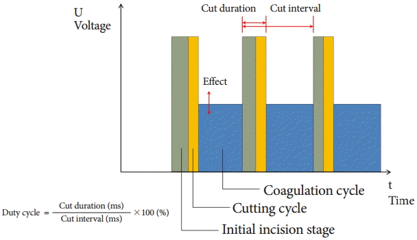
The ESU “endocut” mode has been widely used because of its better diagnostic quality for polypectomy [17]. The characteristic of the endocut mode is the alternating cutting and coagulation current. For more effective cutting or deeper coagulation, the endoscopist can adjust some variables: the coagulation power, the cut duration, and the interval between the previous and next cut (Fig. 5).
Colonoscopic polypectomy has reduced the risk, and thereby, the mortality attributable to colorectal cancer. Mastery of this highly advanced technique requires learning about the available instruments and polypectomy methods. The proper choice of instruments depending on the situation or polyp size is the optimal approach to complete colonoscopic polypectomy.
REFERENCES
1. Baxter NN, Goldwasser MA, Paszat LF, Saskin R, Urbach DR, Rabeneck L. Association of colonoscopy and death from colorectal cancer. Ann Intern Med. 2009; 150:1–8.

2. Ransohoff DF. How much does colonoscopy reduce colon cancer mortality? Ann Intern Med. 2009; 150:50–52.

3. Fyock CJ, Draganov PV. Colonoscopic polypectomy and associated techniques. World J Gastroenterol. 2010; 16:3630–3637.

4. Kim SE, Hong SP, Kim HS, et al. A Korean national survey for colorectal cancer screening and polyp diagnosis methods using web-based survey. Korean J Gastroenterol. 2012; 60:26–35.

5. Lee CK, Shim JJ, Jang JY. Cold snare polypectomy vs. cold forceps polypectomy using double-biopsy technique for removal of diminutive colorectal polyps: a prospective randomized study. Am J Gastroenterol. 2013; 108:1593–1600.

6. Monkemuller KE, Fry LC, Jones BH, Wells C, Mikolaenko I, Eloubeidi M. Histological quality of polyps resected using the cold versus hot biopsy technique. Endoscopy. 2004; 36:432–436.

7. Lee SH, Shin SJ, Park DI, et al. Korean guideline for colonoscopic polypectomy. Clin Endosc. 2012; 45:11–24.

8. Tsai FC, Strum WB. Prevalence of advanced adenomas in small and diminutive colon polyps using direct measurement of size. Dig Dis Sci. 2011; 56:2384–2388.

9. Yamano H, Matsushita H, Yamanaka K, et al. A study of physical Efficacy of different snares for endoscopic mucosal resection. Dig Endosc. 2004; 16(Suppl 1):S85–S88.

10. Iishi H, Tatsuta M, Narahara H, Iseki K, Sakai N. Endoscopic resection of large pedunculated colorectal polyps using a detachable snare. Gastrointest Endosc. 1996; 44:594–597.

11. Miller K, Waye JD. Polyp retrieval after colonoscopic polypectomy: use of the Roth Retrieval Net. Gastrointest Endosc. 2001; 54:505–507.

12. Barlow DE. Endoscopic applications of electrosurgery: a review of basic principles. Gastrointest Endosc. 1982; 28:73–76.

13. Boulay BR, Carr-Locke DL. Current affairs: electrosurgery in the endoscopy suite. Gastrointest Endosc. 2010; 72:1044–1046.

14. Van Gossum A, Cozzoli A, Adler M, Taton G, Cremer M. Colonoscopic snare polypectomy: analysis of 1485 resections comparing two types of current. Gastrointest Endosc. 1992; 38:472–475.

15. Morris ML, Tucker RD, Baron TH, Song LM. Electrosurgery in gastrointestinal endoscopy: principles to practice. Am J Gastroenterol. 2009; 104:1563–1574.

Fig. 1.
The forceps size (the distance between cups) ranges from 5 to 8 mm (left). The diameter of the cup is about 3 mm (right).

Fig. 2.
Forceps polypectomy: colorectal polyp ≤3 mm can be removed by one-bite forceps (left), but 4 to 5 mm polyps need two bites for complete resection.

Fig. 4.
Specific types of snare. (A) Oval snare with spikes. (B) Spiral oval snare. (C) Snareinflator. (D) Rotatable snare.

Fig. 5.
Variables in endocut mode: coagulation power, cut duration, and interval between previous and next cut.

Table 1.
Endoscopic Instruments and Electrosurgery Generator Unit according to Various Polypectomy Techniques


 PDF
PDF Citation
 Print
Print


 XML Download