Abstract
Biliary self-expandable metal stents (SEMS), a group of non-vascular stents, have been used in the palliative management of biliary obstruction around the world. However, there are still unmet needs in the clinical application of biliary SEMS. Comprehensive understanding of the SEMS is required to resolve the drawbacks and difficulties of metal stent development. The basic structure of SEMS, including the materials and knitting methods of metal wires, covering materials, and radiopaque markers, are discussed in this review. What we know about the physical and mechanical properties of the SEMS is very important. With an understanding of the basic knowledge of metal stents, hurdles such as stent occlusion, migration, and kinking can be overcome to develop more ideal SEMS.
Self-expandable metal stents (SEMS) for the treatment of biliary obstruction have been developed by many investigators and medical device companies for several decades since the biliary SEMS were introduced in 1989 [1]. Consequently, many different SEMS with good performance have now become available in the clinical setting. However, there are still unmet clinical needs for biliary SEMS. A deep understanding of the basic structure and mechanical properties of SEMS is needed for the development of improved SEMS.
Regarding the basic structure of tubular-shaped metal stents, the metal wire is an essential material, which gives elasticity and flexibility to the SEMS. The materials used for the wire are stainless steel and nitinol (nickel-titanium) alloys in most SEMS, and recently nitinol has been predominantly used as the material of the stent wire. Nitinol wire is known to have a shape-memory effect [2]. This type of wire can wind into a small diameter and be delivered through a catheter into the bile duct, and it is able to expand to a larger diameter in the bile duct under the warming of body temperature. It is so flexible that a single wire can be braided for an entire stent. For this reason, modification of the stent design or implementation of a new design may be comparatively easy.
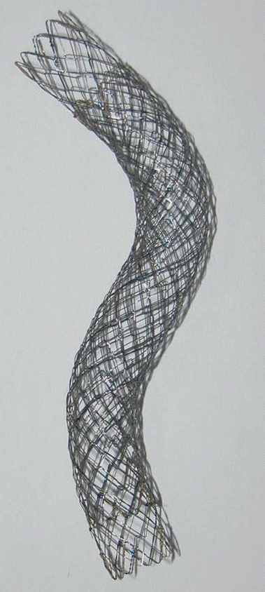
In terms of wire-weaving methods for SEMS, the wire is braided as a wire crossover structure or is cut by laser (Fig. 1). There is also a complicated, wire-braided type of SEMS structure [3]. This type of SEMS consists of two layers of wire-crossover structure with or without an intervening covering membrane. The braided structure of the stent has closed cells and is more resistant to tumor ingrowth or inward tissue hyperplasia compared to the open cell type of stent. Therefore, this type of stent can show longer duration of patency. However, in the open cell type, which is a laser-cut stent structure, the duration of stent patency tends to be shorter [4]. In regard to the shortening ratio of the stent before and after stent deployment, the laser-cut structure is far superior to the braided type; its ratio is zero. This strength is advantageous for proper stent positioning during stent deployment.
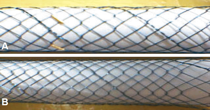
The cell size of the SEMS can be controlled by the wire-weaving method, and as each cell diameter is decreased, stent occlusion caused by tumor ingrowth may theoretically decrease (Fig. 2). However, there has been no definite evidence that tumor ingrowth-induced stent occlusion increases as the cell width increases [5].
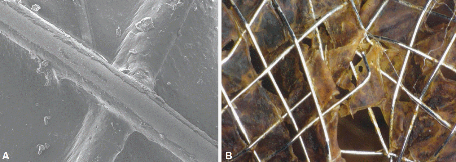
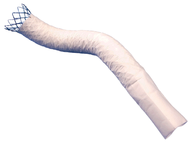
Recently available covering materials used for biliary SEMS around the world include polyurethane, silicone, and expanded polytetrafluoroethylene. Covering membranes have been used in covered SEMS to prevent tumor ingrowth through the cells of the metal stent and to avoid re-intervention [6]. However, whether uncovered or covered SEMS have superior stent patency period remains controversial [7-10]. One of the reasons for this may be because of differences between covering materials in membrane biodurability under long-term exposure to bile juice [11]. The reason that the stent patency period was shorter and the patency rate was lower than expected in some clinical studies of covered SEMS may be due to cracks in the covering membrane with subsequent tumor ingrowth (Fig. 3) [12,13]. However, these covering materials have recently been improved, and all three materials seem to be durable for at least a 6-month period of bile exposure [11]. Meanwhile, biofilm formation with subsequent stent clogging caused by bile sludge (which is one of the unresolved problems for covered SEMS) has been a problem yet to be resolved in the clinical setting.
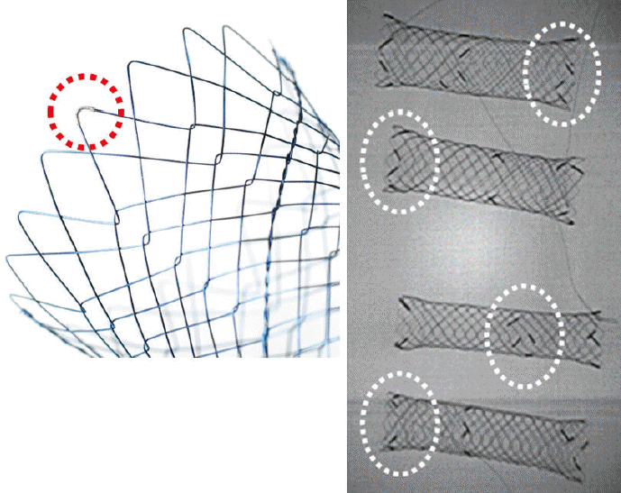
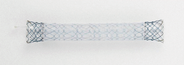
Radiopacity is generally important for SEMS. Increasing the radiopacity of the SEMS could improve not only positioning and deployment control during stent delivery but also monitoring of adequate stent expansion and stent migration after the placement. Therefore, radiopaque markers are applied to SEMS to improve their radiopacity. Radiopaque markers, such as gold, platinum, or tantalum, are attached to the stent wire by microwelding (Fig. 4). The attachment location is usually at both ends of the stent. There are several types of markers used for stents, such as sleeves, rivets, welded inlays, or welded-on end-segments.
The mechanical properties of SEMS that have been previously reported in the literature include radial force (RF), axial force (AF), flexibility, shortening ratio, radiopacity, and trackability [3,14-19]. One of the most important factors is RF because it expands and maintains the lumen at the stricture segment of the bile duct after the SEMS are deployed. The force can be influenced by the wire diameter, cell size (which depends on how close the wire is woven), and the wire-knitting method (braided, specially braided, or laser cut). If RF is low (<4.00 N), SEMS have a risk of migrating within 6 months after the stent placement (odds ratio [OR], 2.23; 95% confidence interval [CI], 1.07 to 4.65; p=0.03) [20].
AF is another important property of SEMS, and measuring the AF of various SEMS was proposed by Isayama et al. [3]. It was suggested to be the recovery force required to keep stents straight as the SEMS bent. AF varies according to the wire-knitted structure of the stents and is influenced by the stent length; AF decreases as the length increases. SEMS are surrounded by cancer tissue; if the AF is strong (>0.40 N), the conformability of the stents in the bile duct may become poor, and both sides of the stent may compress the bile duct wall, the orifice of the cystic duct, or the pancreatic duct orifice. This may cause significant adverse events such as kinking of the stents in the bile duct, cholecystitis (OR, 5.33; 95% CI, 1.74 to 23.27; p=0.002) [21], and pancreatitis (OR, 3.69; 95% CI, 1.19 to 16.2; p=0.022) [22]. Therefore, lower AF confers better conformability and may prevent stent-related complications.
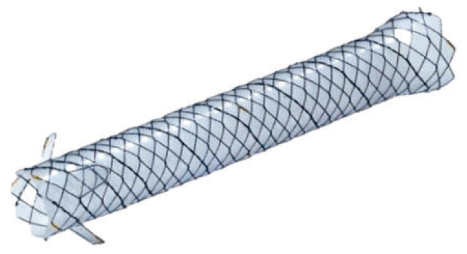
Dedicated, specific structures of SEMS have been developed to overcome drawbacks of the stents in clinical application. Anti-reflux valves were designed to prevent duodenal biliary reflux and achieve longer stent patency (Fig. 5) [24,25]. Drug-eluting covering membranes (which allow the controlled release of anti-cancer drugs such as OK-432 [26] and paclitaxel [27,28]) and special wire-braided, double-layered structures have been developed [29,30] to inhibit tumor ingrowth (Fig. 6). To prevent stent migration, anchoring flaps were recently created on the outer surfaces of SEMS (Fig. 7) [31], and different levels of segmental RF were created by wire-weaving different cell sizes for each segment over the whole length of the SEMS [32] (Fig. 8).
Basic structures and mechanical properties of SEMS are very important in the research and development of SEMS. Clinical data about the stent structures and mechanical properties are informative not only to investigators but also to biliary endoscopists. Comprehensive understanding of SEMS will be useful in resolving unmet clinical needs for biliary SEMS in the near future.
REFERENCES
1. Irving JD, Adam A, Dick R, Dondelinger RF, Lunderquist A, Roche A. Gianturco expandable metallic biliary stents: results of a European clinical trial. Radiology. 1989; 172:321–326.
2. Dotter CT, Buschmann RW, McKinney MK, Rosch J. Transluminal expandable nitinol coil stent grafting: preliminary report. Radiology. 1983; 147:259–260.
3. Isayama H, Nakai Y, Toyokawa Y, et al. Measurement of radial and axial forces of biliary self-expandable metallic stents. Gastrointest Endosc. 2009; 70:37–44.
4. Loew BJ, Howell DA, Sanders MK, et al. Comparative performance of uncoated, self-expanding metal biliary stents of different designs in 2 diameters: final results of an international multicenter, randomized, controlled trial. Gastrointest Endosc. 2009; 70:445–453.
5. Lee JM, Lee SH, Chung KH, et al. Small cell- versus large cell-sized metal stent in endoscopic bilateral stent-in-stent placement for malignant hilar biliary obstruction. Dig Endosc. 2015; 27:692–699.
6. Saito H, Sakurai Y, Takamura A, Horio K. Biliary endoprosthesis using Gore-Tex covered expandable metallic stents: preliminary clinical evaluation. Nihon Igaku Hoshasen Gakkai Zasshi. 1994; 54:180–182.
7. Saleem A, Leggett CL, Murad MH, Baron TH. Meta-analysis of randomized trials comparing the patency of covered and uncovered self-expandable metal stents for palliation of distal malignant bile duct obstruction. Gastrointest Endosc. 2011; 74:321–327.
8. Isayama H, Komatsu Y, Tsujino T, et al. A prospective randomised study of “covered” versus “uncovered” diamond stents for the management of distal malignant biliary obstruction. Gut. 2004; 53:729–734.
9. Telford JJ, Carr-Locke DL, Baron TH, et al. A randomized trial comparing uncovered and partially covered self-expandable metal stents in the palliation of distal malignant biliary obstruction. Gastrointest Endosc. 2010; 72:907–914.
10. Kullman E, Frozanpor F, Soderlund C, et al. Covered versus uncovered self-expandable nitinol stents in the palliative treatment of malignant distal biliary obstruction: results from a randomized, multicenter study. Gastrointest Endosc. 2010; 72:915–923.
11. Bang BW, Jeong S, Lee DH, Lee JI, Lee SC, Kang SG. The biodurability of covering materials for metallic stents in a bile flow phantom. Dig Dis Sci. 2012; 57:1056–1063.
12. Kanasaki S, Furukawa A, Kane T, Murata K. Polyurethane-covered Nitinol Strecker stents as primary palliative treatment of malignant biliary obstruction. Cardiovasc Intervent Radiol. 2000; 23:114–120.
13. Maccioni F, Rossi M, Salvatori FM, Ricci P, Bezzi M, Rossi P. Metallic stents in benign biliary strictures: three-year follow-up. Cardiovasc Intervent Radiol. 1992; 15:360–366.
14. Miura S, Yoshioka T, Furuichi K, Tanaka T, Kichikawa K, Ohishi H. Mechanical properties of biliary metallic stents: an experimental comparison. Nihon Igaku Hoshasen Gakkai Zasshi. 2003; 63:201–209.
15. Dyet JF, Watts WG, Ettles DF, Nicholson AA. Mechanical properties of metallic stents: how do these properties influence the choice of stent for specific lesions? Cardiovasc Intervent Radiol. 2000; 23:47–54.
16. Duda SH, Wiskirchen J, Tepe G, et al. Physical properties of endovascular stents: an experimental comparison. J Vasc Interv Radiol. 2000; 11:645–654.
17. Jedwab MR, Clerc CO. A study of the geometrical and mechanical properties of a self-expanding metallic stent: theory and experiment. J Appl Biomater. 1993; 4:77–85.
18. Stoeckel D, Pelton A, Duerig T. Self-expanding nitinol stents: material and design considerations. Eur Radiol. 2004; 14:292–301.
19. Nuutinen JP, Clerc C, Tormala P. Theoretical and experimental evaluation of the radial force of self-expanding braided bioabsorbable stents. J Biomater Sci Polym Ed. 2003; 14:677–687.
20. Nakai Y, Isayama H, Kogure H, et al. Risk factors for covered metallic stent migration in patients with distal malignant biliary obstruction due to pancreatic cancer. J Gastroenterol Hepatol. 2014; 29:1744–1749.
21. Nakai Y, Isayama H, Kawakubo K, et al. Metallic stent with high axial force as a risk factor for cholecystitis in distal malignant biliary obstruction. J Gastroenterol Hepatol. 2014; 29:1557–1562.
22. Kawakubo K, Isayama H, Nakai Y, et al. Risk factors for pancreatitis following transpapillary self-expandable metal stent placement. Surg Endosc. 2012; 26:771–776.
23. Isayama H, Nakai Y, Hamada T, Yamamoto N, Koike K. Development of an ideal self-expandable metallic stent design. Gastrointest Interv. 2015; 4:46–49.
24. Lee KJ, Chung MJ, Park JY, et al. Clinical advantages of a metal stent with an S-shaped anti-reflux valve in malignant biliary obstruction. Dig Endosc. 2013; 25:308–312.
25. Hu B, Wang TT, Wu J, Shi ZM, Gao DJ, Pan YM. Antireflux stents to reduce the risk of cholangitis in patients with malignant biliary strictures: a randomized trial. Endoscopy. 2014; 46:120–126.
26. Lee DH, Kang SG, Jeong S, et al. Local delivery system of immune modulating drug for unresectable adenocarcinoma: in vitro experimental study and in vivo animal study. Cardiovasc Intervent Radiol. 2006; 29:832–837.
27. Song TJ, Lee SS, Yun SC, et al. Paclitaxel-eluting covered metal stents versus covered metal stents for distal malignant biliary obstruction: a prospective comparative pilot study. Gastrointest Endosc. 2011; 73:727–733.
28. Jang SI, Kim JH, You JW, et al. Efficacy of a metallic stent covered with a paclitaxel-incorporated membrane versus a covered metal stent for malignant biliary obstruction: a prospective comparative study. Dig Dis Sci. 2013; 58:865–871.
29. Isayama H, Kawabe T, Nakai Y, et al. Management of distal malignant biliary obstruction with the ComVi stent, a new covered metallic stent. Surg Endosc. 2010; 24:131–137.
30. Perri V, Boskoski I, Tringali A, et al. Prospective evaluation of the partially covered nitinol “ComVi” stent for malignant non hilar biliary obstruction. Dig Liver Dis. 2013; 45:305–309.
31. Park do H, Lee SS, Lee TH, et al. Anchoring flap versus flared end, fully covered self-expandable metal stents to prevent migration in patients with benign biliary strictures: a multicenter, prospective, comparative pilot study (with videos). Gastrointest Endosc. 2011; 73:64–70.