

 PDF
PDF Citation
 Print
Print


INTRODUCTION
Administration of selective internal radiation with yttrium-90 is an efficient loco-regional treatment method for patients with liver malignancies who are not eligible for surgery. This treatment method has a low gastrointestinal (GI) complication rate, with a reported frequency of 3% to 24% [1]. Following the unavoidable escape of yttrium-90 spherules from the hepatic artery to the stomach and duodenum via aberrant branches, the radiated spherules can cause mucosal damage. Although this condition is resistant to gastric acid suppression, a proton pump inhibitor (PPI) is the first therapeutic option for the treatment of gastroduodenal ulcers caused by radioactive yttrium; PPIs are also used in the treatment of peptic ulcers, depending on different etiologies. In cases accompanied by gastric outlet obstruction (GOO), the risk-to-benefit ratio must be evaluated before gastrectomy or gastric bypass surgery is performed [2].
Self-expandable metallic stents (SEMS) were initially used mainly for the palliative treatment of malignant stenosis of the GI tract [3,4]. Later, they began to be used in the treatment of benign stenosis of the GI tract [5]. The use of SEMS to treat GOO caused by radioactive yttrium treatment has not been previously reported.
We report a case of GOO that developed as a complication after performance of selective internal radiation therapy (SIRT) for treatment of a neuroendocrine tumor that metastasized to the liver in a 58-year-old man. A self-expandable stent was used for treatment.
Go to : 
CASE REPORT
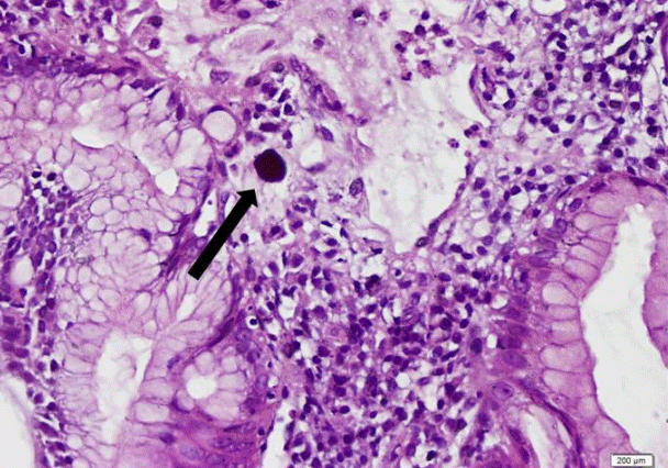
A 58-year-old man with liver metastasis from a neuroendocrine tumor was referred to the nuclear medicine department for treatment with yttrium. Pre-treatment hepatic angiography revealed normal arterial anatomy, and screening with tagged albumin showed the absence of hepatopulmonary shunt. The patient was treated with intra-arterial yttrium-90 microsphere embolization. The patient did not have any GI symptoms before undergoing the procedure; however, he experienced epigastric pain, nausea, and vomiting 3 weeks after undergoing yttrium-90 SIRT. He lost 4 kg weight during this period. Upper GI endoscopy showed a geographic-shaped antropyloric ulcer accompanied by exudation and stricture at the antropyloric region, with strong resistance against the passage of the endoscope through the stricture (Fig. 1A). Multiple biopsies taken from the ulcer margin demonstrated dark microspheres in the deep layer of the lamina propria (yttrium particles), severe reactive glandular atypia, and denudated friable mucosa consistent with radiation injury. No sign of helicobacter pylori infection or malignancy was observed in the biopsy specimens (Fig. 2). The patient had no history of steroid or nonsteroidal anti-inflammatory drug intake. A PPI (esomeprazole 40 mg) was started twice a day for 10 days, but this had no apparent effect on the patient’s nausea, vomiting, and epigastric pain. Therefore, endoscopic balloon dilation was performed with a 15-mm through-the-scope (TTS) balloon (2×60 seconds; Boston Scientific, Watertown, MA USA), and the balloon was easily moved back and forth before it was removed after dilation (Fig. 1B). The patient’s response was not sufficient a week after balloon dilation, so a partially covered TTS (Fig. 1C) (SEMS; Hanarostent, 20 mm/110 mm partially covered SEMS Duodenum/Pylorus Lasso [NCN; M.I. TECH, Seoul, Korea]) was placed under endoscopic (Fig. 1D) and fluoroscopic control. The patient’s oral intake was assessed by using GOO scoring [6]. The pre-stenting GOO scoring was 1, whereas the score on poststenting day 3 was calculated as 3. An endoscopic examination was performed 3 months later, and the stent was retracted by grabbing the stent lasso with grasping forceps (Fig. 1E). During the same endoscopic examination, the ulcer was observed to have significantly healed. Stent migration or complications such as perforation or hemorrhage did not occur. Symptoms such as nausea and vomiting were not present at the follow-up visits at 2, 3, and 6 months after stent removal. Endoscopic examination at the 6-month follow-up visit revealed a lack of both food retention in the stomach and ulceration, and the antropyloric junction was open; further, the patient had gained 10 kg.
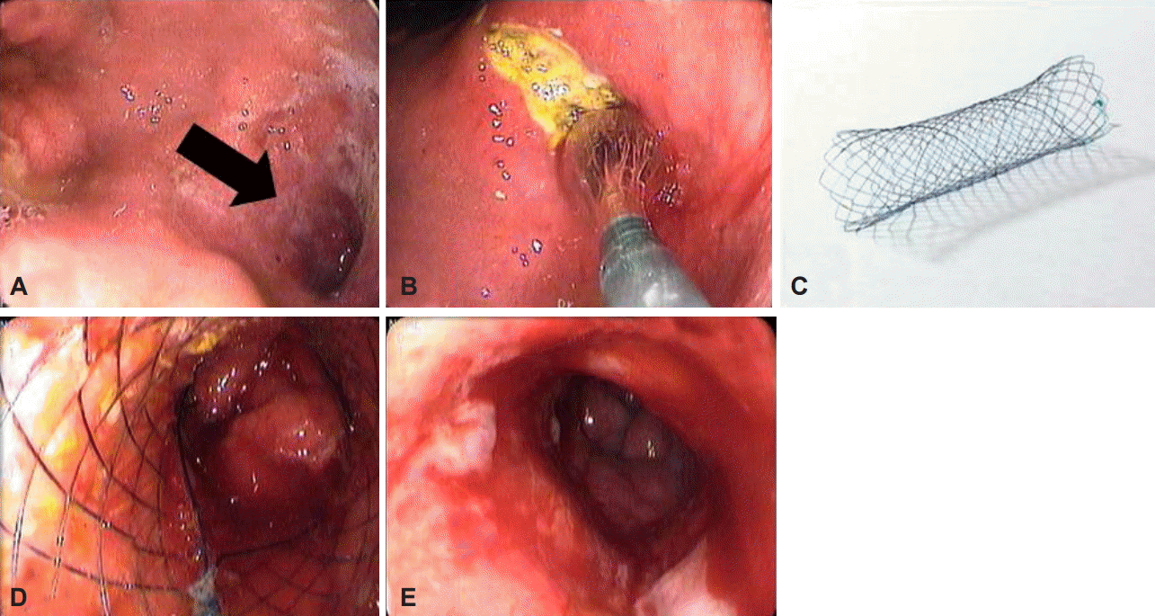 | Fig. 1.(A) Endoscopy shows gastric ulcer and outlet obstruction (arrow), (B) endoscopic view of balloon dilation (15 mm in diameter). (C) Hanarostent, a partially covered self-expendable metallic stent Duodenum/Pylorus Lasso (NCN; M.I. TECH), (D) proximal view of an opened stent after stenting, and (E) endoscopic view after stent removal.
|
Go to :
DISCUSSION
Recently, SIRT has become a popular treatment method for unresectable primary and metastatic hepatic tumors. GI ulceration following hepatic radioembolization is a well-known complication associated with non-target embolization. Despite careful angiographic analysis, the reported frequency of embolization of radioactive spherules to this area is 3% to 24% [1]. Several factors may explain the occurrence of embolization of radioactive spherules. First, small veins that originate from the hepatic artery and supply the GI tract may not be detected in the angiographic examination performed before the procedure. Further, collateral circulation may form during the period between placement of the prophylactic coil and receipt of yttrium treatment. In addition, the embolic effects of the spherules and the contraction effects of the catheter in the vein may cause anterograde escape of spherules [7]. The findings from our patient’s angiographic examination were normal before the procedure.
Embolization-related GI complications, such as gastroduodenal ulcer, cholecystitis, and pancreatitis, are generally observed 2 months after the procedure [8]. In cases with gastric involvement, the principal symptoms are epigastric pain, nausea, and vomiting. GI hemorrhage may also be observed. The spectrum of endoscopic findings may change from erythematous or erosive gastritis to ulceration and/or stricture formation [9]. We identified an antropyloric giant ulcer and stenosis due to edema and scarring during endoscopic examination in our case. At diagnosis, the observation of round-shaped yttrium spherules in biopsy specimens collected from the ulcer region is considered pathognomonic.
The cause of tissue damage appears to be related to radiation rather than to ischemia because the arterial network of the upper GI area is rather wide. In clinical use, such as for Hodgkin’s disease, ionizing radiation has been reported to cause gastritis, gastric and duodenal ulcer, and intestinal obstruction and perforation [10]. However, reports of similar GI toxic effects observed after hepatic chemoembolization or hepatic artery chemotherapy infusion does not support the theory that tissue damage is solely due to ionizing radiation [11].
Treatment of yttrium-90-related GI ulceration is difficult, and no consensus exists regarding the most efficient treatment. The treatment should be individualized according to patient symptoms and localization and diffusiveness of GI involvement. Although high doses of a PPI are recommended, the success rate of this treatment is unsatisfactory (50%) [1,8]. Radiation damage is known to cause excess release of transforming growth factor β1 (TGF-β1), which results in fibrosis and organ failure. Therefore, drugs such as interferon and pentoxifylline, which reduce the effects of TGF-β1, may also be included as treatment options [12]. Early surgery should be considered if abdominal pain and abnormal endoscopic findings persist and if the patient does not respond to medical treatment because delayed surgical intervention can result in fatality. The complication of GOO has been reported in very few cases of gastric or gastroduodenal ulceration. In these rare reported cases, surgery was usually recommended and procedures such as gastrojejunostomy or partial gastrectomy were performed [9,13]. However, in a case series reported by Konda et al. [1], endoscopic balloon dilatation was used in a case with GOO, and stricture persisted after dilation in that patient. Dilatation was also performed once in our case, but recovery of oral intake was not observed; therefore, a partially covered SEMS was subsequently used. Because of the increased morbidity and mortality associated with surgery in patients with malignancy, stenting appears to be a better option for palliation, and it has become the preferred treatment method for palliation of GOO due to both malignant and benign causes. The use of the self-expandable stent for salvage was reported to be efficient and safe for benign pyloric obstruction, whether the condition was naïve or associated with insufficient balloon dilatation. Heo and Jung [5] reported that 3 to 6 months is an adequate stent duration at the stenotic segment.
To our knowledge, this is the first reported case in the literature in which SEMS was used for antropyloric obstruction due to yttrium. The procedure was successful, no complications occurred, and the patient’s oral intake improved. Despite the lack of stent experience with GOO due to SIRT, the consideration of endoscopic treatment options prior to surgery is important for avoiding mortality and morbidity caused by surgery and for the patient’s quality of life. Endoscopic stent placement may be a good therapeutic option for cases with GOO.
Go to :
 XML Download