Abstract
Background/Aims:
Obesity is a much-debated factor with conflicting evidence regarding its association with cecum intubation rates during colonoscopy. We aimed to identify the association between cecal intubation (CI) time and obesity by eliminating confounding factors.
Methods:
A retrospective chart review of subjects undergoing outpatient colonoscopy was conducted. The population was categorized by sex and obesity (body mass index [BMI, kg/m2]: I, <24.9; II, 25 to 29.9; III, ≥30). CI time was used as a marker for a difficult colonoscopy. Mean CI times (MCT) were compared for statistical significance using analysis of variance tests.
Results:
A total of 926 subjects were included. Overall MCT was 15.7±7.9 minutes, and it was 15.9±7.9 and 15.5±7.9 minutes for men and women, respectively. MCT among women for BMI category I, II, and III was 14.4±6.5, 15.5±8.3, and 16.2±8.1 minutes (p=0.55), whereas for men, it was 16.3±8.9, 15.9±8.0, and 15.6±7.2 minutes (p=0.95), respectively.
Colorectal cancer (CRC) is one of the leading causes of malignant neoplasm-related mortality [1,2]. In 2012, CRC was responsible for 694,000 deaths worldwide, accounting for 8.5% of all cancer related deaths [2]. It is estimated that in the United States alone 93,090 new cases of CRC will be diagnosed in 2015 [3]. Obesity is a proven risk factor for CRC [4,5]. It is associated with an increased risk of incident CRC in men of all ages and in women between the ages of 50 to 66 [6]. Obesity, a modifiable risk factor, has also been found to increase the risk of adenoma and advanced adenoma recurrence [7].
Body habitus and cecal intubation (CI) during colonoscopy have a unique relationship. There is evidence to suggest that lean subjects offer a challenge to endoscopists in achieving CI during colonoscopy [8-10]. This has been attributed to the paucity of the visceral fat pad and the smaller size of the abdominal cavity. However, techniques and maneuvers routinely required during the endoscopic examination, such as repositioning and abdominal pressure application, are more difficult to perform on obese patients. It appears there is an optimum body mass index (BMI) that might be considered “just right” for colonoscopy. Obesity is also an independent predictor of inadequate bowel preparation for colonoscopy [11,12], which indirectly contributes to a difficult colonoscopy. In this context, and taking into account that obesity has reached epidemic proportions in America [13] and Europe [14], colonoscopy in obese patients represents a challenging issue for an endoscopist, one that is frequently encountered in daily practice.
Our study aims to delineate the association between BMI and CI difficulty during colonoscopy, after controlling for other confounding variables such as the endoscopist’s experience, quality of bowel preparation, and any risk factors for intra-abdominal adhesions.
To define the association between obesity and CI time during colonoscopy after controlling for confounding factors.
This was a retrospective observational cohort study conducted at a tertiary care teaching hospital. The study protocol was approved by the Institutional Review Board of Einstein Medical Center (IRB no. 4550 EXE). Written informed consent was not required since it was a retrospective chart review study.
Inclusion criteria:
(1) All subjects presenting for an outpatient colonoscopy to our institution over a span of 18 months.
Exclusion criteria:
(1) Poor bowel preparation
(2) Failure of cecum intubation
(3) Personal history of inflammatory bowel disease (IBD)
(4) History of colectomy or abdomino-pelvic surgery (as defined below)
(5) Personal or family history of colon cancer
(6) Procedures performed by fellows
Colonoscopy and anesthesia procedure reports were reviewed to collect data regarding age, sex, weight, height, timing of the procedure (morning vs. afternoon), endoscopist’s experience (fellow vs. attending), past medical history (IBD, CRC), past surgical history (abdomino-pelvic surgery including laparoscopic procedures but excluding umbilical/inguinal hernia repair and transurethral/transvaginal procedures), family history of colon cancer, bowel preparation quality, and CI time. The exclusion criteria were then applied to the initial study population to remove any effect of all the confounding variables. Weight and height was then used to calculate BMI. All data were recorded on Excel spreadsheets without including any identifying information to maintain patient anonymity. The final study population (after applying the exclusion criteria) was then divided into men and women subgroups. Each sex group was sub-categorised based on BMI into three categories: I (non-obese, BMI <25), II (overweight, BMI 25 to 29.9), III (obese, BMI ≥30). Mean CI time (MCT) was then computed for each category. The significance of the difference in CI time values was determined using analysis of variance, which was applied using Stata for mac version 13.0 (Statacorp LP, College Station, TX, USA). A p-value less than 0.05 was used to determine statistical significance.
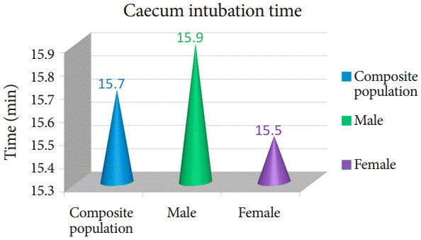
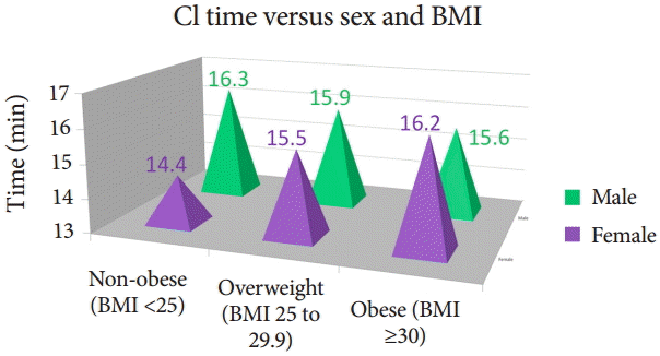
Our study included 926 subjects reporting for outpatient colonoscopy. The mean age of the study population was 58.8 years, and it comprised of 47.7% women and 52.3% men. The demographic characteristics are summarized in Table 1. The distribution of cases across the time of the day, i.e., morning or afternoon, was non-uniform, with 57.8% procedures performed in the morning and 42.2% in the afternoon. MCT for the study population was 15.7±7.9 minutes, with 15.9±7.9 and 15.5±7.9 minutes for men and women, respectively (Fig. 1). The MCT for women in BMI categories I, II, and III was 14.4±6.5, 15.5±8.3, and 16.2±8.1 minutes (p=0.55), respectively (Fig. 2). MCT for men in BMI category I, II, and III was 16.3±8.9, 15.9±8.0, and 15.6±7.2 minutes (p=0.95), respectively (Fig. 2).
Obesity increases the incidence of CRC and colon adenoma, which makes it even more important to screen people with a high BMI. Several methods are available to screen for colon cancer, including fecal occult blood tests and flexible sigmoidoscopy, but the method with the best early colon cancer detection rate is colonoscopy. The number of colonoscopies has increased considerably in the past few years since it became standard practice to offer a CRC screening to all individuals above 50 years or earlier, if so indicated. This trend has sparked a lot of research into the factors that predict a difficult colonoscopy.
The endoscopist’s level of training, quality of bowel preparation, body habitus, subject’s sex, intra-abdominal adhesions secondary to previous surgery, and presence of angulations among large bowel loops predicts the level of difficulty in achieving CI among subjects undergoing colonoscopy [15]. The association between body weight and the technical difficulty in achieving CI during colonoscopy has been a topic of debate. There is conflicting evidence suggesting that both lean and obese subjects present a challenge to the endoscopist during colonoscopy [8,9,15-17]. Obesity has been independently linked to poor bowel preparation, which in turn can also lead to a difficult and prolonged colonoscopy [12].
To truly appreciate the association between body weight and difficult colonoscopy, we need to eliminate the effect of confounding variables. To achieve this, we designed our study to exclude subjects with poor bowel preparation, procedures done by fellows, and subjects with a history of abdominal/pelvic surgery. We used CI time as a surrogate to estimate the degree of difficulty of the colonoscopy.
There was a minimal difference in the MCT between men and women (15.9±7.9 minutes vs. 15.5±7.9 minutes). Interestingly, the MCT had a positive association with BMI for women but had a negative association for men (Fig. 2). Although it failed to achieve statistical significance, it did highlight an important trend. The positive association among women is consistent with the literature regarding the increased difficulty of colonoscopy in obese individuals. The difference between the sexes could be explained by their tendency to accumulate fat at different sites. Men are, in general, more prone to accumulate abdominal fat, whereas women are more prone to accumulate fat in the gluteal region and limbs [18].
Based on the literature review, our study is unique because it attempts to control a lot of confounding variables to delineate the true association between body weight and difficult colonoscopy. Although the absolute difference across different groups based on the BMI is minimal, the consistent trend makes our results interesting and raises new questions that need to be addressed in future studies.
Our study has a few limitations. Being a retrospective single center study, the results cannot be generalized. BMI, although a widely accepted scale for obesity, is not a true measure of intra-abdominal fat [19]. It neither differentiates between fat and muscle, nor between the type and site of fat accumulation [19]. Nagata et al. [16] found that among all of the indices of obesity, subcutaneous fat was the best predictor of CI time. However, since computed tomography scans for abdominal fat measurement are not recommended in routine clinical practice due to the unnecessary radiation exposure and the increased cost of healthcare, we used BMI to estimate obesity. Given the fact that the waist to hip ratio is a better marker of intra-abdominal fat, in light of the above findings we expect the waist to hip ratio to better correlate with CI time. It may be the preferred index of obesity for future studies given its cost-effectiveness and harmlessness [19].
Our study results have important clinical implications. It provides a more accurate and reliable association between obesity and difficult colonoscopy across both sexes. It should also change the endoscopist’s preconceived notions about obese patients undergoing colonoscopy. These results will also assist clinics in developing a more accurate time allotment for colonoscopy; thus, avoiding delays and improving workflow within the endoscopy suite. Larger multicenter trials with better methods to estimate visceral fat are required to confirm our findings.
REFERENCES
1. Bresalier RS. Early detection of and screening for colorectal neoplasia. Gut Liver. 2009; 3:69–80.

2. World Health Organization. Colorectal cancer: estimated incidence, mortality and prevalence Worldwide in 2012 [Internet]. Lyon: World Health Organization;c2016. [cited 2016 Feb 2]. Available from: http://globocan.iarc.fr/Pages/fact_sheets_cancer.aspx?cancer=colorectal.
3. American Cancer Society. Key statistics for colorectal cancer [Internet]. Atlanta: American Cancer Society;c2016. [cited 2016 Feb 2]. Available from: http://www.cancer.org/cancer/colonandrectumcancer/detailedguide/colorectal-cancer-key-statistics.
4. Moghaddam AA, Woodward M, Huxley R. Obesity and risk of colorectal cancer: a meta-analysis of 31 studies with 70,000 events. Cancer Epidemiol Biomarkers Prev. 2007; 16:2533–2547.

5. Moore LL, Bradlee ML, Singer MR, et al. BMI and waist circumference as predictors of lifetime colon cancer risk in Framingham Study adults. Int J Obes Relat Metab Disord. 2004; 28:559–567.

6. Adams KF, Leitzmann MF, Albanes D, et al. Body mass and colorectal cancer risk in the NIH-AARP cohort. Am J Epidemiol. 2007; 166:36–45.

7. Laiyemo AO, Doubeni C, Badurdeen DS, et al. Obesity, weight change, and risk of adenoma recurrence: a prospective trial. Endoscopy. 2012; 44:813–818.

9. Cirocco WC, Rusin LC. Factors that predict incomplete colonoscopy. Dis Colon Rectum. 1995; 38:964–968.

10. Oh SY, Sohn CI, Sung IK, et al. Factors affecting the technical difficulty of colonoscopy. Hepatogastroenterology. 2007; 54:1403–1406.
11. Hsieh YH, Kuo CS, Tseng KC, Lin HJ. Factors that predict cecal insertion time during sedated colonoscopy: the role of waist circumference. J Gastroenterol Hepatol. 2008; 23:215–217.

12. Borg BB, Gupta NK, Zuckerman GR, Banerjee B, Gyawali CP. Impact of obesity on bowel preparation for colonoscopy. Clin Gastroenterol Hepatol. 2009; 7:670–675.

13. Ogden CL, Carroll MD, Kit BK, Flegal KM. Prevalence of obesity in the United States, 2009-2010. NCHS Data Brief. 2012; (82):1–8.
14. Berghöfer A, Pischon T, Reinhold T, Apovian CM, Sharma AM, Willich SN. Obesity prevalence from a European perspective: a systematic review. BMC Public Health. 2008; 8:200.

16. Nagata N, Sakamoto K, Arai T, et al. Predictors for cecal insertion time: the impact of abdominal visceral fat measured by computed tomography. Dis Colon Rectum. 2014; 57:1213–1219.
17. Park HJ, Hong JH, Kim HS, et al. Predictive factors affecting cecal intubation failure in colonoscopy trainees. BMC Med Educ. 2013; 13:5.

18. Scafoglieri A, Clarys JP, Cattrysse E, Bautmans I. Use of anthropometry for the prediction of regional body tissue distribution in adults: benefits and limitations in clinical practice. Aging Dis. 2013; 5:373–393.
19. Harvard T.H.. Chan School of Public Health. From calipers to CAT Scans, ten ways to tell whether a body is fat or lean [Internet]. Boston: Harvard T.H. Chan School of Public Health;c2016. [cited Feb 2]. Available from: http://www.hsph.harvard.edu/obesity-prevention-source/obesity-definition/how-to-measure-body-fatness/.
Table 1.
Demographic Characteristics of the Study Population


 PDF
PDF Citation
 Print
Print


 XML Download