Journal List > Clin Endosc > v.52(2) > 1151273

Yang and Rey: Endoscopic Submucosal Dissection for Colitis-Associated Dysplasia

Abstract

Dysplasia is a precancerous lesion of colorectal cancer in patients with long-standing inflammatory bowel diseases (IBDs), such as ulcerative colitis and Crohn’s disease. Recent guidelines suggest endoscopic resection as a key modality for the treatment of endoscopically resectable dysplasia in patients with colitis. Endoscopic submucosal dissection (ESD) has been suggested as one of the therapeutic options for dysplasia that is potentially resectable but not suitable for the conventional endoscopic mucosal resection technique. Several recent studies supported the feasibility of ESD for the treatment of colitis-associated dysplasia in terms of the en bloc and complete resection rates and the risk of procedure-related complications. However, these studies were performed exclusively in expert centers. Moreover, the local and metachronous recurrence rates were relatively high, and long-term outcome data are still lacking. Endoscopists should be highly skilled in colorectal ESD and have an intensive understanding of not only the lesions but also the conditions of patients with IBDs. Therefore, the decision to perform ESD for colitis-associated dysplasia should be made scrupulously after careful discussion with patients, in collaboration with a multidisciplinary IBD team including physicians, surgeons, and pathologists specialized in IBDs.

INTRODUCTION

The risk of colorectal cancer (CRC) is increased in patients with inflammatory bowel diseases (IBDs) compared with that in the general population [1-3], and dysplasia is considered as a precancerous lesion of CRC in long-standing ulcerative colitis (UC) or Crohn’s disease [4]. Traditionally, total proctocolectomy (TPC) has been regarded as a standard treatment for UC-associated dysplasia [4]. However, since early studies on the feasibility of polypectomy for dysplasia [5,6], subsequent studies have supported the therapeutic role of colonoscopic polypectomy for polypoid dysplasia in patients with colitis. The pooled incidence of CRC after endoscopic resection of polypoid dysplasia is 5.3 cases per 1,000 patient-years in patients with colitis [7], and the incidence of interval CRC was 2.5 cases per 1,000 patient-years in a recent large surveillance study on patients with colitis [8]. Therefore, recent guidelines suggest endoscopic resection as a key modality for the treatment of visible and endoscopically resectable dysplasia in these patients [9-11]. Endoscopic submucosal dissection (ESD) has been proposed as a resection technique for non-polypoid dysplasia [11,12], especially when conventional endoscopic mucosal resection (EMR) is not suitable for en bloc resection. However, only a small number of studies reported the feasibility of ESD for colitis-associated dysplasia to date. Herein, we review the feasibility and limitation of ESD for colitis-associated dysplasia.

ENDOSC PIC DESCRIPTION AND CLASSIFICATION FOR DYSPLASIA

The old terms to describe the gross appearance of colitis-associated dysplasia were notoriously confusing. The term “dysplasia-associated lesion or mass (DALM)” had been coined to describe endoscopically visible dysplasia showing heterogenous appearances, such as single polypoid mass, plaquelike lesions, and multiple polyps, in 1981 [13]. “Flat dysplasia” had been frequently used to describe invisible dysplasia detected by random biopsy in the past [14]. However, as more dysplasias have become visible and characterizable under endoscopy, the need for new and less-confusing classification systems has increased. The recent international consensus statement of the Surveillance for Colorectal Endoscopic Neoplasia Detection and Management in Inflammatory Bowel Disease Patients (SCENIC) suggested a new classification system for dysplasia identified on surveillance colonoscopy [10,11]. First, dysplasia should be divided into visible and invisible dysplasias. Therefore, flat dysplasia is no longer categorized as invisible dysplasia in the SCENIC classification. Second, the other imprecise terms, including DALM, adenoma-lesion or mass, and non-adenoma-like dysplasia, were abandoned. Instead, the Paris classification for sporadic colorectal neoplasia (CRN) was adopted to describe visible dysplasia. Third, two general descriptors for dysplasia, i.e., presence of ulcerations and distinctness of borders, should be considered. Presence of surface ulcerations and indistinct borders are currently suggested as features of endoscopically unresectable dysplasia [11]. Compared with previous descriptions for dysplasia, this new classification can be a more uniformed communication tool among endoscopists.

ENDOSCOPIC FEATURES POTENTIALLY PREDICTIVE OF DEEP SUBMUCOSAL INVASION IN DYSPLASIA

Because of the increased risk of lymph node metastasis, deep submucosal invasive CRC should be treated surgically instead of performing endoscopic resection if it can be predicted precisely [15]. However, the predictive factors of deep submucosal invasion are not well established in patients with long-standing colitis.
In sporadic CRN, type Vn Kudo pit pattern and invasive narrow band image (NBI) patterns, such as Sano type IIIB or Japan NBI expert team type 3, are closely associated with deep submucosal invasion [16-19]. According to a recent meta-analysis on deep submucosal invasion prediction in sporadic CRN, the pooled estimates of sensitivity were 77% for NBI and 81% for magnifying chromoendoscopy, while those of specificity were 98% for NBI and 95% for magnifying chromoendoscopy [20]. Neoplastic pit patterns are deemed to predict dysplasia or neoplasia less accurately in patients with IBDs than in those without. The diagnostic accuracy of neoplastic pit patterns in predicting sporadic CRN reached 98% in a study [21]. Neoplastic pit patterns predicted neoplasia, with sensitivity of 89% and specificity of 86%, in a meta-analysis that included studies based mostly on patients without colitis [22]. Conversely, neoplastic pit patterns could discriminate dysplasia from non-dysplasia, with a relatively low accuracy (62%–73%) [23-25], in patients with colitis, except in an early study that reported excellent diagnostic performance of neoplastic pit patterns (sensitivity of 94%, specificity of 93%, and accuracy of 92% for predicting dysplasia) [26]. Discrimination between carcinoma or high-grade dysplasia and low-grade dysplasia using NBI and magnifying chromoendoscopy is also challenging. A recent study reported sensitivity of 72.2%, specificity of 85.7%, and accuracy of 76% for differentiating carcinoma or high-grade dysplasia from low-grade dysplasia [27]. Given these data, invasive pit or NBI patterns may predict deep submucosal invasion less accurately in colitis-associated dysplasia/CRC than in sporadic CRN, although no comparative study is available yet.
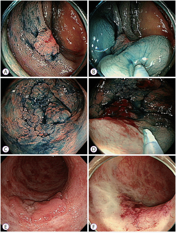
Depression, presence of overlying ulceration, and failure to lift with attempted submucosal injection are suggested as gross features of submucosal invasion in dysplasia [11]. Of these findings, failure to lift with attempted submucosal injection may be often different from the positive non-lifting sign typically observed in deep submucosal invasive CRC in patients without colitis. A classic positive non-lifting sign is originally defined as non-lifting of the lesion with elevated surrounding mucosa after submucosal injection [28]. However, as the background of the lesion often has extensive scar changes, especially in patients with UC, not only the lesion itself, but also the surrounding area may not be lifted (diffuse non-lifting sign; Fig. 1). The diagnostic performance of these three gross features for predicting deep submucosal invasion is still unknown in patients with IBDs and thus needs to be investigated.

TECHNICAL CONSIDERATIONS FOR ESD OF COLITIS-ASSOCIATED DYSPLASIA

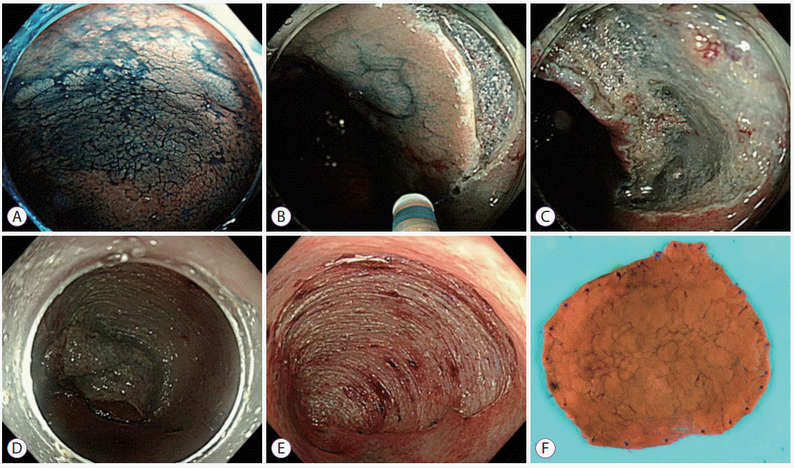
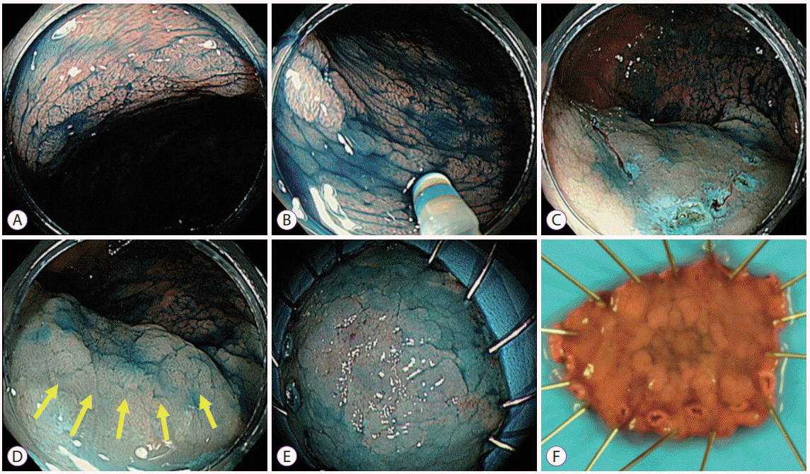
The techniques and equipment for ESD for sporadic CRNs have been described in many publications [29-31], and the basic steps of ESD for sporadic CRNs can also be applied for UC-associated dysplasia (Fig. 2). High-definition endoscope systems and a transparent cap attached to the distal end of the scope are necessary for ESD. Endoknives and electrosurgical units are also essential. Careful observation of the target lesion should be the first step of ESD. Dye chromoendoscopy and/or image-enhanced endoscopy is useful for delineating the margins and characterizing the lesion surfaces. Circumferential diathermic marks before submucosal injection are not generally required during ESD for sporadic CRN [29] because the margins of sporadic CRN are endoscopically distinguishable from the surrounding non-neoplastic mucosa. However, the surrounding mucosa of UC-associated dysplasia frequently shows chronic inflammatory or scar changes [32,33], which the operator may confuse with true dysplasia during a prolonged procedure. Therefore, we recommend making diathermic marks around the margins of dysplasia before injecting a submucosal solution, even if the borders are distinct (Fig. 3). A viscous solution, such as sodium hyaluronate solution, is preferred for submucosal injection, as it can provide a prolonged submucosal cushion [34]. Using an endoknife, a partial mucosal incision should be created at approximately 3–5 mm apart from the delineated margins. Circumferential mucosal incision created at once is not recommended for colorectal ESD because the submucosal solution will leak out easily. Trimming beneath the initial mucosal incision can expose the submucosal layer, and submucosal dissection should be followed. Once the submucosal layer is dissected enough to form a flap or a pocket, the operator can gently press and stretch the submucosal layer using the scope with a transparent cap. With this maneuver, the interface between the proper muscle and submucosal layers is visible via the transparent submucosal layer while submucosal dissection is performed.

HISTOLOGICAL ASSESSMENT OF RESECTED SPECIMENS

The histological features of colitis-associated dysplasia, which are different from those of sporadic colorectal adenoma, can be summarized as follows: unclear delineation between neoplastic and non-neoplastic areas; variety in configuration, size, and diameter of the neoplastic glands; increased infiltration of mononuclear inflammatory cells at the laminar propria; mixtures of the dysplastic and non-dysplastic glands at the lesion surface; and neoplastic proliferation from the base of the gland to the surface [35,36]. These features themselves can confuse the pathologists with true dysplasia and reactive changes and are not actually universally observed in dysplasias. Consequently, the histological diagnosis of dysplasia occasionally shows discrepancies across pathologists, especially between unspecialized and specialized pathologists [37-39]. Current guidelines recommend that the diagnosis of dysplasia should be confirmed by a gastrointestinal (GI) specialist pathologist [9,10].
Similar to the studies on ESD for sporadic CRNs, the currently available studies on ESD for UC-associated dysplasia defined “complete (R0) resection” as en bloc resection with tumor-negative lateral and vertical margins at histology and “curative resection” as R0 resection with superficial (<1,000 mm from the muscularis mucosa) submucosal invasion, no lymphovascular involvement, and good cell differentiation [27,32,33,40]. Therefore, in addition to the grading for dysplasia, both completeness and curativeness should be assessed by a GI specialist pathologist who has expertise in interpreting dysplasia in patients with IBDs.
Conversely, whether biopsies should be obtained from the adjacent mucosa after removing dysplasias is not established yet. Recent studies mostly based on polypoid dysplasia showed that the yield of peri-lesional biopsies ranged from 0.3% to 5% [41-43], and one of them revealed that the diagnostic yield of peri-lesional biopsies from the mucosa not suspicious of dysplasia was 0% [43]. Therefore, peri-lesional biopsies after endoscopic resection of dysplasia may not be mandatory, especially if the peri-lesional mucosa appears normal under endoscopy. However, limited knowledge is currently known regarding the prevalence of invisible or endoscopically unsuspected dysplasia around non-polypoid dysplasia. Therefore, we recommend performing four-quadrant biopsies routinely from the margins of the postprocedural ulceration after ESD, until the yield of peri-lesional biopsies for non-polypoid dysplasias can be determined.

OUTCOMES OF ESD FOR COLITIS-ASSOCIATED DYSPLASIA

Although ESD has been suggested as a resection technique for non-polypoid dysplasia [11,12], its outcomes for non-polypoid dysplasia in patients with colitis were reported in several recent studies. The reported outcomes related to ESD for colitis-associated dysplasia or early CRC include the en bloc and R0 resection rates, curative resection rate, adverse events, local recurrence, and metachronous recurrence (Table 1).

En bloc and complete resection rates

The en bloc and R0 resection rates of ESD for UC-associated dysplasia were reported in several recent studies. Iacopini et al. first reported their experience in ESD for 10 UC-associated dysplasias in nine Italian or Japanese patients [32]. The median size of the lesions was 30 mm (range, 20–50 mm), and the en bloc and R0 resection rates were 80% and 80%, respectively [32]. After this earliest case series, two studies reported the outcomes of ESD for UC-associated dysplasia or early CRC. Suzuki et al. reported the outcome of ESD procedures for 32 lesions (median size = 33 mm) in British or Japanese patients with UC, and their en bloc and R0 resection rates were 97% and 72%, respectively [40]. Around the same time, Kinoshita et al. reported the outcomes of ESD performed in two Japanese referral centers, and the en bloc and R0 resection rates for 25 lesions (mean size=21.6 mm) were 100% and 76%, respectively [27]. According to a very recent study conducted in a single Korean referral center, the en bloc and R0 resection rates for 15 UC-associated dysplasias (median size = 23 mm) were 93.3% and 80%, respectively [33]. These results are comparable with the en bloc resection rates of 89%–92% and complete resection rates of 76%–83% in colorectal ESDs exclusive for sporadic CRNs [44-46]. The curative resection rates of ESD for UC-associated dysplasias ranged from 70% to 80% [27,32,33], which are also comparable with the curative resection rate of ESD for sporadic CRNs (80.9%; 95% confidence interval [CI], 76.0%–85.0%) [46].

Submucosal fibrosis and dissection speed

UC-associated dysplasias may have submucosal fibrosis more frequently than sporadic CRNs, and the incidence of submucosal fibrosis ranged from 66.7% to 100% in previous studies on ESD for dysplasias [32,33,40]. The presence of submucosal fibrosis may slow the dissection speed down, and the median dissection speed of ESD was 0.065 cm2/min in a study reporting a 90% frequency of submucosal fibrosis [32] and 0.083 cm2/min in another study reporting a 97% frequency of submucosal fibrosis [40]. Their dissection speeds seem relatively slower than that of non-granular-type laterally spreading tumor (LST-NG) cases in a previous study on ESD for sporadic CRNs (0.15 cm2/min). However, in a recent Korean study, which reported an incidence of submucosal fibrosis of 66.7%, the dissection speed was 0.13 cm2/min, which is comparable with the dissection speed of ESDs for sporadic LST-NGs [33]. The median procedure time of ESD for dysplasias ranged from 61 to 87 minutes in three studies [32,33,40]. The mean (±standard deviation) procedure time of ESD in another study was 71.7±53.7 minutes [27].

Adverse events

Bleeding and perforation are major complications related to colorectal ESDs, and their incidence during ESD for sporadic CRNs has been reported as 2.7% (95% CI, 2.2%‒3.2%) and 5.2% (95% CI, 4.4%‒6.1%), respectively, in a recent meta-analysis [46]. Among studies on ESD for UC-associated dysplasias, delayed bleeding occurred in two studies, and its incidence was 10% (1/10) in the first study [32] and 3.1% (1/32) in the second study [40]. Conversely, only one study reported a case of perforation (4%, 1/25) [27].

Outcome of surveillance after ESD

The proportion of the patients who underwent colectomy after ESD ranged from 6.7% to 20%, and non-curative resection was the main reason for surgery (Table 2) [27,32,33,40]. The earliest case series reported two cases of local recurrence after ESD for nine dysplasias during a median follow-up duration of 24 months; both of them occurred in the piecemeal resection cases [32]. Metachronous dysplasias developed in three of eight patients (37.5%) during the same follow-up period [32]. Suzuki et al. reported local and metachronous recurrence rates of 3.8% and 11.5%, respectively, during a median follow-up duration of 33 months [40]. Kinoshita et al. reported a local recurrence rate of 0% and a metachronous recurrence rate of 4% during a median follow-up duration of 21 months [27]. However, a single Korean referral center study on ESD for dysplasia with a median follow-up duration of 24.7 months reported that both the local and metachronous recurrence rates were 14.3% [33]. A heterogeneous level of risk for dysplasia or colitic cancer among patients is potentially associated with the wide variation in the metachronous recurrence rates during post-ESD surveillance. Nonetheless, given that the pooled local recurrence rate was 2.0% (95% CI, 1.3%‒3.0%) after ESD for sporadic CRNs in a meta-analysis [46], local recurrence after ESD for UC-associated dysplasias should be regarded more frequent than that after ESD for sporadic CRNs. Additional studies should be conducted to reveal the long-term post-ESD outcomes, such as the recurrence rate and the incidence of post-ESD CRC.

COLECTOMY FOR COLITIS-ASSOCIATED DYSPLASIA

Colectomy is indicated for the treatment of endoscopically unresectable dysplasias in patients with UC [9,11]. However, according to survey-based studies, a considerable proportion of patients with IBDs would refuse colectomy for the treatment of dysplasia [47-49], and the fear of permanent ostomy or complications was the main reason for refusal of surgical treatment [49].
TPC with ileal pouch-anal anastomosis (IPAA) has become a surgical procedure of choice for patients with UC, as the postoperative clinical and functional outcomes were stable during long-term follow-ups [50,51]. However, the mean daytime bowel frequency ranged from 5.7 at 1 year to 6.4 at 20 years [50]. Therefore, patients with UC in remission may not agree with the physician’s recommendation for surgery if they have dysplasia, not cancer. In a Korean multicenter retrospective study [52], 415 patients with UC underwent colon surgery, and most of them (92.2%) underwent TPC. Medical intractability of UC (n=270, 65.1%) was the most common indication of surgery, followed by dysplasia or malignancy (n=52, 12.5%). The overall postoperative complication rate was 34.7%. Ileus (n=21), bleeding (n=16), and anastomotic leakage (n=15) were common early postoperative complications. Pouchitis (n=48) was the most common late complication. In another largescale (n=3,707) study including 2,953 patients with UC and 150 patients with Crohn’s disease, the early and late complication rates in TPC with IPAA were 33.5% and 29.1%, respectively [53]. The authors also reported a perioperative mortality rate of 0.1%.
Therefore, the decision to undergo colectomy for dysplasia should be highly individualized, and the patients’ clinical status, life expectancy, and preferences must be considered [54]. The final decision should be balanced between these factors and the risk of CRC.

FEASIBILITY AND APPROPRIATE CONDITIONS OF ESD FOR COLITIS-ASSOCIATED DYSPLASIA

The procedure-related outcome measures of ESD for colitis-associated dysplasias, such as the en bloc and R0 resection rates and incidence of complications, are comparable with those of ESD for sporadic CRNs. However, such data are based on small studies reported by groups with a high level of expertise [27,32,33,40]. Moreover, the local and metachronous recurrence rates are relatively high, and no long-term outcome data are available yet. Nonetheless, given the functional outcome and risk of complications related to colectomy, ESD can be considered feasible for dysplasia that cannot be removed using the conventional EMR techniques.
The most appropriate conditions for attempting ESD for colitis-associated dysplasia can be suggested as follows (Table 3). First, the peri-lesional mucosa should be in remission endoscopically. Second, the patients’ colitis itself should be medically tractable. Third, any of the endoscopic findings indicating possible invasive cancer should be absent, although the diagnostic performance of invasive pit or vascular patterns, surface ulcerations, and the non-lifting sign has not been determined yet. Fourth, an expert endoscopist highly skilled in both colorectal ESD and surveillance colonoscopy for patients with colitis should assess the lesion and perform ESD. The endoscopists in the currently available studies already had expertise in colorectal ESD before performing ESD for colitis-associated dysplasia [27,32,33,40]. However, the qualification needed for performing ESD of colitis-associated dysplasia among endoscopists is not currently available. A recent Korean study reported that the first ESD for colitis-associated dysplasia had been attempted after approximately 200 cases of ESD experiences [33]. Fifth, a GI pathologist specialized in interpreting the histology of dysplasia should be available. Finally, both an IBD physician and an IBD surgeon should be available to provide proper management after curative or non-curative ESDs, respectively. Therefore, we strongly suggest that ESD for colitis-associated dysplasia be performed by an ESD expert endoscopist in a center with a well-organized IBD team.

CONCLUSIONS

ESD has been suggested as one of the therapeutic options for endoscopically resectable dysplasia in patients with colitis. Recent studies performed by expert centers support the feasibility of ESD for the treatment of colitis-associated dysplasia in terms of the en bloc and R0 resection rates and the risk of procedure-related complications. The relatively high incidence of colectomy-related complications justifies selecting ESD as a primary treatment for endoscopically resectable dysplasia that cannot be effectively treated using the conventional EMR technique. However, the local and metachronous recurrence rates after ESD for colitis-associated dysplasia are relatively high, and long-term therapeutic outcome data are still lacking. In addition, the lesion and patient characteristics suitable for performing ESD are not well established in colitis-associated dysplasia. Therefore, the therapeutic decision for colitis-associated dysplasia, which potentially needs ESD, should be made after individualized and multidisciplinary approach in close collaboration with a multidisciplinary IBD team. The lesion characteristics, patients’ general medical conditions and preferences, and status of colitis must be taken into consideration. After curative ESD, meticulous colonoscopic surveillance is mandatory to monitor for local and metachronous recurrences of dysplasia. Further studies should be conducted to establish the long-term therapeutic outcomes of ESD, such as the incidence of post-ESD CRC and the risk of colectomy after ESD.

Notes

Conflicts of Interest:The authors have no financial conflicts of interest.

REFERENCES

1. Jess T, Rungoe C, Peyrin-Biroulet L. Risk of colorectal cancer in patients with ulcerative colitis: a meta-analysis of population-based cohort studies. Clin Gastroenterol Hepatol. 2012; 10:639–645.
crossref
2. Colman RJ, Rubin DT. Histological inflammation increases the risk of colorectal neoplasia in ulcerative colitis: a systematic review. Intest Res. 2016; 14:202–210.
crossref
3. Zhiqin W, Palaniappan S, Raja Ali RA. Inflammatory bowel disease-related colorectal cancer in the Asia-Pacific region: past, present, and future. Intest Res. 2014; 12:194–204.
crossref
4. Velayos F, Kathpalia P, Finlayson E. Changing paradigms in detection of dysplasia and management of patients with inflammatory bowel disease: is colectomy still necessary? Gastroenterology. 2017; 152:440–450.e1.
5. Rubin PH, Friedman S, Harpaz N, et al. Colonoscopic polypectomy in chronic colitis: conservative management after endoscopic resection of dysplastic polyps. Gastroenterology. 1999; 117:1295–1300.
crossref
6. Engelsgjerd M, Farraye FA, Odze RD. Polypectomy may be adequate treatment for adenoma-like dysplastic lesions in chronic ulcerative colitis. Gastroenterology. 1999; 117:1288–1294. discussion 1488-1491.
crossref
7. Wanders LK, Dekker E, Pullens B, Bassett P, Travis SP, East JE. Cancer risk after resection of polypoid dysplasia in patients with longstanding ulcerative colitis: a meta-analysis. Clin Gastroenterol Hepatol. 2014; 12:756–764.
crossref
8. Mooiweer E, van der Meulen-de Jong AE, Ponsioen CY, et al. Incidence of interval colorectal cancer among inflammatory bowel disease patients undergoing regular colonoscopic surveillance. Clin Gastroenterol Hepatol. 2015; 13:1656–1661.
crossref
9. Annese V, Daperno M, Rutter MD, et al. European evidence based consensus for endoscopy in inflammatory bowel disease. J Crohns Colitis. 2013; 7:982–1018.
crossref
10. Laine L, Kaltenbach T, Barkun A, McQuaid KR, Subramanian V, Soetikno R. SCENIC international consensus statement on surveillance and management of dysplasia in inflammatory bowel disease. Gastroenterology. 2015; 148:639–651.e28.
crossref
11. American Society for Gastrointestinal Endoscopy Standards of Practice Committee, Shergill AK, Lightdale JR, et al. The role of endoscopy in inflammatory bowel disease. Gastrointest Endosc. 2015; 81:1101–1121. e1-e13.
crossref
12. Soetikno R, Subramanian V, Kaltenbach T, et al. The detection of nonpolypoid (flat and depressed) colorectal neoplasms in patients with inflammatory bowel disease. Gastroenterology. 2013; 144:1349–1352. 1352.e1-e6.
crossref
13. Blackstone MO, Riddell RH, Rogers BH, Levin B. Dysplasia-associated lesion or mass (DALM) detected by colonoscopy in long-standing ulcerative colitis: an indication for colectomy. Gastroenterology. 1981; 80:366–374.
crossref
14. Itzkowitz SH, Harpaz N. Diagnosis and management of dysplasia in patients with inflammatory bowel diseases. Gastroenterology. 2004; 126:1634–1648.
crossref
15. Lee SH, Shin SJ, Park DI, et al. Korean guideline for colonoscopic polypectomy. Clin Endosc. 2012; 45:11–24.
crossref
16. Kudo S, Rubio CA, Teixeira CR, Kashida H, Kogure E. Pit pattern in colorectal neoplasia: endoscopic magnifying view. Endoscopy. 2001; 33:367–373.
crossref
17. Kudo S, Lambert R, Allen JI, et al. Nonpolypoid neoplastic lesions of the colorectal mucosa. Gastrointest Endosc. 2008; 68(4 Suppl):S3–S47.
crossref
18. Ikematsu H, Matsuda T, Emura F, et al. Efficacy of capillary pattern type IIIA/IIIB by magnifying narrow band imaging for estimating depth of invasion of early colorectal neoplasms. BMC Gastroenterol. 2010; 10:33.
crossref
19. Sumimoto K, Tanaka S, Shigita K, et al. Diagnostic performance of Japan NBI Expert Team classification for differentiation among noninvasive, superficially invasive, and deeply invasive colorectal neoplasia. Gastrointest Endosc. 2017; 86:700–709.
crossref
20. Backes Y, Moss A, Reitsma JB, Siersema PD, Moons LM. Narrow band imaging, magnifying chromoendoscopy, and gross morphological features for the optical diagnosis of T1 colorectal cancer and deep submucosal invasion: a systematic review and meta-analysis. Am J Gastroenterol. 2017; 112:54–64.
crossref
21. Wada Y, Kashida H, Kudo SE, Misawa M, Ikehara N, Hamatani S. Diagnostic accuracy of pit pattern and vascular pattern analyses in colorectal lesions. Dig Endosc. 2010; 22:192–199.
crossref
22. Li M, Ali SM, Umm-a-OmarahGilani S, Liu J, Li YQ, Zuo XL. Kudo’s pit pattern classification for colorectal neoplasms: a meta-analysis. World J Gastroenterol. 2014; 20:12649–12656.
crossref
23. Hata K, Watanabe T, Motoi T, Nagawa H. Pitfalls of pit pattern diagnosis in ulcerative colitis-associated dysplasia. Gastroenterology. 2004; 126:374–376.
crossref
24. Bisschops R, Bessissow T, Dekker E, et al. Pit pattern analysis with high-definition chromoendoscopy and narrow-band imaging for optical diagnosis of dysplasia in patients with ulcerative colitis. Gastrointest Endosc. 2017; 86:1100–1106.e1.
crossref
25. Carballal S, Maisterra S, López-Serrano A, et al. Real-life chromoendoscopy for neoplasia detection and characterisation in long-standing IBD. Gut. 2018; 67:70–78.
crossref
26. Kiesslich R, Fritsch J, Holtmann M, et al. Methylene blue-aided chromoendoscopy for the detection of intraepithelial neoplasia and colon cancer in ulcerative colitis. Gastroenterology. 2003; 124:880–888.
crossref
27. Kinoshita S, Uraoka T, Nishizawa T, et al. The role of colorectal endoscopic submucosal dissection in patients with ulcerative colitis. Gastrointest Endosc. 2018; 87:1079–1084.
crossref
28. Uno Y, Munakata A. The non-lifting sign of invasive colon cancer. Gastrointest Endosc. 1994; 40:485–489.
crossref
29. Saito Y, Otake Y, Sakamoto T, et al. Indications for and technical aspects of colorectal endoscopic submucosal dissection. Gut Liver. 2013; 7:263–269.
crossref
30. Yang DH, Jeong GH, Song Y, et al. The feasibility of performing colorectal endoscopic submucosal dissection without previous experience in performing gastric endoscopic submucosal dissection. Dig Dis Sci. 2015; 60:3431–3441.
crossref
31. Toyonaga T, Nishino E, Man IM, East JE, Azuma T. Principles of quality controlled endoscopic submucosal dissection with appropriate dissection level and high quality resected specimen. Clin Endosc. 2012; 45:362–374.
crossref
32. Iacopini F, Saito Y, Yamada M, et al. Curative endoscopic submucosal dissection of large nonpolypoid superficial neoplasms in ulcerative colitis (with videos). Gastrointest Endosc. 2015; 82:734–738.
crossref
33. Yang DH, Kim J, Song EM, et al. Outcomes of ulcerative colitis-associated dysplasia patients referred for potential endoscopic submucosal dissection. J Gastroenterol Hepatol. 2019 Feb 6 [Epub]. https://doi.org/10.1111/jgh.14623.
crossref
34. Fujishiro M, Yahagi N, Kashimura K, et al. Comparison of various submucosal injection solutions for maintaining mucosal elevation during endoscopic mucosal resection. Endoscopy. 2004; 36:579–583.
crossref
35. Vieth M, Neumann H. Current issues in inflammatory bowel disease neoplasia. Histopathology. 2015; 66:37–48.
crossref
36. Langner C, Magro F, Driessen A, et al. The histopathological approach to inflammatory bowel disease: a practice guide. Virchows Arch. 2014; 464:511–527.
crossref
37. Allende D, Elmessiry M, Hao W, et al. Inter-observer and intra-observer variability in the diagnosis of dysplasia in patients with inflammatory bowel disease: correlation of pathological and endoscopic findings. Colorectal Dis. 2014; 16:710–718. discussion 718.
crossref
38. Eaden J, Abrams K, McKay H, Denley H, Mayberry J. Inter-observer variation between general and specialist gastrointestinal pathologists when grading dysplasia in ulcerative colitis. J Pathol. 2001; 194:152–157.
crossref
39. Odze RD, Goldblum J, Noffsinger A, Alsaigh N, Rybicki LA, Fogt F. Interobserver variability in the diagnosis of ulcerative colitis-associated dysplasia by telepathology. Mod Pathol. 2002; 15:379–386.
crossref
40. Suzuki N, Toyonaga T, East JE. Endoscopic submucosal dissection of colitis-related dysplasia. Endoscopy. 2017; 49:1237–1242.
crossref
41. Ten Hove JR, Mooiweer E, Dekker E, et al. Low rate of dysplasia detection in mucosa surrounding dysplastic lesions in patients undergoing surveillance for inflammatory bowel diseases. Clin Gastroenterol Hepatol. 2017; 15:222–228.e2.
crossref
42. Krugliak Cleveland N, Huo D, Sadiq F, et al. Assessment of peri-polyp biopsy specimens of flat mucosa in patients with inflammatory bowel disease. Gastrointest Endosc. 2018; 87:1304–1309.
crossref
43. Lahiff C, Mun Wang L, Travis SPL, East JE. Diagnostic yield of dysplasia in polyp-adjacent biopsies for patients with inflammatory bowel disease: a cross-sectional study. J Crohns Colitis. 2018; 12:670–676.
crossref
44. Patel N, Patel K, Ashrafian H, Athanasiou T, Darzi A, Teare J. Colorectal endoscopic submucosal dissection: systematic review of mid-term clinical outcomes. Dig Endosc. 2016; 28:405–416.
crossref
45. Akintoye E, Kumar N, Aihara H, Nas H, Thompson CC. Colorectal endoscopic submucosal dissection: a systematic review and meta-analysis. Endosc Int Open. 2016; 4:E1030–E1044.
crossref
46. Fuccio L, Hassan C, Ponchon T, et al. Clinical outcomes after endoscopic submucosal dissection for colorectal neoplasia: a systematic review and meta-analysis. Gastrointest Endosc. 2017; 86:74–86.e17.
crossref
47. Baars JE, Siegel CA, van’t Spijker A, Markus T, Kuipers EJ, van der Woude CJ. Inflammatory bowel disease-patients are insufficiently educated about the basic characteristics of their disease and the associated risk of colorectal cancer. Dig Liver Dis. 2010; 42:777–784.
crossref
48. Siegel CA, Schwartz LM, Woloshin S, et al. When should ulcerative colitis patients undergo colectomy for dysplasia? Mismatch between patient preferences and physician recommendations. Inflamm Bowel Dis. 2010; 16:1658–1662.
crossref
49. Lopez A, Collet-Fenetrier B, Belle A, Peyrin-Biroulet L. Patients’ knowledge and fear of colorectal cancer risk in inflammatory bowel disease. J Dig Dis. 2016; 17:383–391.
crossref
50. Hahnloser D, Pemberton JH, Wolff BG, Larson DR, Crownhart BS, Dozois RR. Results at up to 20 years after ileal pouch-anal anastomosis for chronic ulcerative colitis. Br J Surg. 2007; 94:333–340.
crossref
51. Heikens JT, de Vries J, Goos MR, Oostvogel HJ, Gooszen HG, van Laarhoven CJ. Quality of life and health status before and after ileal pouchanal anastomosis for ulcerative colitis. Br J Surg. 2012; 99:263–269.
crossref
52. Yoon YS, Cho YB, Park KJ, et al. Surgical outcomes of Korean ulcerative colitis patients with and without colitis-associated cancer. World J Gastroenterol. 2015; 21:3547–3553.
crossref
53. Fazio VW, Kiran RP, Remzi FH, et al. Ileal pouch anal anastomosis: analysis of outcome and quality of life in 3707 patients. Ann Surg. 2013; 257:679–685.
54. Beaugerie L, Itzkowitz SH. Cancers complicating inflammatory bowel disease. N Engl J Med. 2015; 372:1441–1452.
crossref

Fig. 1.
(A, B) A classic non-lifting sign was observed in sporadic deep submucosal invasive cancer. The lesion was not lifted; however, the surrounding mucosa was elevated after submucosal injection. The final histology of this lesion was SM3 adenocarcinoma. (C, D) A classic non-lifting sign was observed in colitis-associated dysplasia. The dysplasia itself was not lifted; however, the surrounding mucosa was lifted after submucosal injection. Its colectomy specimen revealed an adenocarcinoma invading the proper muscle (T2 stage). (E, F) A diffuse non-lifting sign was observed in colitis-associated dysplasia located on the extensive scar changes. Neither the dysplasia nor the surrounding mucosa was lifted after submucosal injection. The final histology of this lesion was lowgrade dysplasia.
ce-2019-047f1.tif
Fig. 2.
Representative example of endoscopic submucosal dissection for colitis-associated dysplasia. (A) Large, non-ulcerated Paris type O-IIa dysplasia with a distinct border in the rectum. (B) Mucosal incision was performed after submucosal injection. (C) Mild but diffuse submucosal fibrosis and submucosal fat deposition. (D-F) The colonoscope was changed into a gastroscope to expose the submucosal layer more effectively, and en bloc resection was achieved. The final histology revealed low-grade dysplasia (42×40 mm in size, with clear lateral and vertical margins).
ce-2019-047f2.tif
Fig. 3.
(A, B) Diathermic marks were made; the border of the dysplasia was clearly distinguishable under dye chromoendoscopy. (C, D) After submucosal injection, the border of the lesion became less distinguishable. (E, F) Endoscopic submucosal dissection was performed along the diathermic marks, and en bloc complete resection was possible. The final histology was low-grade dysplasia (15×10 mm in size, with clear lateral and vertical margins).
ce-2019-047f3.tif
Table 1.
Characteristics of the Lesions and Procedure-Related Outcomes of the Studies for the Endoscopic Submucosal Dissection of the Dysplasia or Early Colorectal Cancer in the Patients having Ulcerative Colitis
Study Number of cases Size of the lesions, mm, median (range) or mean±SD Submucosal fibrosis ESD time, minutes, median (range) or mean±SD En bloc resection R0 resection Delayed bleeding Perforation
Iacopini et al. (2015) [32] 10 15 (10–20) 90% 63 (45–130) 80% 80% 10% (1/10) 0
Suzuki et al. (2017) [40] 32 33 (12–73) 97% 87 (6–290) 97% 79% 3.1% (1/32) 0
Kinoshita et al. (2018) [27] 25 21.6±12.8 100% 71.7±53.7 100% 76% 0 4% (1/25)
Yang et al. (2019) [33] 15 23 (12–48) 66.7% 61 (12–160) 93.3% 80% 0 0

ESD, endoscopic submucosal dissection; SD, standard deviation.

Table 2.
The Therapeutic Outcomes of Endoscopic Submucosal Dissection for the Colitis-Associated Dysplasia
Study Rate and reason of colectomy after ESD Local recurrence Metachronous recurrence Follow-up, mo, median (range)
Iacopini et al. (2015) [32] 10% (1/10) 22.2% (2/9) 37.5% (3/8) 24 (6–72)
 T1 cancer with vascular invasion (n=1)
Suzuki et al. (2017) [40] 12.5% (4/32) 3.8% (1/26) 11.5% (3/26) 33 (6–76)
 T1 cancer (n=2)
 Invisible dysplasia (n=1)
 Patient’s preference for treating visible dysplasia (n=1)
Kinoshita et al. (2018) [27] 20% (5/25) 0 5% (1/20) 21 (8–80)
 T1 cancer with non-curative resection (n=5)
Yang et al. (2019) [33] 6.7% (1/15) 14.3% (2/14) 14.3% (2/14) 24.7 (5.2–64.8)
 Missed synchronous T1 cancer detected during ESD (n=1)

ESD, endoscopic submucosal dissection.

Table 3.
Suggestion for the Required Conditions for Attempting Endoscopic Submucosal Dissection for Colitis-Associated Dysplasia
Suggested requirements
Colitis Peri-lesional mucosa should be in endoscopic remission (Mayo subscore 0–1 or UCEIS 0–2)
The coliits should be medically tractable
The patient should be at least in clinical remission and preferably in endoscopic and histologic remission
Dysplasia Should satisfy all of the following conditions
 Distinct border
 No surface ulceration
 Absence of non-lifting sign which precludes any further submucosal dissection
 Absence of endoscopic features suggesting invasive cancer
Endoscopist Should be highly-skilled in colorectal ESD
Should have expertise in the surveillance colonoscopy for effective monitoring after ESD
Pathologist Should have expertise in the histologic interpretation of the dysplasia in the colitic patients
IBD physician Should be available to provide the proper management and surveillance after curative ESD
IBD surgeon Should be available to provide the proper management after non-curative ESD

ESD, endoscopic submucosal dissection; IBD, inflammatory bowel disease; UCEIS, ulcerative colitis endoscopic index of severity.

TOOLS
Similar articles