

 PDF
PDF Citation
 Print
Print


INTRODUCTION
Colorectal cancer is a major cause of cancer deaths throughout the world, with an increasing incidence [1]. Despite significant advances in the treatment of colorectal cancer, the main strategy for the management of this disease is still early detection and resection. One of the major roles of colonoscopy is the management of colorectal cancer, including both diagnosis and treatment, and colorectal polypectomy decreases mortality [2].
Advanced endoscopic resection techniques for the treatment of superficial neoplasms, such as snare polypectomy, endoscopic mucosal resection (EMR), and endoscopic submucosal dissection (ESD) are now available as minimally invasive curative treatments for many patients with early-stage colorectal cancer. Accurate pretreatment diagnosis using high-definition magnifying endoscopy with image-enhanced endoscopy, such as narrow-band imaging (NBI) and blue light imaging (BLI) is available for the selection of the most appropriate treatment strategy and determining the line of resection prior to endoscopic resection. However, these advancements in endoscopy can be applied only when neoplastic lesions are detected at an early stage.
Go to : 
ENDOSCOPIC RESECTION OF SUPERFICIAL COLORECTAL NEOPLASM
Curative endoscopic resection of superficial tumors is limited to mucosal lesions and tumors with shallow submucosal invasion (<1,000 μm). The quality of endoscopic resection is determined by careful evaluation of the specimen after resection. An en bloc resection with negative margins is always expected to achieve a curative resection. Although most local recurrences due to positive lateral margins can be managed by additional endoscopic resection during follow-up colonoscopy, a positive vertical margin spoils the clinical significance of endoscopic resection itself. To obtain a curative resection, an R0 resection (en bloc resection with negative margins), no lymphovascular invasion, budding grade 1, and no deep submucosal invasion (>1,000 μm) are required [3]. Notably, the presence or absence of these factors is assessed with pathological evaluation of the submucosal layer to determine the necessity for additional surgical intervention. Therefore, the resected specimen ideally contains a thick submucosal layer. A large superficial tumor is usually treated with ESD. To achieve a high-quality ESD, an en bloc specimen with a thick submucosal layer is required.
Go to :
THE POCKET-CREATION METHOD
To accomplish an efficacious and safe ESD throughout the alimentary tract, we developed the pocket-creation method (PCM; Fig. 1), which is routinely used for gastric, colorectal, and duodenal ESD in our institution [4-6]. The standard approach is antegrade rather than retroflexed maneuvering. A key element of the PCM is a submucosal pocket creation with a minimal mucosal incision. Following the minimal mucosal incision, a circumferential incision is not made; however, a submucosal dissection is performed to create a pocket. The tip of the endoscope enters the pocket through the minimally incised mucosa. After entering the pocket, prudent dissection by identifying the deep submucosal layer is repeated to extend the pocket. The PCM facilitates selection of the exact plane of dissection and avoids dissecting at a superficial submucosal level that has plentiful blood vessels and adipose tissue. Dissection of the deep submucosal layer is a key element to obtain a good pathological specimen with a thick submucosal layer. During the procedure, additional injection of sodium hyaluronate just above the muscularis provides clear visualization of the muscularis through the endoscope hood. The volume of intraluminal gas is minimized to stabilize the maneuvering of the endoscope. During the last stage of the PCM, the pocket is opened after the complete dissection under the tumor. Consideration of the direction of gravity and a step-by-step mucosal incision with opening of the pocket from the inside to the luminal side should be kept in mind to avoid making the procedure difficult.
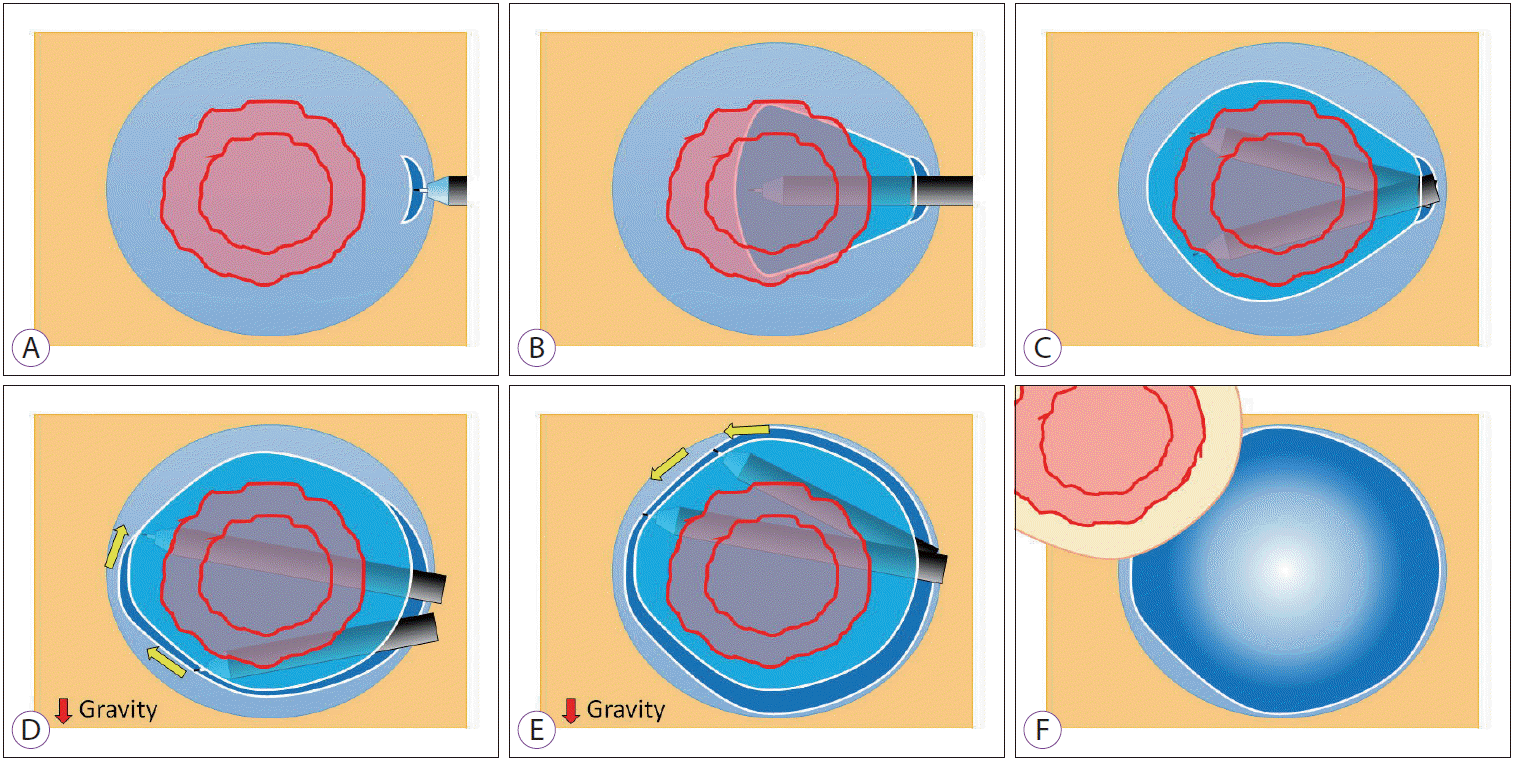
The PCM has four advantages as follows: (1) the injected solution is not dispersed because of a minimal incision; (2) both traction and countertraction are simultaneously obtained because the transparent hood stretches the submucosal tissue in the limited space; (3) a vertical approach toward the muscularis can be changed to a horizontal approach; and (4) the influence of cardiopulmonary movement is less because of the synchronization between the endoscope and the pocket (Fig. 2). Unlike various techniques that require special devices such as clip-and-line, S-O clip, and external forceps to obtain traction [7-9], the PCM does not require any special devices except a transparent hood and electrosurgical knives. We use a small-caliber-tip transparent hood to obtain clear visualization in the pocket [10]. Use of sodium hyaluronate is recommended to maintain a long-lasting submucosal cushion [6]. The PCM can be performed as far as the endoscope reaches because it does not require special devices. Therefore, it can be applied for the ESD of duodenal lesions, which has been considered the most difficult ESD procedure [5]. The PCM is not only useful for conquering difficulties such as duodenal ESD or a vertical approach but also for resecting routine lesions more easily using this universally applicable strategy.
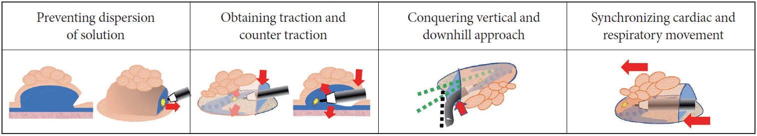 | Fig. 2.Advantages of the pocket-creation method (PCM). Minimal mucosal incision prevents dispersion of injected solution resulting in long-lasting maintenance of mucosal elevation. In the pocket, both traction and countertraction are obtained, and the tip of the endoscope is stabilized. Even in difficult locations, PCM enables stable submucosal dissection. A stabilized endoscope in the pocket synchronizes cardiac and respiratory movement.
|
Go to :
COLD SNARE POLYPECTOMY
Cold snare polypectomy (CSP) has been widely disseminated to resect sub-centimeter colorectal polyps. We previously reported the efficacy and safety of CSP in comparison with conventional polypectomy, the so-called hot snare polypectomy (HSP). CSP significantly shortens the procedure time and shows a trend to decrease delayed bleeding as compared with HSP [11]. CSP enables omission of HSP-specific procedures, such as placing a disposable electrode on the patient and performing electrocauterization at the time of polypectomy. Furthermore, CSP does not use cauterization that may extend areas of ulceration and injure submucosal arteries at the polypectomy site [12,13].
Go to :
EARLY DETECTION AND RESECTION
By the early twenty-first century, minimally invasive resection methods, such as CSP, EMR, and ESD have been all widely used [12]. The importance of early detection of gastrointestinal tract tumors has been emphasized over the last 20 years because minimally invasive endoscopic resection can only be used for premalignant or early-stage malignancies. Therefore, early detection is the most important first step to maximize the usefulness of recent advances in endoscopic resection techniques. However, the detection of small, flat-shaped, or faded-color lesions remains difficult. New image-enhanced endoscopic techniques facilitate the recognition of slight differences by enhancing the color contrast in a bright field of view.
Go to :
CHARACTERISTICS OF LINKED-COLOR IMAGING
Linked color imaging (LCI) is a unique image-enhanced endoscopic technique that uses a novel multi-light technology developed by Fujifilm Company (Tokyo, Japan). LCI is available for both the laser endoscopic system (LASEREO) and the light-emitting diode endoscopic system (ELUXEO). LCI has the potential to increase the detection rate of adenomas and early colorectal cancers by enhancing the color differences between the neoplastic mucosa and the surrounding normal mucosa. As with previously developed technologies such as NBI and BLI, LCI enhances an image using pre-processing of narrow-band light irradiation followed by post-imaging signal processing. In the post-processing phase, acquired images are separated into blue, green, and red colors, which are then reallocated to enhance the color differentiation in an appropriate balance. Using this combination of colors, the red and white lesions become redder and whiter in LCI mode, respectively (Fig. 3) [14].
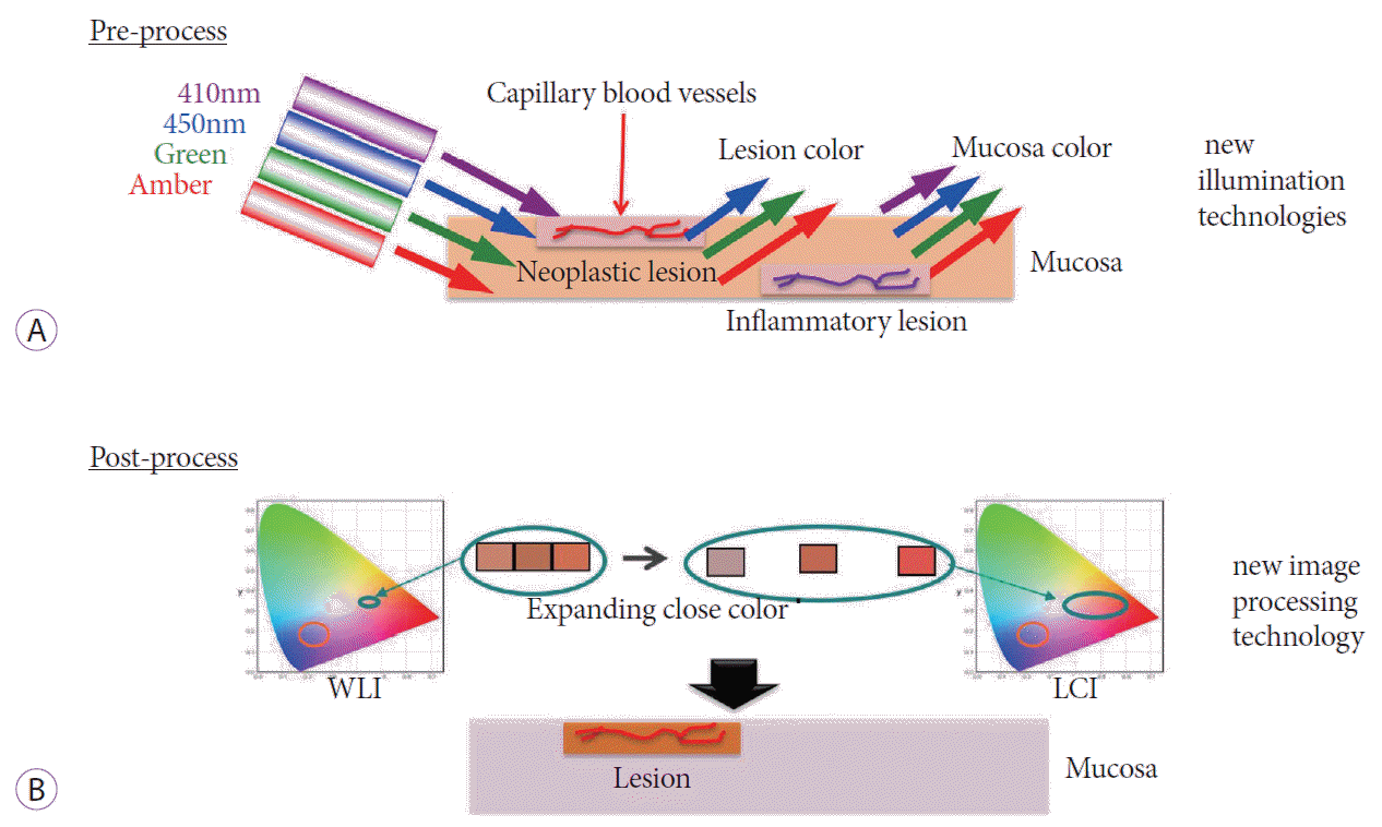 | Fig. 3.(A) Pre-processing illumination technology of linked-color imaging (LCI): 410-nm violet light can penetrate only a short distance from the mucosal surface and is easily absorbed by hemoglobin. Typically, the dilated microvasculature is concentrated in the superficial mucosa in neoplastic lesions and in the deep mucosa in inflammatory lesions. Therefore, the 410-nm violet light is absorbed by neoplastic lesions but is reflected by inflammatory lesions. (B) The post-processing technology of LCI: color contrast is enhanced to make the red and white lesions become redder and whiter, respectively. WLI, white light imaging.
|
Compared with white light imaging (WLI), LCI enhances differences in the red color tone by advanced post-processing steps. The color correction limited to the red region emphasizes the contrast of the red color. The red color leads to an especially improved detection of inflammation with accurate delineation. We reported that LCI facilitates the detection of early gastric cancer by enhancing color contrast [15].
However, the most striking feature of LCI is its ability to display the color of early neoplastic lesions as distinct from inflammatory changes that both have similar “redness” when imaged with WLI. Using LCI, the “redness” in areas of inflammatory change becomes purple, while neoplastic lesions remain red. This feature comes from the effect of 410-nm laser light in LASEREO and the violet light emitting diode in ELUXEO. The 410-nm or violet light can penetrate only a short distance from the mucosal surface and is specifically absorbed by hemoglobin. In neoplastic lesions, dilated microvasculature is concentrated in the superficial mucosa, and 410-nm violet light is absorbed by hemoglobin in the concentrated vasculature (Fig. 3). Therefore, neoplastic areas of the mucosa look red on LCI imaging. In inflammatory mucosa, the dilated microvasculature is concentrated in the deep mucosa, and 410-nm violet light does not reach the vasculature and is reflected by the superficial layer without absorption (Fig. 3). Therefore, the mucosa in areas of inflammation looks violet on LCI imaging. Both neoplastic lesions and inflammatory mucosa have a similar level of “redness” when imaged with WLI. Consequently, LCI can distinguish neoplastic lesions from inflammatory mucosa. LCI provides good color contrast between neoplastic lesions and the surrounding mucosa by producing a bright and natural image, which is suitable for the detection of flat neoplastic lesions at a distant view.
Go to :
EARLY DETECTION OF COLORECTAL SUPERFICIAL LESION WITH LCI
The miss rate for colorectal polyps is a great concern, even for well-trained colonoscopists. Heresbach et al. reported that a miss rate for colorectal polyps of 9%–28% was frequently associated with lesions in the left colon and sessile- or flat-shaped polyps [16]. Leufkens et al. also reported that a miss rate for colorectal polyps of 25% was significantly more frequently associated with adenomas in the right colon than in the left colon [17]. Singh et al. reported that the mortality rate in patients with proximal colon cancer was not decreased with colonoscopy [18]. Therefore, current routine colonoscopy may not be sufficiently sensitive to decrease the mortality of patients with right colon cancer, and colonoscopists should make every effort to increase the polyp detection rate in the right colon.
To improve the polyp detection rate in the right colon, some novel endoscopic equipment and add-on devices have been developed, including a transparent hood, balloon colonoscopy, wide-angle colonoscopy, and third-eye colonoscopy. Matsushita et al. reported that use of a transparent hood provided a 15% additional rate of polyp detection as compared with not using it [19]. Use of a balloon colonoscope (G-EYE endoscope; Smart Medical Systems, Ra’anana, Israel) had a significantly lower adenoma miss rate than conventional colonoscopy (7.5 % vs. 44.7 %, p<0.001) [20]. A recent randomized trial reported that full-spectrum endoscopy had a significantly lower adenoma miss rate than standard forward-viewing colonoscopy (7 % vs. 41 %, p<0.001) [21]. A prospective, multicenter, randomized-controlled trial reported that third-eye colonoscopy increased the adenoma detection rate by expanding the visual field behind the folds [22]. Despite these greatly improved polyp detection rates, special endoscopic equipment or other add-on devices are required to use these techniques.
NBI and BLI are greatly useful to investigate known lesions by clearly visualizing their architecture and vascular pattern. However, these technologies may not be appropriate for screening endoscopy from a distant view because of insufficient light intensity to detect small or flat lesions in the stomach or colon, which have wide lumens [23]. The color tones produced by NBI and BLI are quite different from WLI. Therefore, these narrow-band technologies are unlikely to be used from the beginning of observation, without WLI.
By contrast, the color tone produced by LCI is similar to white light, and LCI is as bright as or even brighter than WLI. Therefore, LCI can be used from the beginning of observation without needing to also use WLI. To increase the polyp detection rate, LCI should be used from the beginning before detecting suspicious lesions with other modalities. LCI is expected to have the potential to improve colorectal polyp detection rates, especially in the right colon and flat-shaped polyps. Fujimoto et al. analyzed the color differences among WLI, BLI, BLI-bright, and LCI using still endoscopic images containing a sessile serrated adenoma/polyp [24]. Polyp detection and color differences were highest when using LCI, compared with the other image-enhanced technologies [24]. A clearer visualization of the vascular network when using LCI allows endoscopists to avoid damaging submucosal vessels with resultant bleeding by selecting a less vascularized area during submucosal injection [25]. A multicenter, randomized study from China reported a higher polyp detection rate with LCI colonoscopy than with WLI colonoscopy (91% vs. 73%, p<0.001) [26]. We previously reported a patient with a laterally spreading tumor of the rectum that was clearly visualized as a bright red area on LCI, even at a distant view [14]. A study from Japan using endoscopic images reported that the visibility score of colorectal non-granular flat tumors imaged with LCI was significantly higher than those imaged using WLI or BLI-bright [27]. A study conducted using recorded video images reported that the polyp visibility scores when using LCI were significantly higher than those when using WLI for both expert and non-expert endoscopists [28]. Screening colonoscopy with LCI improves the detection rate of small, flat-shaped, and/or faded-color colorectal lesions by enhancing the color contrast in a bright field of view.
Chromoendoscopy is a viable option to detect flat or small lesions using WLI at a distant view, but scattering indigo-carmine dye through the working channel is itself a time-consuming procedure. Unlike applying indigo-carmine dye, using the LCI mode requires just the push of a button. Taken together, BLI is effective for characterizing lesions with magnification, and LCI is good for early detection of small, flat, or faded-color neoplastic lesions. LCI can detect smaller colorectal polyps than WLI, which may increase the use of CSP and decrease the need for ESD or surgical resection (Figs. 4-8).
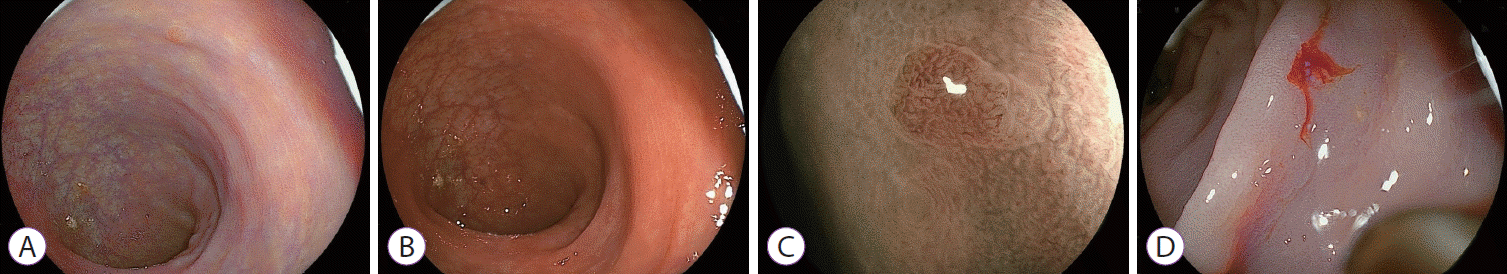
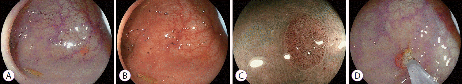 | Fig. 4.small sessile sigmoid colon polyp. (A) Easily detected by linked-color imaging. (B) Difficult detection by white-light imaging. (C) Blue laser imaging scan with magnification shows meshed capillaries compatible with a benign neoplastic lesion (JNET type 2A). (D) Cold-snare polypectomy.
|
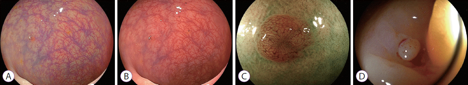
 | Fig. 5.A small sessile sigmoid colon polyp. (A) Easy detection by linked-color imaging. (B) Difficult detection by white-light imaging. (C) Blue laser imaging scan with magnification shows meshed capillaries (JNET type 2A). (D) Cold-snare polypectomy.
|
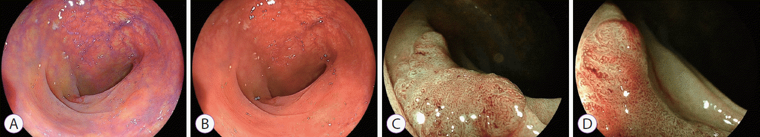
 | Fig. 6.A small sessile rectal polyp. (A) Easy detection by linked-color imaging. (B) Difficult detection by white-light imaging. (C) Blue laser imaging scan with magnification shows meshed capillaries (JNET type 2A). (D) Cold-snare polypectomy.
|
 | Fig. 7.Early colon cancer in the transverse colon. (A) Detectable using linked-color imaging. (B) Difficult detection by white light imaging. (C, D) Blue laser imaging scan with magnification shows irregular capillaries and an irregular surface pattern (JNET type 2B) that suggests superficial carcinoma.
|
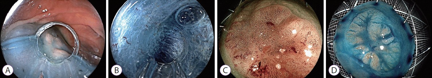 | Fig. 8.Endoscopic submucosal dissection of a laterally spreading tumor of non-granular pseudo-depressed type (the lesion in Fig. 7), using the pocket-creation method. (A) Mucosal elevation using sodium hyaluronate. (B) Submucosal dissection in the pocket. (C) Blue laser imaging scan of the resected specimen shows irregular capillaries (JNET type 2B). (D) Chromoendoscopy image with indigocarmine dye spray of the resected specimen shows en bloc resection. Pathology: Well-differentiated tubular adenocarcinoma, 25×21 mm in size, and intramucosal cancer with no lymphatic and venous invasions, and negative resection margins. A complete en bloc resection.
|
Go to :
CONCLUSIONS
Endoscopic therapeutic procedures have progressed considerably. We can now select appropriate endoscopic resection techniques such as CSP, HSP, EMR, and ESD according to the size and nature of the neoplastic lesions. To make the most of these therapeutic techniques, endoscopic diagnosis has become more important than before. Of all the diagnostic procedures, detection is the most important first step.
LCI is a promising technology that has the potential to increase the adenoma detection rate during colonoscopy. LCI can enhance the color contrast of early colorectal neoplasms compared with the surrounding normal mucosa to improve the detection rate. To increase the detection rate, LCI should be used from the outset for endoscopic observation. Early detection leads to minimally invasive treatment such as CSP and ESD. The PCM facilitates colorectal ESD regardless of size, shape, and location, and consequently achieves a high success rate of complete en bloc resection. Early detection of superficial colorectal tumors and appropriate selection of endoscopic resection technique can result in decreasing colorectal cancer death rates and maintaining a good quality of life for patients.
Go to :
 XML Download