Introduction
Case report
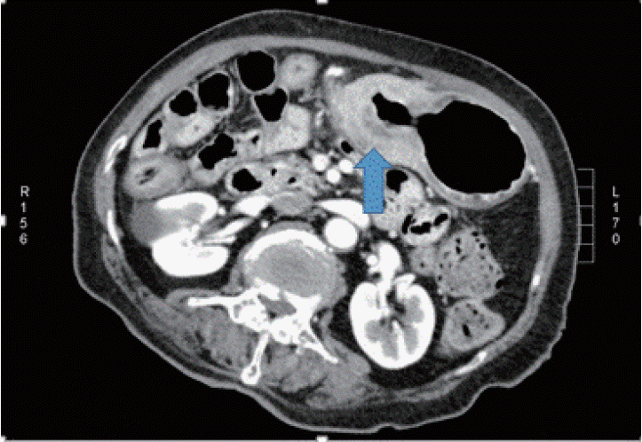 | Fig. 1.Abdominal computed tomography showing diffuse thickening of the gastric wall extending from the body to the antrum (arrow). |
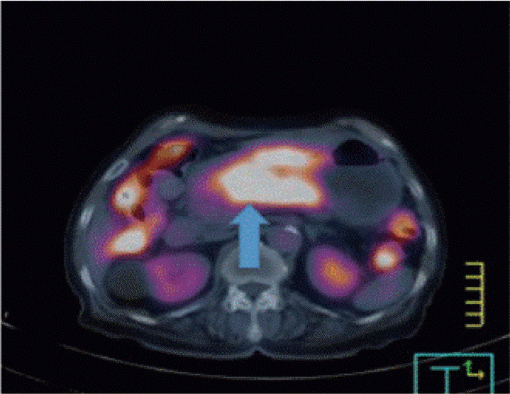 | Fig. 2.Positron emission tomography-computed tomography of the chest, abdomen, and pelvis showing fluorodeoxyglucose uptake along the gastric antrum and body (arrow), with a maximum standardized uptake value of 7.76. |
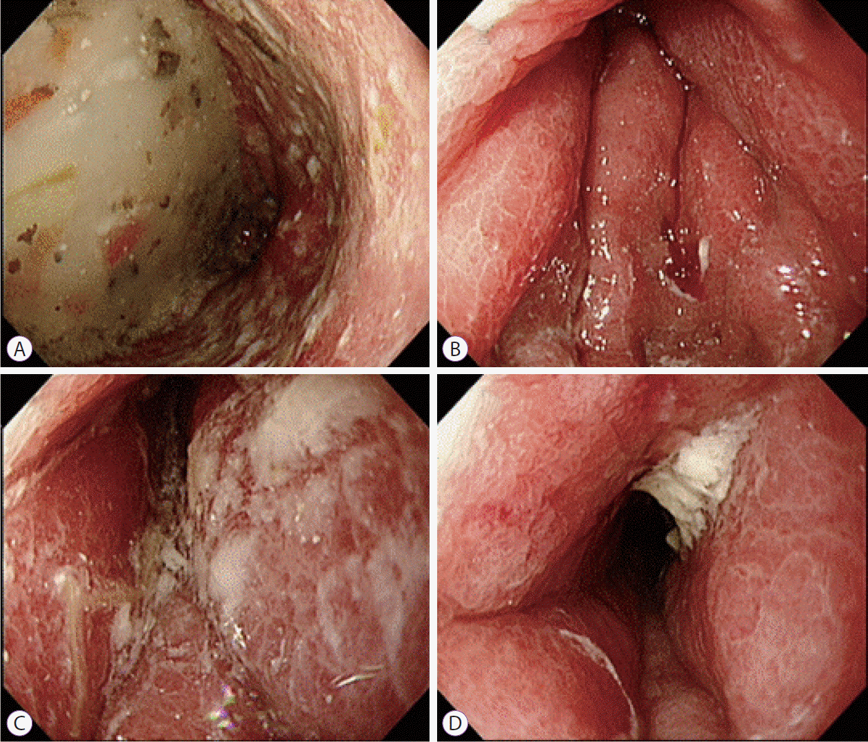 | Fig. 3.Esophagogastroduodenoscopy showing diffuse circumferential thickening and poor distension of the gastric walls from the antrum to the gastric body. |
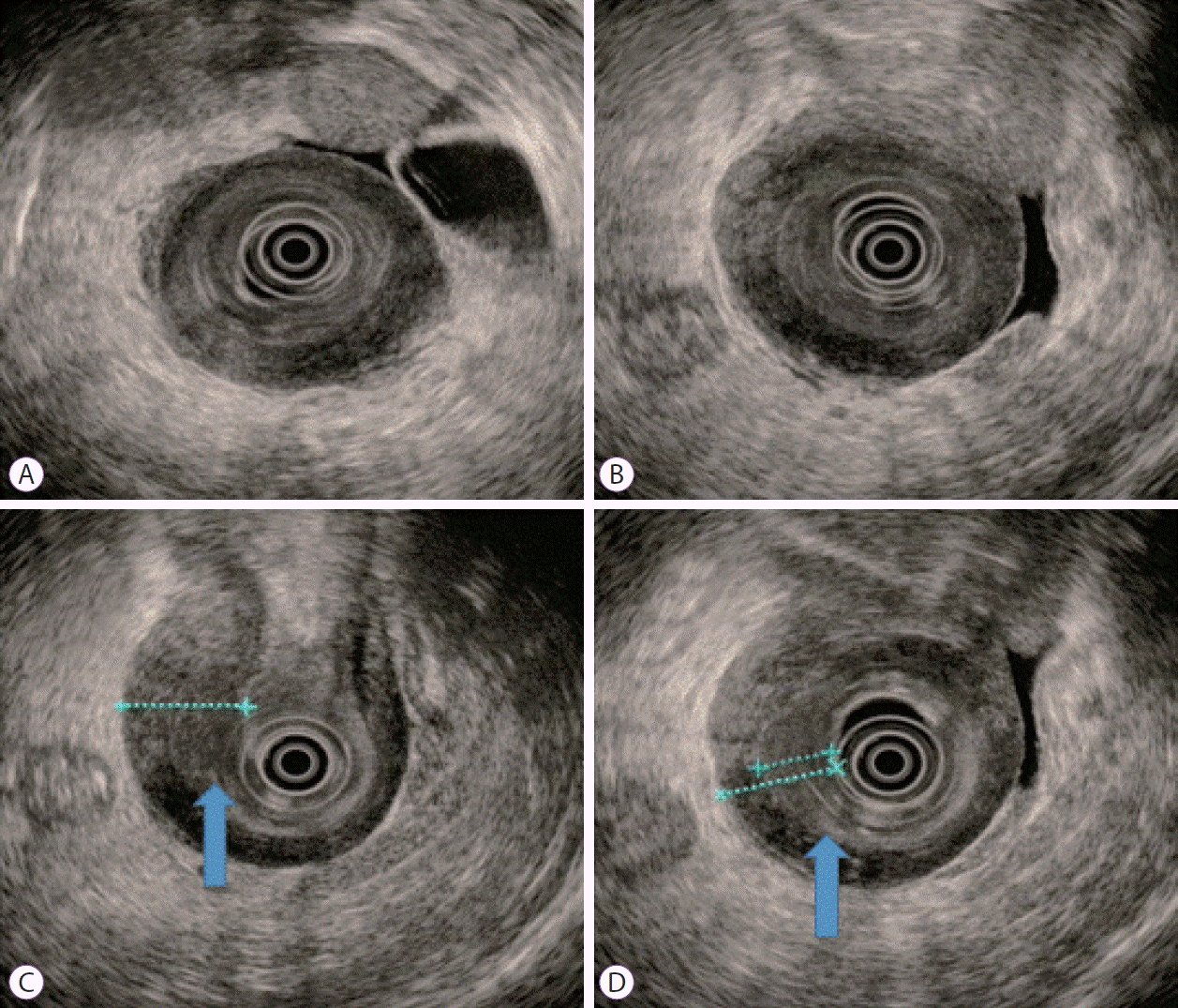 | Fig. 4.Conventional endoscopic ultrasound (EUS) images. A gastric infiltrating tumor diagnosed using EUS (A, B). EUS showing homogeneous, slightly hypoechogenic thickening of the submucosal layer (12 mm) and intact muscularis propria layer shows reactive wall thickening (C, arrow). The thinnest part of the mucosal lesion was selected for the biopsy after EUS localization (D, arrow). |
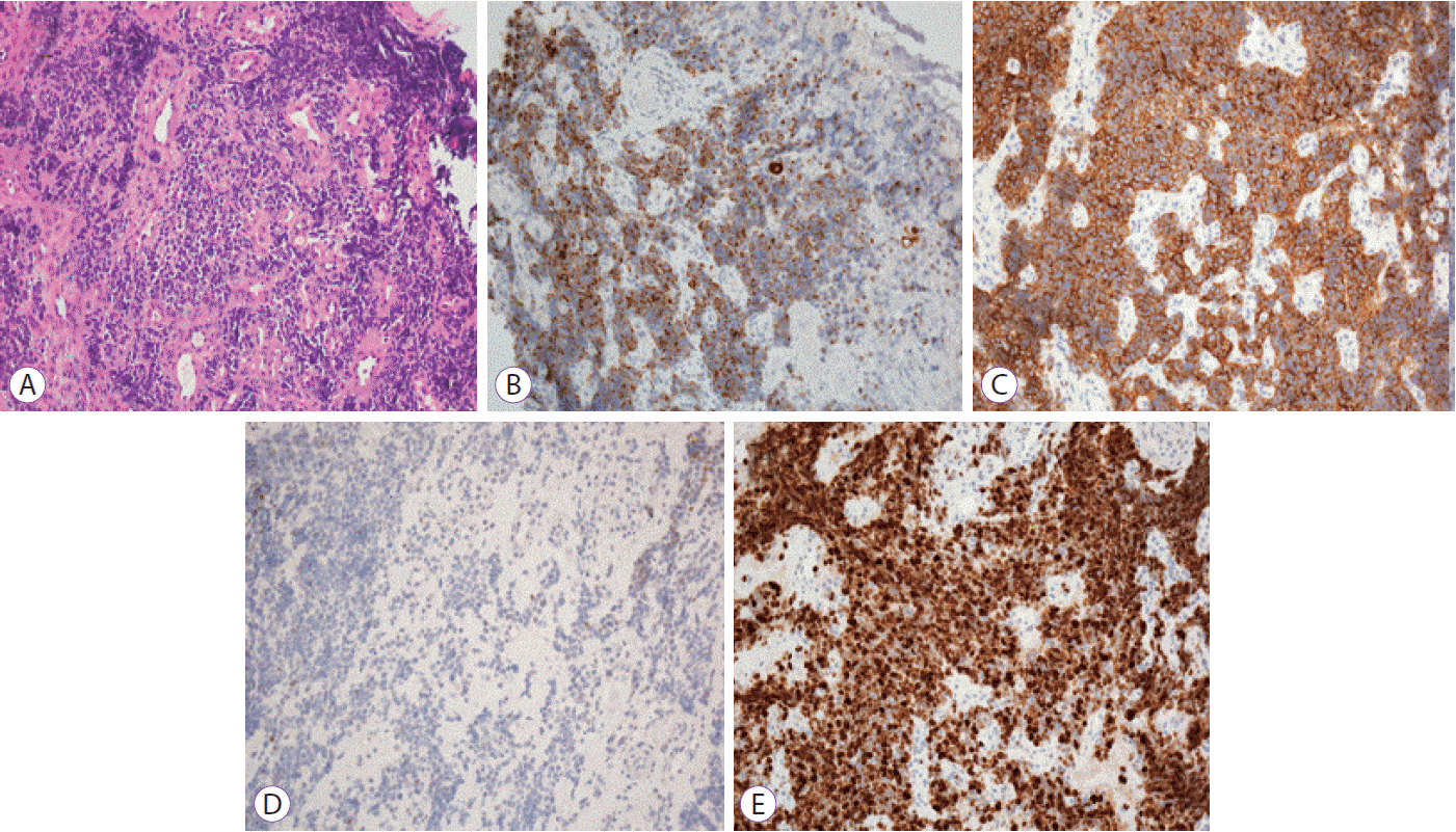 | Fig. 5.Microscopic findings of the gastric tissue. Hematoxylin and eosin stained biopsy specimens show small tumor cells with irregular nuclei and scant cytoplasm (A). Tumor cells stained positive for cytokeratin (B) and CD56 (C), while they were negative for thyroid transcription factor-1 staining (D). Detection of a strong Ki-67 reactivity was observed (E). All the images were captured at ×200 magnification. |


 PDF
PDF Citation
 Print
Print


 XML Download