This article has been
cited by other articles in ScienceCentral.
Abstract
Background/Aims
Colorectal polyps are a common cause of lower gastrointestinal bleeding in children. Our aim was to study the causes of isolated lower gastrointestinal bleeding and to analyze the characteristics of the colorectal polyps found in our cohort.
Methods
We retrospectively reviewed colonoscopic procedures performed between 2007 and 2015. Children with isolated lower gastrointestinal bleeding were included in the study.
Results
A total of 185 colonoscopies were performed for isolated lower gastrointestinal bleeding. The median patient age was 8 years, and 77 patients (41.6%) were found to have colonic polyps. Normal colonoscopy findings were observed and acute colitis was detected in 77 (41.6%) and 14 (7.4%) patients, respectively. Single colonic polyps and 2–3 polyps were detected in 73 (94.8%) and 4 (5.2%) patients with polyps, respectively. Of the single polyps, 69 (94.5%) were juvenile polyps, among which 65 (94.2%) were located in the left colon.
Conclusions
Single left-sided juvenile polyps were the most common cause of isolated lower gastrointestinal bleeding in our study. It was rare to find multiple polyps and polyps proximal to the splenic flexure in our cohort. A full colonoscopy is still recommended in all patients in order to properly diagnose the small but significant group of patients with pathologies found proximal to the splenic flexure.
Go to :

Keywords: Colitis, Colonic polyps, Colonoscopy, Juvenile polyps, Rectal bleeding
INTRODUCTION
Lower gastrointestinal bleeding is a common reason for colonoscopy in children [
1]. The common etiologies of lower gastrointestinal bleeding in children include anal fissures, ulcerative colitis, and colonic polyps. Etiologies of painless isolated lower gastrointestinal bleeding with normal bowel movements may be secondary to colonic polyps and Meckel’s diverticulum.
Colonic polyps in children are mainly solitary benign juvenile polyps requiring endoscopic removal to prevent possible sequelae [
1]. The peak age of diagnosis of juvenile polyps is between 2 and 5 years, with a male predominance [
1,
2]. Juvenile polyps represent 70%–80% of colonic polyps found in children, and 60%–70% of them are solitary [
3-
5]. Solitary juvenile polyps tend to be located in the rectosigmoid colon; however, 35% have been reported to be located in the proximal colon [
6-
9].
Much of the information in the literature on colonoscopic findings in lower gastrointestinal bleeding involved children with a board range of complaints. Our aim was to assess the findings on colonoscopies performed for isolated lower gastrointestinal bleeding in otherwise healthy children.
Go to :

MATERIALS AND METHODS
We retrospectively reviewed all colonoscopic procedures performed between October 2007 and October 2015 in patients 0–18 years old at the Institute of Gastroenterology, Nutrition and Liver Diseases, Schneider Children’s Medical Center of Israel.
Only patients with isolated lower gastrointestinal bleeding were included in our study. Children with characteristics of inflammatory bowel disease such as diarrhea, arthritis, aphthous stomatitis, perianal disease, weight loss, or increased serum inflammatory markers were excluded. In addition, patients with a known polyposis syndrome or other underlying diseases that may affect the bowel, such as graft versus host disease, were also excluded. Abdominal pain and constipation (without an apparent anal fissure) were considered nonspecific symptoms and were not excluded.
The recorded information included patient demographics, clinical presentation, laboratory test results, colonoscopy findings, histology, complications, and completeness of the endoscopic procedure. Colonoscopy was considered to be complete if it included an examination of the cecum or terminal ileum. Anemia was defined by age and sex [
10].
Statistical analysis of data was performed using the SPSS 24 software program. Frequency and proportion were used to describe categorical characteristics. Median values and interquartile range (IQR) were used to describe quantitative characteristics. Scale variables were analyzed using a t-test for parametric and the Mann-Whitney test for nonparametric distributed variables. Categorical outcomes were compared using the chi-square test or the Fisher exact test. A p-value of <0.05 was considered statistically significant. The study was approved by the Rabin Medical Center Institutional Review Board (no. 14-0268).
Go to :

RESULTS
A total of 388 colonoscopies for lower gastrointestinal bleeding were evaluated. Of them, 203 colonoscopies were excluded from the study because of other complaints in addition to lower gastrointestinal bleeding. These complaints included a history of chronic disease (80) including inflammatory bowel disease and a post-organ transplant status; intermittent diarrhea presenting with hypoalbuminemia, anemia, and elevated inflammatory markers (83); intermittent diarrhea with hypoalbuminemia (20); perianal disease (2); arthritis (2); weight loss and anemia (8); constipation with anal fissures (7); and Blue rubber bleb nevus syndrome (1). A total of 185 colonoscopies met our criteria for isolated lower gastrointestinal bleeding during the study period. All of these were complete colonoscopies. The median patient age was 8 years (IQR, 4.9–12.5 years), and 88 (47.6%) were female patients.
A total of 77 of 185 patients (41.6%) with isolated lower gastrointestinal bleeding were found to have colonic polyps. Normal colonoscopy findings, acute (transient) colitis, and ulcerative colitis were observed in 77 (41.6%), 14 (7.4%), and 6 (3.2%) patients, respectively. Other infrequent findings are summarized in
Table 1.
Table 1.
Infrequent Colonoscopic Findings in Our Cohort
|
Finding |
Number (%) |
|
Hemorrhoids |
2 (1%) |
|
Perianal fissures |
3 (1.6%) |
|
Reactive lymphoid hyperplasia |
1 (1%) |
|
Single erosion in the ileocecal valve |
1 (0.5%) |
|
Eosinophilic colitis |
1 (0.5%) |
|
Prolapse of the rectal mucosa |
1 (0.5%) |
|
Prominent vessel in the rectosigmoid |
1 (0.5%) |
|
Unspecified inflammatory stenosis in the left colon |
1 (0.5%) |

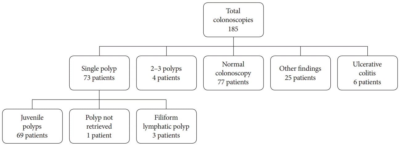
Among the 77 patients found to have colonic polyps, single colonic polyps were detected in 73 (94.8%) and 2–3 polyps were found in 4 (5.2%) patients (
Fig. 1). Sixty-nine (94.5%) of the single polyps were juvenile polyps (
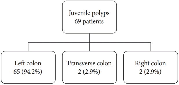
Fig. 1), of which 65 (94.2%) were located in the left colon, 2 (2.9%) in the transverse colon, and 2 (2.9%) in the ascending colon (
Fig. 2). One patient was noted to have a polyp in the left colon that could not be retrieved by the endoscopist for pathologic evaluation. Three patients had a single filiform lymphatic polyp that was located in the left colon. Of the 73 patients with a single polyp, 69 (95.5%) had the polyp in the left colon. All 4 patients with multiple polyps were found to have juvenile polyps. In 3 of the 4 patients, the polyps were located in the left colon; in the fourth patient, 2 polyps were noted in the left colon and 1 was found in the cecum. Of the 77 patients with colonic polyps, 72 (93.5%) had only left-sided colonic polyps.
 | Fig. 1.Endoscopic and histologic findings in our cohort. 
|
 | Fig. 2.Distribution of juvenile polyps in the colon. 
|
Children with polyps had a median age of 5.5 years (IQR, 3.9–8.1 years), whereas those without polyps had a median age of 10.9 years (IQR, 5.8–15.7 years,
p=0.0001;
Table 2). Hemoglobin results from before the colonoscopy were available for 165 of the 185 children. Children with polyps were found to have a lower hemoglobin level (mean 12.1±1.4 mg/dL) than those without polyps (mean 12.59±1.45 mg/dL) (
p=0.037). Anemia was detected in 58% of the children with polyps and in 42% of those without polyps. However, this difference was not statistically significant (Pearson chi-square test,
p=0.053).
Table 2.
Epidemiological, Demographic, and Clinical Characteristics of the Patients
|
Patients with polyps (n=77) |
Patients without polyps (n=108) |
p-value |
|
Age: median (IQR) |
5.5 yr (3.95–8.1 yr) |
10.9 yr (5.8–15.7 yr) |
0.0001 |
|
Female: n (%) |
41 (43.2%) |
47 (53.2%) |
0.192 |
|
Hemoglobin: g/dL (SD) |
12.13±1.45 |
12.6±1.45 |
0.037 |
|
Albumin: mg/dL (SD) |
4.2±0.92 |
4.44±0.42 |
0.073 |
|
Constipation |
3 (3.9%) |
9 (8.3%) |
0.365 |
|
Anemiaa)
|
40 (58%) |
41 (42.7%) |
0.053 |
|
Hypoalbuminemiaa)
|
2 (3.9%) |
2 (2.4%) |
0.635 |

The only complication noted in this cohort was a postpolypectomy bleeding event that was effectively treated with the application of a hemostatic clip in 1 patient.
Go to :

DISCUSSION
Our study investigated children who underwent colonoscopy for isolated lower gastrointestinal bleeding with no diarrhea. The findings in our cohort provide additional data to the limited literature, as follows: (1) The most common abnormal finding was colorectal polyps (noted in 42% of the patients). (2) The vast majority of these were single juvenile polyps located in the left colon. (3) Only 4 children had multiple polyps, with no patient having more than 3 polyps. Of these, only 1 child had a polyp proximal to the splenic flexure. (4) Of the patients with a single polyp, only 4 children had a polyp proximal to the splenic flexure. Lastly, overall, only 5 of all 77 polyps (6.5%) were located proximal to the splenic flexure.
The findings of our cohort stand in contrast to the study by Fox et al., who reported that 60.9% of juvenile polyps were single and 66.6% of these polyps were located in the left colon while the remaining 33.3% were located proximal to the splenic flexure [
11]. A less pronounced difference was noted when our results were compared with the study by Poddar et al., who demonstrated 76% solitary polyps with 85% of these being located in the rectosigmoid [
12]. These studies differ from ours in that they reviewed all colonoscopies that demonstrated polyps without regard to the clinical presentation and included patients with polyposis syndromes. A recent study reported that only 19.5% of patients with painless lower gastrointestinal bleeding had polyps, and that when patients with an additional complaint of abdominal pain with no change in bowel movements were included in the analysis, only 14.8% were found to have polyps [
13]. In contrast, our study included patients undergoing colonoscopy for isolated lower gastrointestinal bleeding, and we believe that excluding patients with polyposis syndromes and those with lower gastrointestinal bleeding associated with other complaints is likely a major reason for the different results obtained in our study.
Multiple studies have reported that juvenile polyps were more common in male patients [
3,
4,
7,
13]. Our study, however, did not demonstrate a statistically significant difference between male and female patients.
Fox et al. reported neoplasia in 3.9% of their cohort [
11]. All of these cases involved patients with more than 5 polyps [
10]. In our study, there were no cases of neoplasia and no patients with more than 3 polyps. This difference may be explained by the difference in the composition of the cohorts and the absence of patients with more than 3 polyps in our study. Overall, this indicates that when colonoscopy is performed for painless lower gastrointestinal bleeding, neoplasia is rarely, if at all, found.
Of interest, there were 6 (3.2%) patients with ulcerative colitis in our study. Three of them had proctitis. This rate is lower than that reported in a recent study that analyzed patients with painless rectal bleeding with or without abdominal pain but with no change in bowel movements, in which inflammatory bowel disease was present in 10.7% of the patients [
13].
Currently, pancolonoscopy is the recommended method for the evaluation of recurrent painless rectal bleeding in children [
14]. All patients in this study had complete colonoscopy. As result, we found a small but significant minority (6.5%) of patients with polyps proximal to the splenic flexure.
The main limitation of this study stems primarily from its retrospective design that did not allow complete data collection for several variables. Despite this limitation, our pediatric isolated lower gastrointestinal bleeding cohort study is one of the few studies that assessed the findings on colonoscopies performed for this indication.
In conclusion, we found that single left-sided juvenile polyps are the most common cause of isolated lower gastrointestinal bleeding in children. It was rare to find multiple polyps and polyps proximal to the splenic flexure in our cohort. It is an accepted practice to perform a full colonoscopy in patients presenting with isolated lower gastrointestinal bleeding. Despite the large majority of a left-sided pathology noted in our study, a full colonoscopy is still recommended in order to properly diagnose the small but significant group of patients with pathologies found proximal to the splenic flexure.
Go to :


 PDF
PDF Citation
 Print
Print


 XML Download