INTRODUCTION
The International Study Group of Liver Surgery has defined bile duct leak (BDL) as “fluid with an increased bilirubin concentration in the abdominal drain or in the intra-abdominal fluid or as the need for radiologic intervention because of biliary fluid collection or re-laparotomy resulting from bile peritonitis” [
1]. Bile leak is commonly associated with surgeries (such as cholecystectomy, liver transplantation, partial hepatectomy, and hydatid cyst excision). However, it may also occur secondary to various necro-inflammatory conditions involving the liver. The most frequent cause of bile leak is iatrogenic, and cholecystectomy is the most common procedure associated with this condition. Bile leak occurs in 0.1%–0.5% of patients who underwent open cholecystectomy [
2,
3]. Approximately 0.5%–2% of patients who underwent laparoscopic cholecystectomy can develop bile leak, with the majority arising from the cystic duct stump [
4-
6]. The clinical presentation of bile leak varies from asymptomatic drainage of bile to life-threatening conditions including biliary peritonitis. Thus, bile leak can be associated with significant morbidity and can lead to death if left untreated.
The communication between the biliary tree and amoebic liver abscess is observed in up to 27% of cases, and it commonly presents with jaundice and has a longer duration [
7]. In patients with a hydatid cyst, intrabiliary rupture of the cyst can lead to cystobiliary communication, which may result in postoperative bile leak in up to 28.6% of cases [
8].
The management of bile leak has been radicalized after the development of endoscopic retrograde cholangiopancreatography (ERCP), which is important for both the diagnosis and the management of symptomatic bile leak. Endoscopic therapy aims to promote bile leak healing by reducing the pressure gradient across the sphincter of Oddi, thereby increasing the flow of bile into the duodenum [
9]. Numerous studies have shown that the success rate of endoscopic treatment modalities ranges from 85% to 100% in post-cholecystectomy bile leaks [
10-
12], and the complication rate is extremely low. However, only few studies assessed the efficacy of endoscopic therapies in bile leak with other etiologies, such as liver abscess and hepatic hydatid cyst.
Thus, this study aimed to evaluate the efficacy and outcomes of endoscopic management for bile leak with various etiologies.
Go to :

MATERIALS AND METHODS
Study design and patient selection
We conducted a single-center retrospective analysis of patients with symptomatic bile leak who were referred for endoscopic management at the department of gastroenterology in a tertiary care hospital between April 2016 and April 2019. All patients who underwent ERCP for bile leak were included.
Exclusion criteria
Patients who were managed conservatively or who underwent percutaneous biliary drainage or surgery before ERCP were excluded from the study.
Treatment protocol
All patients at our center underwent laparoscopic cholecystectomy. Patients who had cholecystectomy were diagnosed with bile leak based on the presence of bile salts and pigments in the subhepatic drain placed intraoperatively due to suspected biliary injury or the presence of fluid collection on postoperative imaging that was performed secondary to symptoms. Similarly, in patients with liver abscess and a hydatid cyst or those who underwent hydatid surgery, bile leak was diagnosed based on the presence of bile in the percutaneous drain. After diagnosis, the patients were provided with standard medical care, including antibiotic treatment, IV fluid therapy, pigtail drainage of localized collections, and nutritional support. Based on the anatomical findings of contrast-enhanced computed tomography scan and/or magnetic resonance cholangiopancreatography, ERCP was performed. Informed consent regarding the nature of the procedure and its potential benefits and associated complications was obtained from all patients. The procedure was performed while the patient was in the left lateral or supine position. Based on the general assessment findings, all procedures were conducted under either moderate sedation or general anesthesia. Three endoscopists with experience in performing at least 300 ERCPs performed the procedure using a side-viewing duodenoscope (TJF-150/TJF-180; Olympus Optical Co., Tokyo, Japan). Guidewire-assisted cannulation of the common bile duct (CBD) was performed, followed by contrast injection, which showed the leak site. Endoscopic placement of a guidewire in the bile duct was carried out, and the proximal end of the guidewire was placed above the site of the leak, as confirmed on fluoroscopy. Then, a stent was placed over the guidewire. Patients with calculi in the CBD underwent extraction under the same setting. Amsterdam plastic biliary stents with a size ranging from 7 to 10 Fr and length ranging from 10 to 15 cm (Cook Medical, Bloomington, IN, USA) were placed based on cholangiogram findings.
The Strasberg classification (
Fig. 1) [
13] was used to categorize bile duct injury after cholecystectomy. Based on the extent of involvement, the bile duct injury was classified as major (complete or partial involvement of the CBD, common hepatic duct, or major segmental ducts at the porta hepatis) and minor (leak from the cystic duct, subvesical duct of Luschka, or gallbladder fossa) [
14]. Patients were followed up for clinical improvement, and drainage catheters were removed after no drain output for 3 days. Repeat cholangiogram was performed at 6 weeks to identify any residual leak. Re-stenting was performed if there was any residual leak.
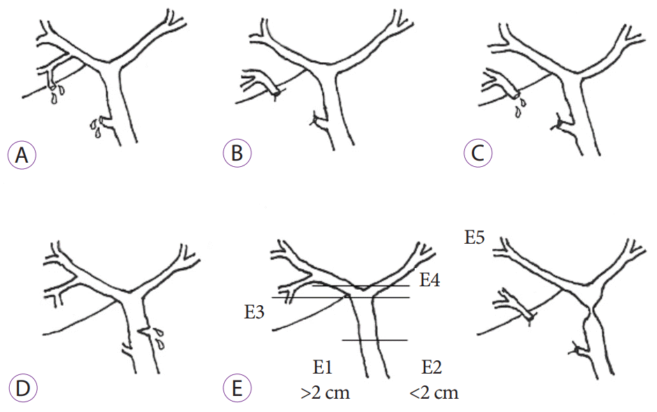 | Fig. 1.Strasberg classification [ 13]. (A) bile leak from the cystic duct or liver bed without injury, (B) partial occlusion of the biliary tree, most commonly from the aberrant right hepatic duct, (C) bile leak from the duct (aberrant right hepatic duct) without communication with the common bile duct, (D) lateral injury of the biliary system without loss of continuity, and (E) circumferential injury of the biliary tree with loss of continuity: (E1) transected main bile duct with a stricture >2 cm from the hilum, (E2) transected main bile duct with a stricture <2 cm from the hilum, (E3) stricture of the hilum with communication between the right and left ducts, (E4) stricture of the hilum with separation of the right and left ducts, and (E5) stricture of the main bile duct and the right posterior sectorial duct. 
|
Outcome measures
The primary outcome of the study was complete improvement of symptoms without extravasation of the contrast medium during subsequent ERCP conducted after 6 weeks.
The secondary outcomes included in-hospital mortality and differences in outcome with regard to the timing of ERCP, duration of hospital stay, and requirement of repeat stent exchange after 6 weeks.
Statistical analysis
Continuous variables were presented as median and range, and categorical variables were presented as frequencies and percentages. Significant differences were compared using Fisher’s exact test. Statistical Package for the Social Sciences software, version 16.0 (Armonk, NY, USA), was used for statistical analyses.
Go to :

RESULTS
In total, 71 patients (male participants, n=43 [60.5%]; mean age, 46±15 years) were included in our study. The etiologies of bile leak were post-cholecystectomy injury in 34 (47.8%), liver abscess in 20 (28.1%), post-hydatid cyst surgery in 11 (15.4%), injury during other surgeries in 5 (7.0%), and trauma in 1 (1.4%) patient.
Table 1 shows the demographic characteristics of the patients and the etiologies and clinical presentations. Bile leak was more common in male patients who underwent cholecystectomy (19 [56%] of 34) and in those with liver abscess (17 [85%] of 20). In our study, patients with percutaneous bile drainage (97.1%) were commonly diagnosed with bile leak. Abdominal pain (46.4%) was the most common symptom, followed by jaundice in 32.3%, fever in 25.3% and ascites in 5.6%. One patient with liver abscess coughed up bile-stained sputum (biliptysis) and was found to have a bilio-bronchial fistula.
Table 1.
|
n=71 |
|
Age, yr |
46±15 |
|
Male, n (%) |
43 (60.5) |
|
Etiologies, n (%) |
|
|
Post-cholecystectomy |
34 (47.8) |
|
Liver abscess |
20 (28.1) |
|
Hydatid cyst surgery |
11 (15.4) |
|
Other surgeries |
5 (7.0) |
|
Trauma |
1 (1.4) |
|
Clinical features, n (%) |
|
|
Bile in drain output |
69 (97.1) |
|
Abdominal pain |
33 (46.4) |
|
Jaundice |
23 (32.3) |
|
Fever |
18 (25.3) |
|
Interval between diagnosis and ERCP, n (%) |
|
|
Within one day |
15 (21.1) |
|
Within 2–3 days |
19 (26.7) |
|
>3 days |
37 (52.1) |

In total, 15 (21.1%) patients underwent ERCP within a day after presentation, 19 (26.7%) underwent within 2–3 days, and 37 (52.1%) underwent after 3 days. Most patients who underwent ERCP within a day had also underwent cholecystectomy, and those in the other groups underwent ERCP usually after 2 days. The early diagnosis of bile leak after cholecystectomy was attributed to intraoperative drain placement in suspected patients (draining bile alone). In contrast, in patients with abscess, the drainage of necrotic contents by pigtail insertion, with bile may delay the diagnosis.
The details of the ERCP procedure and findings are given in
Table 2. Successful CBD cannulation was achieved in all patients with bile leak. The right hepatic duct (RHD) or right biliary system was the most common leak site, accounting for 45.1% (32/71) of cases, and it is also the most common site in all etiologies except for post-cholecystectomy in which the cystic duct stump was the most frequently involved site (23 [67.6%] of 34). Overall, six (8.4%) patients, two of whom underwent cholecystectomy, had bile leak due to injury in the left hepatic duct (LHD). However, this finding is not common and has not been reported in previous studies. Three (4.2%) patients had choledocholithiasis and underwent stone extraction under the same setting (
Figs. 2-
5). One patient who underwent distal gastrectomy had complete transection of CBD with extravasation of the contrast medium on cholangiogram. Hence, the patient underwent sphincterotomy and then surgery. Stents with a diameter of 7–10 Fr and a length of 10–15 cm were used. The 10 Fr × 10 cm stent was most commonly used. The complications included post-ERCP pancreatitis in four patients and post-sphincterotomy bleeding in one patient. These conditions were managed conservatively.
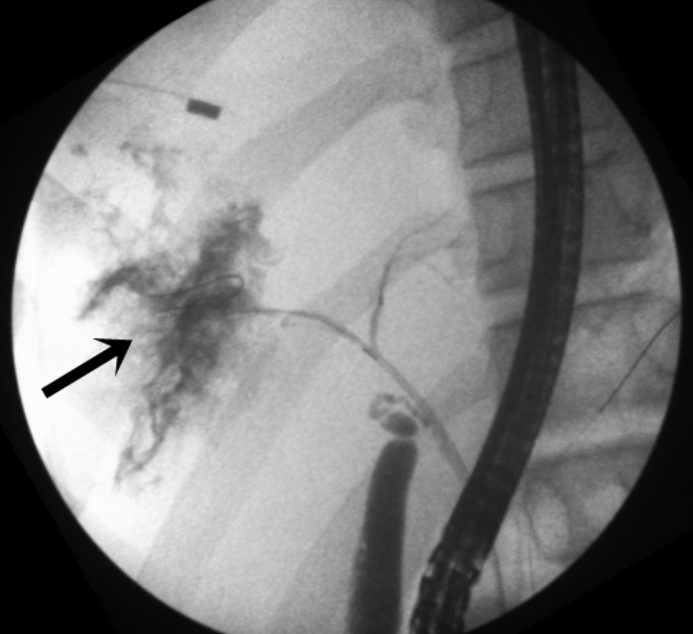 | Fig. 2.Leak from the right hepatic duct (arrow) in a patient with liver abscess. 
|
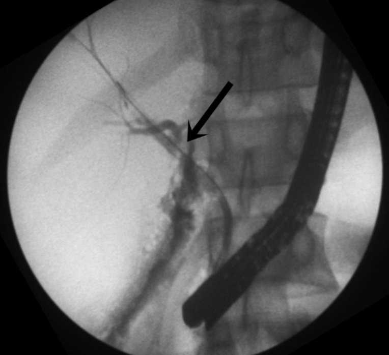 | Fig. 3.Large bile duct injury at the common hepatic duct level with left hepatic duct draining through a leak (arrow). 
|
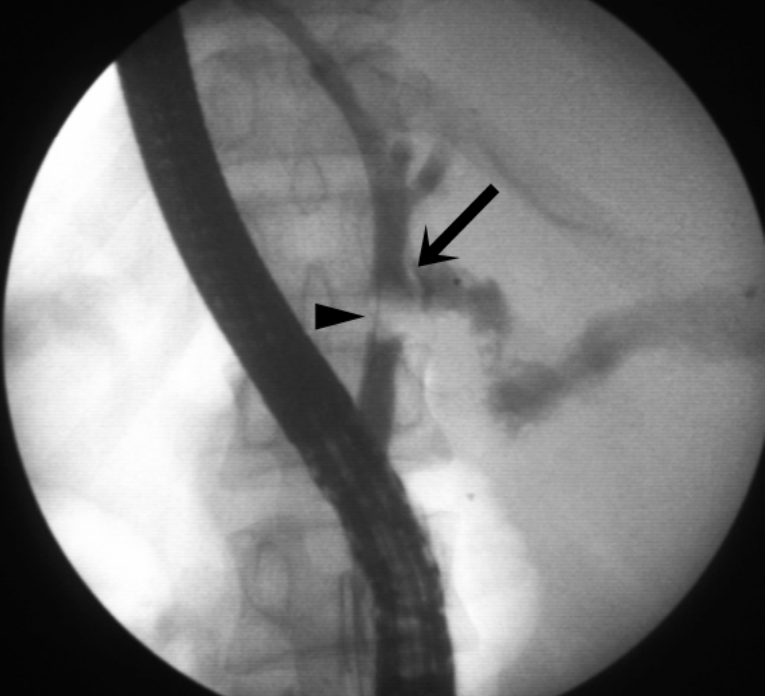 | Fig. 4.Cystic duct stump leak (arrow) with calculus in the proximal common bile duct (arrowhead). 
|
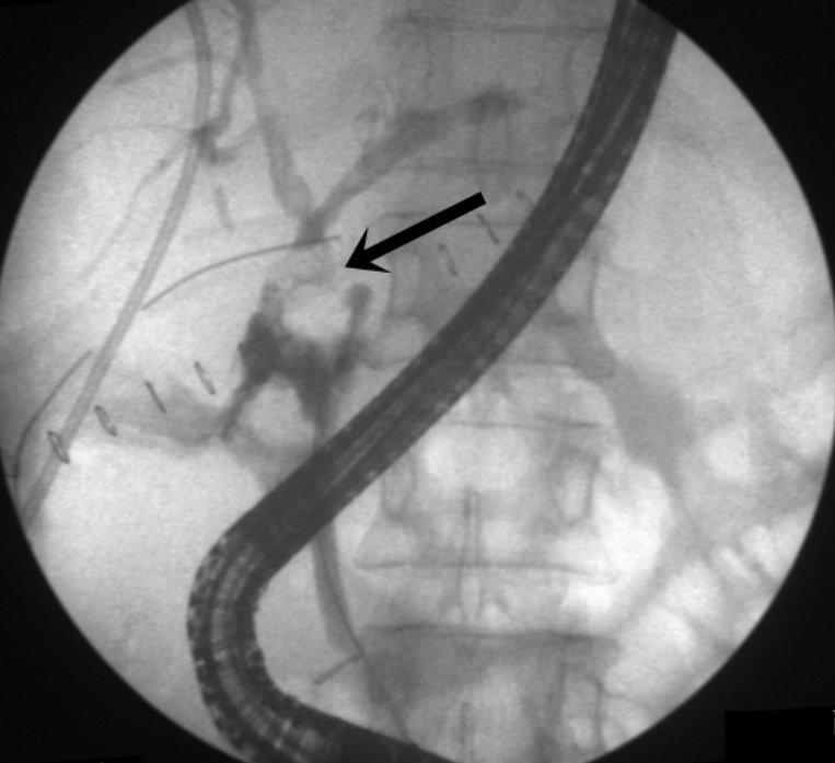 | Fig. 5.Bile duct injury at the common hepatic duct (arrow) with axis deviation at the level of injury with opacification in both the right and left hepatic ducts. 
|
Table 2.
Details of Endoscopic Retrograde Cholangiopancreatography Findings
|
n=71 |
|
Successful CBD cannulation |
71 (100%) |
|
Site of bile leak on ERCP, n (%) |
|
|
Cystic duct stump |
23 (32.4) |
|
CBD |
7 (9.8) |
|
CHD |
3 (4.2) |
|
RHD |
23 (32.4) |
|
LHD |
6 (8.4) |
|
Right aberrant duct |
2 (2.8) |
|
Smaller peripheral branches |
7 (9.8) |
|
Other findings, n (%) |
|
|
CBD stones |
3 (4.2) |
|
Size of stent used, n (%) |
|
|
7 Fr × 10 cm |
2 (2.8) |
|
7 Fr × 12 cm |
12 (17.1) |
|
7 Fr × 15 cm |
3 (4.2) |
|
10 Fr × 10 cm |
46 (65.7) |
|
10 Fr × 12 cm |
7 (10) |
|
Complications, n (%) |
|
|
Pancreatitis |
4 (5.6) |
|
Post-ERCP bleeding |
1 (1.4) |

Table 3 shows the details of the bile duct injuries in the post-surgical group. Among the post-surgical patients, 23 (58.9%) presented with class A injury, 2 (5.1%) with class C injury, 13 (33.3%) with class D injury, and 1 (2.5%) with class E injury based on the Strasberg classification. Major bile duct injury was observed in 14 (35.8%) and minor bile duct injuries in 25 (64.1%) patients.
Table 3.
Surgical Bile Duct Injuries
|
n=39 |
|
Strasberg classification, n (%) |
|
|
A (cystic duct stump) |
23 (58.9) |
|
B |
0 |
|
C (right aberrant duct injury) |
2 (5.1) |
|
D |
13 (33.3) |
|
RHD |
5 |
|
LHD |
3 |
|
CBD |
5 |
|
E (complete transection of CBD) |
1 (2.5) |
|
Major bile duct injury, n (%) |
14 (35.8) |
|
Minor bile duct injury, n (%) |
25 (64.1) |

Table 4 shows the primary and secondary outcomes in patients who underwent endoscopic therapy. The primary outcome of complete symptomatic improvement without extravasation of the contrast medium on repeat cholangiogram after 6 weeks was achieved in 65 (91.5%) of 71 patients. Because there was no symptom improvement, five (7.0%) patients required CBD stent exchange and one stent exchange. Four patients were asymptomatic but had persistent leaks on repeat ERCP. Only one patient required surgery due to complete transection of CBD. There was no bile leak-related mortality in our study population. The median length of hospital stay was 10 (range, 6–25) days. Patients who underwent ERCP after 1, within 2–3, and after 3 days of diagnosis did not have significant difference in terms of outcome (
p=0.120). Moreover, there was no significant difference in outcome in relation to the size of the stent used (
p=1).
Table 4.
Outcomes of Endoscopic Management
|
Outcomes |
n=71 |
|
Primary outcome achieved |
65 (91.5%) |
|
Requirement for surgery |
1 (1.4%) |
|
Repeat CBD stenting required |
5 (7.0%) |
|
In-hospital mortality (%) |
0 |
|
Length of hospital stay (median, range) |
10 days (6–25) |

Table 5 shows differences in the outcome of endoscopic management for bile leak between the surgical and non-surgical groups. Although bile leak can develop after hydatid cyst surgery, its cause is usually necro-inflammatory rather than iatrogenic. This was observed in the non-surgical group, as most patients had communication between the biliary radicle and the cyst on imaging. The primary outcome was achieved in 35 (89.7%) of 39 patients in the surgical group and in 30 (93.7%) of 31 patients in the non-surgical group. There was no significant difference between the two groups with regard to the primary outcome and need for surgery or repeat CBD stenting. The median length of hospital stay was significantly longer in the non-surgical group (11.5 days) than in the surgical group (8 days) (
p=0.0001) (
Fig. 6).
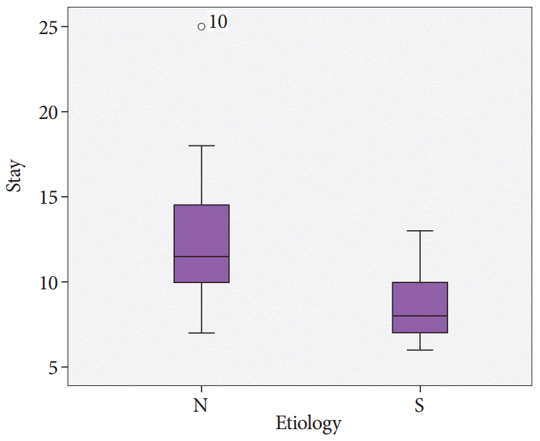 | Fig. 6.Duration of hospital stay of the non-surgical (N) and surgical (S) groups, showing a significant difference between the groups (p=0.0001). 
|
Table 5.
Difference in Outcome between Subgroups
|
Outcomes |
Surgical group |
Non-surgical group |
p-value |
|
n=39 |
n=32 |
|
Primary outcome achieved |
35 |
30 |
0.683 |
|
Requirement for surgery |
1 |
0 |
1.000 |
|
Repeat CBD stenting required |
3 |
2 |
1.000 |
|
In-hospital mortality |
0 |
0 |
1.000 |
|
Median length of hospital stay |
8 days (6–13) |
11.5 days (7–25) |
0.0001 |

Go to :

DISCUSSION
To the best of our knowledge, this is the largest study conducted in India that compares different etiologies of bile duct injuries and their outcomes. The technical success of ERCP with sphincterotomy and CBD stent placement was achieved in all patients, except for one who required surgery. The incidence of complications from ERCP was similar to that of ERCP conducted for other indications. There was no mortality due to complications associated with bile leak.
Males accounted for 60% of all patients and 85% of those with liver abscess. A possible explanation for this might be the high alcohol intake among men, as alcohol suppresses the function of Kupffer cells. Furthermore, a previous study conducted in India showed that 72% of patients with liver abscess were alcoholics [
15].
The cystic duct stump was the most common leak site in post-cholecystectomy patients, followed by the RHD. An interesting finding of our study was that LHD injury was observed in two post-cholecystectomy patients, and this condition has not been reported in previous studies. Only one case report of LHD injury due to clipping without section of the duct has been reported [
16]. Retained CBD stones or strictures can promote bile leak due to increased pressure in the CBD, as observed in three patients with CBD stones. However, none presented with CBD stricture.
Bile leak can be managed with endoscopic sphincterotomy with or without stent placement. However, stent placement in the biliary system does not only seal the leakage point but also further reduces CBD pressure. Then, this decreases the upstream pressure and facilitates early healing of the leaking point while preventing injury related to secondary ductal stenosis. There is no conclusive evidence showing whether the placement of the stent or nasobiliary drain across the site of leak is required or beneficial, and this may not be possible in most patients with peripheral parenchymal bile leaks [
17].
The incidence of pancreatitis after ERCP is approximately 9.7%, with a mortality rate of 0.7% [
18]. In our study, ERCP-related complications were observed in 7.1% of patients, and the rate is comparable to that of ERCP conducted for other indications.
Sandha et al. have assessed 207 patients with post-cholecystectomy bile leaks who were managed with either biliary sphincterotomy alone for low-grade leaks (identified only after intrahepatic opacification) or who underwent stent placement for high-grade leaks (observed before intrahepatic opacification) [
10]. Among patients with low-grade leaks without any risk factors such as biliary stricture, coagulopathy, and severe sepsis, sphincterotomy alone led to symptom improvement in 91% of patients who required stent placement or surgery. Therefore, in the absence of risk factors, patients with low-grade bile leak could be managed with sphincterotomy alone [
10].
Abbas et al. conducted a large-scale study in the USA, which included 1,028 patients with post-cholecystectomy bile leak who underwent endoscopic therapy, and results showed that the failure rates were lower in stent monotherapy with or without sphincterotomy (3% and 4%, respectively) than in sphincterotomy monotherapy (11%) [
11]. No difference was observed in patient outcomes and adverse events with regard to the timing of ERCP after the development of bile leak [
11].
A similar study was conducted on 27 patients with bile leak in India. Of these patients, 23 (92%) were managed with sphincterotomy with stent placement and 2 (8%) with sphincterotomy alone. The primary outcome, which is symptom improvement without bile leak on subsequent ERCP, was achieved in 92.8% of the patients. The outcome was similar to the that of our study [
19].
Sharma et al. showed that the median healing duration of fistulas in all patients (
n=38) with liver abscess communicating with the bile ducts who were managed endoscopically was 6 (range, 4–40) days [
14]. In this study, a nasobiliary drain was placed in 47% of patients [
14]. The placement of a nasobiliary tube is useful in repeat cholangiogram, as it can identify fistula healing, thereby avoiding the need for second ERCP and preventing its associated complications. However, this technique is not frequently used, as it is associated with patient discomfort and the risk of inadvertent removal.
Bile leak associated with a hydatid cyst can be attributed to the cyst communicating with the bile duct or can develop after surgery. In the study conducted by Tuxun et al., the risk of bile leak after hydatid surgery is approximately 6.24% [
20]. In fact, prophylactic endoscopic sphincterotomy is performed to prevent postoperative bile leak in hydatid liver disease. In a randomized controlled study conducted in Egypt, preoperative endoscopic sphincterotomy significantly reduced the incidence rate of bile leak (11.1% vs. 40.7%) and duration of fistula closure [
21].
In another prospective study conducted by Sharma et al. on patients with a hydatid cyst communicating with the bile duct, sphincterotomy with insertion of a nasobiliary drain (
n=6) or sphincterotomy with biliary stenting (
n=22) led to fistula healing in all patients after a median period of 11 (range, 5–45) days [
22]. The removal of nasobiliary drainage catheters and stents was performed 8–45 days after stent placement [
22].
Patients who were diagnosed with bile leak intraoperatively during cholecystectomy or those suspected with biliary injury should immediately undergo drain placement and ERCP. However, our analysis did not show any significant difference in the outcome in relation to the timing of ERCP after the onset of bile leak between the two groups.
In a study conducted by Adler et al., 518 patients with post-cholecystectomy bile leaks were classified into three groups according to the timing of ERCP after bile leak (within 1 day, on day 2 or 3, and after 3 days) [
23]. No significant difference was observed in terms of the success rate of BDL for ERCPs (91.2%, 90%, and 88.5%, respectively,
p=0.77) and the rate of adverse effects between the three groups (21.1%, 22.9%, and 24.6%, respectively,
p=0.81) [
23]. The authors showed that patients with bile leak can undergo ERCP on an elective rather than emergent basis.
The impact of stent diameter and length on treatment success was not clearly elucidated [
24], and the current study obtained a similar result. However, for refractory leaks or leaks from damage to the CBD or proximal hepatic ducts, which are often associated with larger defects, stents with a large diameter or multiple stents are placed to fill the bile duct lumen and to cross the site of leakage for the potential tamponade effect with the additional benefit of preventing subsequent stricture at the injury site.
Unlike the previous practice involving surgical management, endoscopic therapy has emerged as the first-line management for bilio-bronchial fistula [
25]. In our study, endoscopic therapy alone led to symptom improvement without the need for any surgery. In a previous trial, fistula closure with coil and glue has been performed in patients who do not meet the criteria for ERCP with CBD stenting [
26].
No previous study has compared the outcome of bile leak between the surgical and non-surgical groups. In the current study, there was no significant difference between the surgical and non-surgical groups in terms of primary and secondary outcomes except for the duration of hospital stay, which was significantly longer in the non-surgical group than in the surgical group. However, this difference was attributed to the nature of the disease itself, as necro-inflammatory conditions require longer hospitalization. To the best of our knowledge, this is the largest study on the management of bile duct injuries in India. The functional success of endotherapy for bile leaks after 6 weeks with repeat cholangiogram was assessed, and this is considered the strength of our study.
However, the current study had major limitations. First, it was retrospective in nature. Second, the sample size was small. In addition, data regarding the interval between ERCP and the removal of drainage catheter were not available. Hence, studies with a prospective study design and a larger sample size would have shown a better outcome.
Endoscopic therapy plays an important role in the management of symptomatic bile leak with various etiologies, and the outcomes are similar to those of post-cholecystectomy bile leaks. Based on previous beliefs, delayed diagnosis of bile leak is associated with higher morbidity and risk of surgery. However, the current study shows that even bile leaks diagnosed at a later stage can be managed endoscopically, and the outcomes did not significantly differ between those who received early or late management.
Go to :


 PDF
PDF Citation
 Print
Print


 XML Download