Journal List > Clin Endosc > v.53(3) > 1151083

Jain, Lee, and Rajala: Atraumatic Splenic Hemorrhage as a Rare Complication of Pancreatitis: Case Report and Literature Review

Abstract

Splenic hemorrhage (hematoma and rupture) is a rare complication of pancreatitis. In this article, we present a rare case of spontaneous splenic rupture as a complication of acute pancreatitis. A literature review was also completed to describe the patient characteristics, associated pancreatitis etiology, clinical presentations, risk factors, diagnostic and treatment modalities, and outcomes.

INTRODUCTION

Acute pancreatitis is one of the leading causes of hospitalization with a gastrointestinal condition in the United States [1]. The incidence of acute pancreatitis ranges from 13 to 45 per 100,000 individuals per year. Over 270,000 hospital admissions annually are attributed to acute pancreatitis, which have increased by 15% over the past 10 years [1,2]. Gallstones remain the most common cause of acute pancreatitis, followed by alcohol consumption. These two causes account for 70% of cases [3]. Other causes include hypertriglyceridemia, medications, autoimmune diseases, surgical procedure (e.g., endoscopic retrograde cholangiopancreatography), trauma, infection, and genetic and idiopathic factors. Although most patients with acute pancreatitis have mild, self-limited diseases, 20% to 30% can have a severe course with life-threatening complications. Local complications include fluid collection (e.g., acute peripancreatic fluid collection, pancreatic pseudocysts, acute necrotic collection, and walled-off pancreatic necrosis) with or without infection and vascular conditions (e.g., splenic vein thrombosis and splenic pseudo-aneurysm); conversely, systemic complications include systemic inflammatory response syndrome (SIRS) and subsequent organ failures [4-6].
Spontaneous splenic hemorrhage (e.g., rupture and subcapsular hematoma) is rare yet an important complication of pancreatitis. The exact incidence and epidemiology have not been clearly defined in the literature. Given the anatomic proximity between the pancreatic tail and splenic hilum, the direct erosion of pseudocysts, extravasation of pancreatic enzymes, localized portal hypertension due to splenic vein thrombosis, and adhesions from recurrent pancreatitis are the proposed pathophysiology of splenic manifestations [7-9]. As the clinical presentations of splenic hemorrhage can be nonspecific, imaging studies, especially computed tomography (CT), are warranted for differentiation from other etiologies [10]. Hemodynamically stable patients can be managed conservatively with percutaneous drainage and splenic artery embolization [11]. In contrast, surgical intervention with splenectomy is required for hemodynamically unstable patients [12]. Early recognition and intervention are critical for optimal patient outcomes.
In this literature review, we present a rare case of spontaneous splenic rupture as a complication of acute pancreatitis and provide a review of the literature to describe the patient characteristics, associated pancreatitis etiology, clinical presentations, risk factors, diagnostic and treatment modalities, and outcomes.

CASE PRESENTATION

A 49-year-old woman presented with a 3-day history of fever, generalized malaise, and worsening of severe, diffuse, acute-onset abdominal pain that was greatest felt in the left upper quadrant.
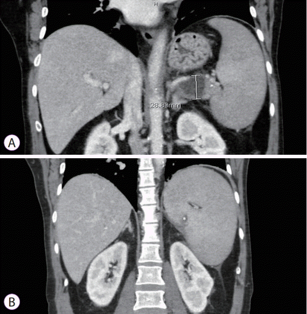
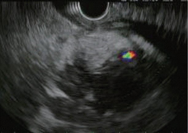
She had a history of three hospitalizations and one short observation stay for acute pancreatitis over a 4-month period. Her symptoms were classic for acute pancreatitis: abdominal pain with radiation to the back, elevated lipase level, and radiologic findings of acute pancreatitis involving the tail of the pancreas. She was treated conservatively following these events and recovered uneventfully, except for the persistence of intermittent left upper quadrant abdominal pain. The diagnosis was considered idiopathic, as she reported no significant alcohol consumption, trauma, new culprit medications, laboratory test result abnormalities, or imaging findings to suggest gallstone pancreatitis, hypercalcemia, or hypertriglyceridemia. On her second admission (approximately 8 weeks since the first admission), imaging revealed new peripancreatic (tail) fluid collection abutting the spleen and gastric fundus (Fig. 1A). On the third admission (approximately 6 weeks since the second admission), imaging revealed new acute splenic vein thrombosis, gastric varices, and splenomegaly in addition to the known peripancreatic fluid collection (Fig. 1B). The patient was started on anticoagulation with low-molecular-weight heparin bridged to a direct-acting oral anticoagulant (DOAC) after hematologist consultation. The international normalized ratio on DOACs ranged from 2.1 to 3.4. Diagnostic endoscopic ultrasound (3 weeks since the third admission and 3 days before the current admission) showed a 3-cm heterogeneous fluid collection in the pancreatic tail with an irregular ill-defined margin extending to the splenic hilum consistent with the known finding (Fig. 2). No mass lesion was identified. The pancreatic parenchyma in the head and body of the pancreas was within normal limits. The common bile duct and pancreatic duct were non-dilated with no filling defects. Several benign-appearing 1-cm lymph nodes were noted.
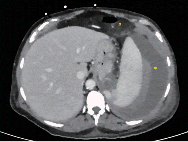
On arrival to the emergency department, the patient was hypotensive at a blood pressure of 75/45 mm Hg and tachycardic at a heart rate of 120 beats/min. The laboratory test results were remarkable for the lipase level (147 IU/L), white blood cell count (15×103 /μL), Hb level (10 g/dL), creatinine level (1.6 mg/dL), and beta-hemolytic non-group A or B streptococcus bacteremia presence. Abdominal and pelvic CT showed acute-onset chronic pancreatitis with fluid collection along the pancreatic tail (not organized) that extended into the postero-inferior aspect of the spleen and known thrombosis of the splenic vein with portosystemic collaterals, gastric varices, and mild splenomegaly. She was initially treated with fluid resuscitation and vasopressor and antibiotic administration with initial improvement in clinical status. However, she developed acute shortness of breath, worsening abdominal pain (progressed to diffuse, severe, and constant pain), and distension the following day. Repeat CT showed splenic rupture with moderate hemoperitoneum and perisplenic hematoma (Fig. 3). Emergency laparotomy with splenectomy was performed successfully with an uncomplicated recovery. Pathology revealed multiple lacerated spleen fragments.

METHODS OF LITERATURE REVIEW

An extensive English-language literature search was performed until November 2018 in PubMed, Cochrane Database, MEDLINE, and Google Scholar to identify case reports and case series employing the following keywords: pancreatitis, splenic rupture, and splenic hematoma. Cases related to pseudo-aneurysm rupture, previous procedure or surgery, and abdominal trauma were excluded. The search yielded 19 case reports [13-31] and 5 case series [32-36]. However, we included one case with splenic artery pseudo-aneurysm, as it did not rupture and therefore was not considered as the cause of splenic hematoma [23].
Patient demographics, comorbidities, social history, clinical presentations, diagnostic modalities, pancreatitis etiology and complications, splenic rupture timing and management, and spleen histology were reviewed and are summarized in Table 1. For the review, a total of 28 cases, including our case, were considered appropriate to be included. We summarized the available literature in addition to our case in this article.

SUMMARY OF LITERATURE REVIEW

Patient characteristics

The mean age of the patients was 42.71 years. The majority (78.6%) were men. Out of the 28 cases, 89.3% (25/28) presented with acute pancreatitis [13,15-19,21-29,31-36], while 10.7% (3/28) presented with chronic pancreatitis [14,20,30]. Among the acute pancreatitis cases, 20% (5/25) demonstrated known medical history or imaging evidence of chronic pancreatitis [19,22,32,33,35]. More than half of the cases (53.6%, 15/28) had prior episodes of pancreatitis; six cases had one prior episode [14,16,20,29,32,36]; one case had two prior episodes [18]; our case had three prior episodes; and seven cases had multiple (exact number not defined by individual study authors) prior episodes [21,24,25,34-36].

Pancreatitis etiology

The leading etiology of pancreatitis complicated by splenic rupture was alcohol consumption (67.9%, 19/28) [13-16,18,19,21,23,26-30,33-36]. Of the remaining cases, 7.1% (2/28) were idiopathic [36]; 3.6% (1/28) were related to Crohn’s disease [17]; and 21.4% (6/28) had no specified etiology [20,22,24,25,31,32].

Clinical presentations

The majority of the patients (96.4%, 27/28) presented with a chief complaint of abdominal pain, most commonly at the upper abdomen [20]. Other common symptoms included nausea or vomiting (21.4%) [13,27,29,30,35], pain in the left chest or hypochondrium pain (17.9%) [15,19,32-34], pain radiating to the left shoulder (14.3%) [14,16,32,36], and referred back pain (10.7%) [18,27,33]. There were 23 (82.1%) cases of splenic rupture causing hemoperitoneum [13-17,21-30,32-36] and 5 (17.9%) cases of confined splenic hematoma without rupture [18-20,31,36]. Further, 64.3% (18/28) of the cases had splenic rupture or subcapsular hematoma on presentation [14-20,22,24,26,28,29,32-36]. The other patients developed rupture or hematoma during hospitalization mostly within 1 week (32.1%, 9/28) [13,21,23,25,27,30,36], while one patient developed hematoma after 1 month of presentation [31]. However, the timing of splenic rupture or subcapsular hematoma can greatly vary if previous episodes of acute pancreatitis are considered as starting points.

Risk factors

Among the cases, 60.7% (17/28) demonstrated fluid collections around the pancreas [13,16,19,20,22,24,26,28-31,33-36], 76.5% (13/17) of which had collections in the tail of the pancreas adjacent to the spleen [16,19,22,24,28,30,31,33-36]. Three cases had no specified location of fluid collections [13,20,29]. Among the patients with fluid collections, 76.5% (13/17) had well-defined pseudocy sts [16,19-22,24,26,28-30,34-36]. Three cases, including our case, showed acute peripancreatic fluid collections without a definite margin [31,33], and one case demonstrated acute necrotic collection [13]. One case had an infected pseudocyst verified on presentation [31], and another case showed infected peripancreatic fluid collection after surgery [13]. Splenic vein thrombosis was present in 21.4% (6/28) of the cases [13,17,20,23,36]. More than half of the cases (71.4%, 20/28) had either chronic pancreatitis or history of acute pancreatitis. In total, 92.8% (26/28) of the cases had one of the risk factors for splenic rupture.

Diagnosis and management

Splenic rupture and subcapsular hematoma were diagnosed using CT or ultrasound, except in three cases. For these cases, diagnostic laparotomy was performed under high suspicion for splenic rupture based on abnormal physical examination results (abdominal fluid thrill), vital signs (hemodynamical instability), laboratory test results (elevated amylase level and leukocytosis), and chest X-ray results (elevated left hemidiaphragm, basal lung collapse, and pleural effusion) [28,29,32].
Most of the rupture cases (95.7%, 22/23) were managed by laparotomy with or without splenectomy [13-16,21-30,32-36], except for one case that was treated conservatively [17]. Most patients underwent emergency laparotomy, except for two who received splenic artery embolization for stabilization before elective surgery [23,36]. All hematoma cases without rupture were treated with conservative management [19,20,36], percutaneous drainage [18,31], or splenic artery embolization [20].

Outcomes

Among the 23 cases of splenic rupture, most (78.3%, 18/23) had a full recovery without complications. There were one case of mortality from postoperative pancreatitis and multi-organ failure [15] and four cases with significant complications [13,27,29,35]. The first case was complicated by infected peripancreatic fluid collection after surgery, which was successfully treated with percutaneous drainage [13]. The second case was complicated by pancreatic necrosis, which prompted pancreatic necrosectomy with good recovery [27]. In the third case, the patient returned after 4 months with a large pancreatic cyst that required marsupialization [29]. In the last case, the surgery was complicated by suppuration in the spleen cavity [35]. Further follow-up was not provided in the study. All hematoma cases without rupture (100%, 5/5) were treated conservatively and recovered without mortality or morbidity.

Histology

Eleven cases had available histopathological results [14,15,26-28,30,33-35]. The most common finding was capsular rupture of the spleen [14,26,27,33,35]. One case showed subcapsular hematoma of the spleen without capsular rupture [35]. Three cases had normal spleen histology, suggesting no underlying splenic etiology for the ruptures [15,27,34]. In three cases, histopathology demonstrated fluid collections, especially formation of pseudocysts [26,35]. Two cases revealed signs of recurrent pancreatitis [25] and chronic fibrosing pancreatitis [30]. Adhesion between the pancreas and the hilum of the spleen was also noted in two cases [26,35]. Signs of peptic enzyme digestion in the hilum were also documented in two cases [28,35].

DISCUSSION

Atraumatic splenic rupture and hematoma are rare complications of pancreatitis but can cause detrimental consequences and even a fatal outcome. In this review of the literature, the patient characteristics, associated pancreatitis etiology, clinical presentations, related risk factors, diagnostic and treatment modalities used, and outcomes were identified.
The majority of the patients in this study were middle-aged men who presented with acute pancreatitis. The leading etiology was alcohol consumption (67.9%); however, idiopathic pancreatitis was also a common etiology (21.4%). There are multiple different scoring methods to estimate the severity of pancreatitis, including Ranson’s score, Acute Physiology and Chronic Health Evaluation II score, SIRS score, and Bedside Index of Severity in Acute Pancreatitis score. These scoring systems are complex and not routinely used. Most case reports did not assess severity as scores nor use uniform information to evaluate such. Therefore, the correlation between the severity of pancreatitis and spontaneous splenic rupture could not be investigated in this study. The common chief complaints included upper abdominal pain, left chest or hypochondrium pain, left shoulder pain, and referred back pain. Associated nausea or vomiting was also common. These presenting symptoms are not specific to acute pancreatitis nor splenic rupture. Laboratory tests and imaging studies are required to rule out other etiologies, including peptic ulcer disease, left-sided pneumonia, aortic dissection, and acute myocardial infarction.
Although the pathophysiology of splenic complications is incompletely understood, the close anatomical proximity between the spleen and the pancreas has been considered as a major contributor to splenic complications during the course of pancreatitis. The splenic artery and vein and pancreatic tail, which are all located in the lienorenal ligament, enter the splenic hilum. This close anatomical association may lead to splenic involvement (e.g., intra-splenic abscess, pseudocyst formation, infarction, hemorrhage, and rupture) with pancreatitis [9]. The direct erosion of pseudocysts or extravasation of pancreatic enzymes from a fluid collection is considered as one of the potential mechanisms of splenic rupture [7]. In fact, the majority of the patients demonstrated fluid collections around the pancreas, especially in the tail of the pancreas adjacent to the spleen. Most of the fluid collections had well-defined pseudocysts. Splenic vein thrombosis was also shown to be a common risk factor associated with splenic rupture in this study. Venous outflow obstruction caused by this thrombosis may induce localized hypertension and congestion within the splenic parenchyma [8]. More than 70% of the cases had either chronic pancreatitis or prior episodes of pancreatitis. Adhesions from recurrent pancreatitis are also one of the proposed mechanisms of splenic complications. Although there were multiple risk factors suggested in this study, risk factor calculations are not possible with our limited number of patients.
In most cases, splenic rupture or hematoma was diagnosed using contrast-enhanced CT. Results can be obtained rapidly, and the extent of splenic hemorrhage along with other complications, such as fluid collection or vascular involvement, can be assessed [10,37]. More than 90% of the ruptured cases were managed with laparotomy with splenectomy. All hematoma cases were treated with conservative management and minimally invasive procedures, including percutaneous drainage or splenic artery embolization. In this study, most patients fully recovered without complications. However, there were one mortality and three significant complications reported among the 28 cases (14.3%), which cannot be overlooked. A high degree of clinical suspicion for splenic hemorrhage is required for prompt diagnosis and appropriate management, which can lead to a better outcome.

CONCLUSIONS

Splenic hemorrhage is a rare but life-threatening complication of pancreatitis. The exact incidence is unknown, and our findings are based on case reports and case series, including 28 patients. The patients in this study were predominantly men with recurrent episodes of alcoholic pancreatitis. The presence of pancreatic fluid collection, splenic vein thrombosis with left-sided portal hypertension, and pancreatitis involving the tail of the pancreas are potential risk factors for splenic hemorrhage among these patients. However, risk factor calculations are not possible in our limited number of patients. A high degree of clinical suspicion for splenic hemorrhage for prompt diagnosis is required to achieve optimal outcomes. Contrast-enhanced CT remains the most comprehensive and sensitive diagnostic modality for splenic rupture. Explorative laparotomy with splenectomy remains the first-line management method for patients with splenic rupture; conversely, splenic embolization and supportive care remain the preferred strategy for patients with splenic hematoma. Prompt recognition and intervention for this adverse event have maintained the associated mortality low.

Notes

Conflicts of Interest: The authors have no financial conflicts of interest.

Author Contributions

Conceptualization: Deepanshu Jain, Byeori Lee

Data curation: DJ, BL

Formal analysis: DJ, BL

Supervision: Michael Rajala

Validation: MR

Writing-original draft: DJ, BL

Writing-review&editing: MR

REFERENCES

1. Peery AF, Crockett SD, Murphy CC, et al. Burden and cost of gastrointestinal, liver, and pancreatic diseases in the United States: update 2018. Gastroenterology. 2019; 156:254–272.e11.
crossref
2. Yadav D, Lowenfels AB. The epidemiology of pancreatitis and pancreatic cancer. Gastroenterology. 2013; 144:1252–1261.
crossref
3. Forsmark Ch E, Vege SS, Wilcox CM. Acute pancreatitis. N Engl J Med. 2017; 376:598–599.
crossref
4. Greenberg JA, Hsu J, Bawazeer M, et al. Clinical practice guideline: management of acute pancreatitis. Can J Surg. 2016; 59:128–140.
crossref
5. Ahmed M, Aziz MU, Mansoor MA, Anwar S. Vascular complications in cases of acute pancreatitis - CT scan based study. J Pak Med Assoc. 2016; 66:977–989.
6. Zerem E. Treatment of severe acute pancreatitis and its complications. World J Gastroenterol. 2014; 20:13879–13892.
crossref
7. Chaudhary A, Dhar P, Sachdev A, Agarwal AK. Surgical management of pancreatic necrosis presenting with locoregional complications. Br J Surg. 1997; 84:965–968.
crossref
8. Xu W, Qi X, Chen J, Su C, Guo X. Prevalence of splanchnic vein thrombosis in pancreatitis: a systematic review and meta-analysis of observational studies. Gastroenterol Res Pract. 2015; 2015:245460.
crossref
9. Fishman EK, Soyer P, Bliss DF, Bluemke DA, Devine N. Splenic involvement in pancreatitis: spectrum of CT findings. AJR Am J Roentgenol. 1995; 164:631–635.
crossref
10. Mortelé KJ, Mergo PJ, Taylor HM, Ernst MD, Ros PR. Splenic and perisplenic involvement in acute pancreatitis: determination of prevalence and morphologic helical CT features. J Comput Assist Tomogr. 2001; 25:50–54.
crossref
11. Heider R, Behrns KE. Pancreatic pseudocysts complicated by splenic parenchymal involvement: results of operative and percutaneous management. Pancreas. 2001; 23:20–25.
crossref
12. Werner J, Hartwig W, Hackert T, Büchler MW. Surgery in the treatment of acute pancreatitis--open pancreatic necrosectomy. Scand J Surg. 2005; 94:130–134.
13. Hernani BL, Silva PC, Nishio RT, Mateus HC, Assef JC, De Campos T. Acute pancreatitis complicated with splenic rupture: a case report. World J Gastrointest Surg. 2015; 7:219–222.
crossref
14. Sharada S, Olakkengil S, Rozario AP. Occult splenic rupture in a case of chronic calcific pancreatitis with a brief review of literature. Int J Surg Case Rep. 2015; 14:95–97.
15. Debnath J, Sonkar S, Sharma V, Chatterjee S, Srivastava V, Khanna SP. Spontaneous rupture of spleen masquerading as acute pancreatitis. Am J Emerg Med. 2014; 32:394.e1–e3.
crossref
16. Cengiz F, Yakan S, Enver İ. A rare cause of acute abdomen: splenic hematoma and rupture resulting from pancreatitis. Ulus Cerrahi Derg. 2013; 29:81–83.
crossref
17. Mujtaba G, Josmi J, Arya M, Anand S. Spontaneous splenic rupture: a rare complication of acute pancreatitis in a patient with Crohn’s disease. Case Rep Gastroenterol. 2011; 5:179–182.
crossref
18. Tseng CW, Chen CC, Chiang JH, Chang FY, Lin HC, Lee SD. Percutaneous drainage of large subcapsular hematoma of the spleen complicating acute pancreatitis. J Chin Med Assoc. 2008; 71:92–95.
crossref
19. Patel VG, Eltayeb OM, Zakaria M, Fortson JK, Weaver WL. Spontaneous subcapsular splenic hematoma: a rare complication of pancreatitis. Am Surg. 2005; 71:1066–1069.
crossref
20. Sawrey M, Hughes RG. An interesting cause of collapse in a patient with chronic pancreatitis. BMJ Case Rep. 2013; 2013.
crossref
21. Huang TY, Chao YC. Spontaneous splenic rupture in recurrent acute pancreatitis. Am J Emerg Med. 2002; 20:493–494.
crossref
22. Kuramitsu T, Komatsu M, Ono T, et al. Ruptured subcapsular giant hematoma of the spleen as a complication of chronic pancreatitis. Intern Med. 1995; 34:564–568.
crossref
23. Zhou Q, Shah C, Arthus JM, Vingan H, Agola J. Atraumatic splenic rupture precipitated by splenic vein thrombosis. Radiol Case Rep. 2016; 11:86–89.
crossref
24. Gandhi V, Philip S, Maydeo A, Doctor N. Ruptured subcapsular giant haematoma of the spleen--a rare complication of acute pancreatitis. Trop Gastroenterol. 2010; 31:123–124.
25. Katsanos KH, Theodorou J, Katopodis KP, Siamopoulos KC. Spontaneous splenic rupture complicating pancreatitis in a chronic hemodialysis patient. Clin Nephrol. 2004; 61:448–449.
crossref
26. Adelekan MO, Nasmyth DG, Joglekar VM. Acute pancreatitis presenting as a case of splenic rupture. Hosp Med. 2003; 64:432–433.
crossref
27. Toussi HR, Cross KS, Sheehan SJ, Bouchier-Hayes D, Leahy AL. Spontaneous splenic rupture: a rare complication of acute pancreatitis. Br J Surg. 1996; 83:632.
crossref
28. Catanzaro FP, Abiri M, Allegra S. Spontaneous rupture of spleen and pleural effusion complicating pancreatitis. R I Med J. 1968; 51:328–329.
29. Labree RH, Fuller J, Boman PG. Spontaneous rupture of the spleen after acute pancreatitis: report of a case. J Am Med Assoc. 1960; 172:816–817.
30. Moori P, Nevins EJ, Wright T, Bromley C, Rado Y. A case of a chronic pancreatic pseudocyst causing atraumatic splenic rupture without evidence of acute pancreatitis. Case Rep Surg. 2016; 2016:2192943.
crossref
31. Vyborny CJ, Merrill TN, Reda J, Geurkink RE, Smith SJ. Subacute subcapsular hematoma of the spleen complicating pancreatitis: successful percutaneous drainage. Radiology. 1988; 169:161–162.
crossref
32. Moore PG, Gillies JG, James OF, Saltos N. Occult ruptured spleen--two unusual clinical presentations. Postgrad Med J. 1984; 60:171–173.
crossref
33. Jha AK, Pokharna R, Nijhawan S, et al. Atraumatic rupture of spleen. Trop Gastroenterol. 2011; 32:238–240.
34. Torricelli P, Coriani C, Marchetti M, Rossi A, Manenti A. Spontaneous rupture of the spleen: report of two cases. Abdom Imaging. 2001; 26:290–293.
crossref
35. Scherer K, Kramann B. Rupture of the spleen by penetration of pancreatic pseudocysts. Eur J Radiol. 1987; 7:67–69.
36. Patil PV, Khalil A, Thaha MA. Splenic parenchymal complications in pancreatitis. JOP. 2011; 12:287–291.
37. Habib E, Elhadad A, Slama JL. [Diagnosis and treatment of spleen rupture during pancreatitis]. Gastroenterol Clin Biol. 2000; 24:1229–1232.

Fig. 1.
(A) Peripancreatic tail fluid collection. (B) Splenic vein thrombosis.
ce-2019-087f1.tif
Fig. 2.
Fluid collection in the pancreatic tail.
ce-2019-087f2.tif
Fig. 3.
Splenic rupture with hemoperitoneum (#) and perisplenic hematoma (*).
ce-2019-087f3.tif
Table 1.
Summary of Reported Splenic Hemorrhage Cases
Study Age/Gender Pancreatitis characteristics/Risk factors Diagnosis/Treatment and outcome
Hernani et al. (2015) [13] Brazil 30/M 1) Type: acute 1) Splenic rupture
2) Etiology: alcohol 2) Timing: on hospital day 6
3) Prior acute pancreatitis: No 3) Management: laparotomy with splenectomy
4) Chronic pancreatitis: No 4) Outcome: complicated by infected peripancreatic collection with subsequent drainage and full recovery
5) Fluid collection: acute necrotic collection
6) SVT: Yes
7) Splenomegaly: No
Sharada et al. (2015) [14] India 25/M 1) Type: chronic 1) Splenic rupture
2) Etiology: alcohol 2) Timing: on presentation
3) Prior acute pancreatitis: Yes (1 episode) 3) Management: laparotomy with splenectomy
4) Chronic pancreatitis: Yes 4) Outcome: full recovery
5) Fluid collection: No
6) SVT: No
7) Splenomegaly: Yes
Debnath et al. (2014) [15] India 45/M 1) Type: acute 1) Splenic rupture
2) Etiology: alcohol 2) Timing: on presentation
3) Prior acute pancreatitis: No 3) Management: laparotomy with splenectomy
4) Chronic pancreatitis: No 4) Outcome: complicated by postoperative pancreatitis, multi-organ failure, and eventually death
5) Fluid collection: No
6) SVT: No
7) Splenomegaly: No
Cengiz et al. (2013) [16] Turkey 38/F 1) Type: acute 1) Splenic rupture
2) Etiology: alcohol 2) Timing: on the presentation
3) Prior acute pancreatitis: Yes (1 episode) 3) Management: laparotomy with splenectomy
4) Chronic pancreatitis: No 4) Outcome: full recovery
5) Fluid collection: pseudocyst at the tail
6) SVT: No
7) Splenomegaly: No
Mujtaba et al. (2011) [17] USA 37/M 1) Type: acute 1) Splenic rupture
2) Etiology: Crohn’s disease 2) Timing: on presentation
3) Prior acute pancreatitis: No 3) Management: conservative management with transfusion and fluid resuscitation
4) Chronic pancreatitis: No
5) Fluid collection: No 4) Outcome: full recovery
6) SVT: Yes
7) Splenomegaly: No
Tseng et al. (2008) [18] Taiwan 32/M 1) Type: acute 1) Subcapsular splenic hematoma
2) Etiology: alcohol 2) Timing: on presentation
3) Prior acute pancreatitis: Yes (2 episodes) 3) Management: percutaneous drainage for 4 weeks
4) Chronic pancreatitis: No 4) Outcome: full recovery
5) Fluid collection: No
6) SVT: No
7) Splenomegaly: No
Patel et al. (2005) [19] USA 44/M 1) Type: acute 1) Subcapsular splenic hematoma
2) Etiology: alcohol 2) Timing: on presentation
3) Prior acute pancreatitis: No 3) Management: conservative management
4) Chronic pancreatitis: Yes 4) Outcome: full recovery
5) Fluid collection: pseudocyst at the tail
6) SVT: No
7) Splenomegaly: No
Sawrey et al. (2013) [20] UK 55/M 1) Type: chronic 1) Intracapsular splenic hematoma
2) Etiology: N/A 2) Timing: on presentation
3) Prior acute pancreatitis: Yes (1 episode) 3) Management: splenic artery embolization
4) Chronic pancreatitis: Yes 4) Outcome: in recovery
5) Fluid collection: pseudocyst
6) SVT: Yes
7) Splenomegaly: No
Huang et al. (2002) [21] Taiwan 25/M 1) Type: acute 1) Splenic rupture
2) Etiology: alcohol 2) Timing: on hospital day 3
3) Prior acute pancreatitis: Yes 3) Management: laparotomy with splenectomy
4) Chronic pancreatitis: No 4) Outcome: full recovery
5) Fluid collection: No
6) SVT: No
7) Splenomegaly: No
Kuramitsu et al. (1995) [22] Japan 63/M 1) Type: acute 1) Splenic rupture
2) Etiology: N/A 2) Timing: splenic hematoma on presentation. Ruptured 1 month after
3) Prior acute pancreatitis: No
4) Chronic pancreatitis: Yes 3) Management: laparotomy without splenectomy
5) Fluid collection: pseudocyst at the tail 4) Outcome: full recovery
6) SVT: No
7) Splenomegaly: No
Zhou et al. (2016) [23] USA 59/M 1) Type: acute 1) Splenic rupture
2) Etiology: alcohol 2) Timing: on hospital day 2
3) Prior acute pancreatitis: No 3) Management: splenic artery embolization first, followed by laparotomy with splenectomy
4) Chronic pancreatitis: No
5) Fluid collection: No 4) Outcome: full recovery
6) SVT: Yes
7) Splenomegaly: No
Gandhi et al. (2010) [24] India 35/M 1) Type: acute 1) Splenic rupture
2) Etiology: N/A 2) Timing: on presentation
3) Prior acute pancreatitis: Yes (multiple episodes) 3) Management: laparotomy with distal pancreatectomy and splenectomy
4) Chronic pancreatitis: No
5) Fluid collection: multiple pseudocysts at the tail 4) Outcome: full recovery
6) SVT: No
7) Splenomegaly: No
Katsanos et al. (2004) [25] Greece 65/M 1) Type: acute 1) Splenic rupture
2) Etiology: N/A 2) Timing: on hospital day 6
3) Prior acute pancreatitis: Yes (multiple episodes) 3) Management: laparotomy with splenectomy
4) Chronic pancreatitis: No 4) Outcome: full recovery
5) Fluid collection: No
6) SVT: No
7) Splenomegaly: No
Adelekan et al. (2003) [26] Not defined 31/F 1) Type: acute 1) Splenic rupture
2) Etiology: alcohol 2) Timing: on presentation
3) Prior acute pancreatitis: No 3) Management: laparotomy with splenectomy and distal pancreatectomy
4) Chronic pancreatitis: No
5) Fluid collection: small pseudocyst in retroperitoneum 4) Outcome: full recovery
6) SVT: No
7) Splenomegaly: Yes
Toussi et al. (1996) [27] Ireland 52/F 1) Type: acute 1) Splenic rupture
2) Etiology: alcohol 2) Timing: on hospital day 7
3) Prior acute pancreatitis: No 3) Management: laparotomy with splenectomy
4) Chronic pancreatitis: No 4) Outcome: complicated by necrotic pancreas with a large cyst, which was managed with laparotomy with pancreatic necrosectomy. Full recovery after
5) Fluid collection: No
6) SVT: No
7) Splenomegaly: No
Catanzaro et al. (1968) [28] USA 33/M 1) Type: acute 1) Splenic rupture
2) Etiology: alcohol 2) Timing: on presentation
3) Prior acute pancreatitis: No 3) Management: laparotomy with splenectomy
4) Chronic pancreatitis: No 4) Outcome: full recovery
5) Fluid collection: pseudocyst at the tail
6) SVT: No
7) Splenomegaly: No
Labree et al. (1960) [29] USA 45/M 1) Type: acute 1) Splenic rupture
2) Etiology: alcohol 2) Timing: on presentation
3) Prior acute pancreatitis: Yes (1 episode) 3) Management: laparotomy without splenectomy, drainage inserted
4) Chronic pancreatitis: No
5) Fluid collection: small pseudocyst 4) Outcome: complicated by a large pancreatic cyst, which was marsupialized. Full recovery after
6) SVT: No
7) Splenomegaly: No
Moori et al. (2016) [30] UK 29/M 1) Type: chronic 1) Splenic rupture
2) Etiology: alcohol 2) Timing: on hospital day 2
3) Prior acute pancreatitis: No 3) Management: laparotomy with splenectomy and distal pancreatectomy
4) Chronic pancreatitis: Yes
5) Fluid collection: pseudocyst at the tail 4) Outcome: full recovery
6) SVT: No
7) Splenomegaly: No
Vyborny et al. (1988) [31] USA 58/M 1) Type: acute 1) Subcapsular splenic hematoma
2) Etiology: N/A 2) Timing: 1 month after initial presentation
3) Prior acute pancreatitis: No 3) Management: percutaneous drainage
4) Chronic pancreatitis: No 4) Outcome: full recovery
5) Fluid collection: infected pseudocyst in head and neck, and multiple small peripancreatic collections at the tail
6) SVT: No
7) Splenomegaly: No
Moore et al. (1984) [32] Australia 42/M 1) Type: acute 1) Splenic rupture
2) Etiology: N/A 2) Timing: on presentation
3) Prior acute pancreatitis: Yes (1 episode) 3) Management: laparotomy with splenectomy
4) Chronic pancreatitis: Yes 4) Outcome: full recovery
5) Fluid collection: No
6) SVT: No
7) Splenomegaly: No
Jha et al. (2011) [33] India 45/M 1) Type: acute 1) Splenic rupture
2) Etiology: alcohol 2) Timing: on presentation
3) Prior acute pancreatitis: No 3) Management: laparotomy with splenectomy
4) Chronic pancreatitis: Yes 4) Outcome: full recovery
5) Fluid collection: fluid along the pancreatic tail
6) SVT: No
7) Splenomegaly: No
Torricelli et al. (2001) [34] Italy 67/M 1) Type: acute 1) Splenic rupture
2) Etiology: alcohol 2) Timing: on presentation
3) Prior acute pancreatitis: Yes (multiple episodes) 3) Management: laparotomy with splenectomy
4) Chronic pancreatitis: No 4) Outcome: full recovery
5) Fluid collection: pseudocyst at the tail
6) SVT: No
7) Splenomegaly: No
Scherer et al. (1987) [35] Germany 45/M 1) Type: chronic 1) Splenic rupture
2) Etiology: alcohol 2) Timing: on presentation
3) Prior acute pancreatitis: Yes (multiple episodes) 3) Management: laparotomy with splenectomy and distal pancreatectomy
4) Chronic pancreatitis: Yes
5) Fluid collection: pseudocyst at the tail 4) Outcome: full recovery
6) SVT: No
7) Splenomegaly: Yes
32/M 1) Type: acute 1) Splenic rupture
2) Etiology: alcohol 2) Timing: on presentation
3) Prior acute pancreatitis: Yes (multiple episodes) 3) Management: laparotomy with splenectomy and distal pancreatectomy
4) Chronic pancreatitis: No
5) Fluid collection: pseudocyst at the tail 4) Outcome: complicated by suppuration in the spleen cavity
6) SVT: No
7) Splenomegaly: Yes
Patil et al. (2011) [36] UK 47/M 1) Type: acute 1) Splenic rupture
2) Etiology: alcohol 2) Timing: On hospital day 2
3) Prior acute pancreatitis: Yes (multiple episodes) 3) Management: Laparotomy with splenectomy
4) Chronic pancreatitis: No 4) Outcome: Full recovery
5) Fluid collection: No
6) SVT: No
7) Splenomegaly: No
23/F 1) Type: acute 1) Splenic rupture
2) Etiology: alcohol 2) Timing: on hospital day 7
3) Prior acute pancreatitis: No 3) Management: splenic artery embolization, followed by laparotomy with excision of splenic tissue
4) Chronic pancreatitis: No
5) Fluid collection: pseudocyst at the tail 4) Outcome: full recovery
6) SVT: Yes
7) Splenomegaly: No
45/F 1) Type: acute 1) Subcapsular hematoma
2) Etiology: idiopathic 2) Timing: on presentation
3) Prior acute pancreatitis: Yes (1 episode) 3) Management: conservative management
4) Chronic pancreatitis: No 4) Outcome: full recovery
5) Fluid collection: No
6) SVT: No
7) Splenomegaly: No

N/A, not applicable; SVT, splenic vein thrombosis.

TOOLS
Similar articles