This article has been
cited by other articles in ScienceCentral.
Abstract
Superior mesenteric artery syndrome (SMAS) causes compression and partial or complete obstruction of the duodenum, resulting in abdominal pain, nausea, vomiting, and weight loss. If conservative therapy fails, the patient is typically referred for enteral feeding or laparoscopic gastrojejunostomy.
The last few years have seen increasing use of endoscopic ultrasound-guided gastrojejunostomy (EUS-GJ) for gastric obstruction indications. EUS-GJ involves the creation of a gastric bypass via an echoendoscope in cases in which the small intestine can be punctured under ultrasonographic visualization, resulting in an incision-free, efficient, and safe procedure.
In this case report, we present the first case of SMAS treated using a reverse EUS-GJ, and describe the steps and advantages of the procedure in this particular case.
Keywords: Endoscopic ultrasound, Gastrojejunostomy, Superior mesenteric artery syndrome
INTRODUCTION
Palliation of gastric outlet obstruction (GOO) is typically performed with an enteral stent [
1]. Unfortunately, placement of an uncovered metal stent is contraindicated in benign diseases and has been associated with mid-term failure related to intimal hyperplasia. The introduction of lumen-apposing metal stents (LAMSs) has provided an interesting alternative by bypassing the site of obstruction [
2].
Superior mesenteric artery syndrome (SMAS) is a rare disease that induces major malnutrition. When conservative therapy fails, enteral feeding or laparoscopic gastrojejunostomy (LAP-GJ) can be offered to bypass the site of obstruction.
Recently, several groups have reported on performing endoscopic ultrasound-guided gastrojejunostomy (EUS-GJ) for indications such as GOO [
2,
3]. EUS-GJ is a gastrojejunal bypass procedure that involves creating a gastrojejunal fistulous tract using a LAMS under EUS guidance. A direct comparison of EUS-GJ with LAP-GJ for GOO showed similar success rates, although the LAP-GJ group presented with a higher rate of complications [
3]. In this case report, a novel technique is described, which can be offered whenever the small intestine is reached by an echoendoscope.
CASE REPORT
A 32-year-old woman presented with a 7-month history of progressive abdominal discomfort, post-prandial nausea, poor appetite, emesis, and inability to maintain per-oral diet. She reported unintentional weight loss of 15.9 kg. She weighed 42.4 kg with a body mass index of 14 kg/m2 at the time of presentation. The results of laboratory tests (including liver function tests) and an upper endoscopy were unremarkable. However, computed tomography imaging of the abdomen and pelvis showed compression of the second part of the duodenum by the superior mesenteric artery, consistent with SMAS (possibly idiopathic). Initial conservative medical treatment with nutritional supplementation and anti-emetics was administered for 3 weeks; however, she continued to lose weight. Consequently, EUS-GJ was offered as an alternative to LAP-GJ for the treatment of SMAS.
EUS-GJ involves the creation of a gastrojejunal bypass under ultrasound guidance. The procedure requires accessing the jejunum from the stomach to create a fistulous tract and then deploying a LAMS across it. [
3] However, for this SMAS case, we used a reverse EUS-guided technique in which the stomach was accessed from the jejunum to create the bypass (
Supplementary Video 1).
After 3 days of clear liquid diet, an upper endoscopy was performed, which showed extensive residual fluid and food debris in the stomach that did not clear with suctioning. An extrinsic stenosis was noted in the second part of the duodenum with dilation of the stomach proximal to this obstruction. A nasogastric tube (NGT) was placed for gastric decompression, and the endoscopy was postponed to permit gastric clearance.
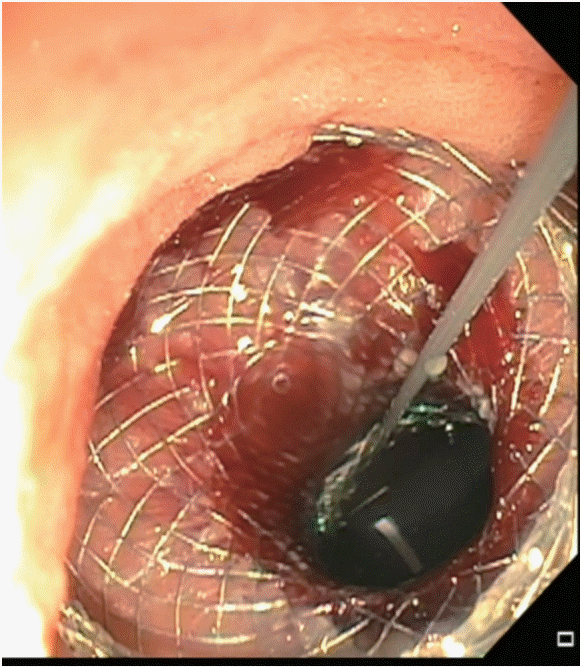
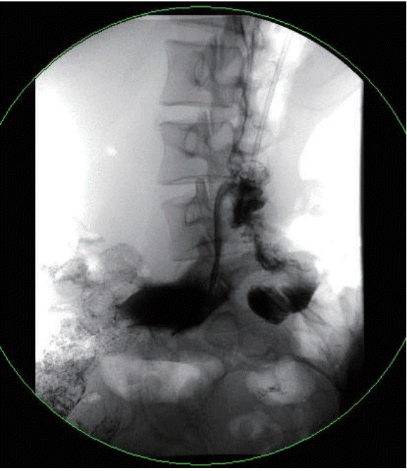
After 24 h of NGT decompression, a linear echoendoscope (TGF-UC180J; Olympus, Center Valley, PA, USA) was inserted into the stomach, which was intentionally filled with water, and then advanced into the jejunum. After the water-filled stomach was identified under EUS guidance, the stomach was accessed using a 19-gauge fine-needle aspiration needle (Cook Endoscopy, Winston-Salem, NC, USA) to create a fistulous tract. Contrast injection confirmed the access location within the stomach, and a 0.035-in hydrophilic wire was advanced through the needle and coiled within the stomach. A cautery-enhanced 15-mm LAMS (AXIOS; Boston Scientific, Natick, MA, USA) was inserted into the stomach over the wire, and the distal end was deployed under EUS guidance. After pulling the LAMS to appose the stomach wall with the jejunal wall, the proximal end was deployed under endoscopic visualization. A 15-m controlled radial expansion balloon was used to dilate the lumen of the stent to its full diameter. The LAMS was visualized through the echoendoscope, confirming that the distal end of the stent was in the stomach (
Fig. 1). A 10 Fr × 10 cm double-pigtail plastic stent (Boston Scientific) was placed across the LAMS (
Fig. 2). At 2 months follow-up, the patient reported tolerating a stent diet, resulting in a 4.5 kg weight gain.
DISCUSSION
SMAS is characterized by compression of the third portion of the duodenum due to narrowing of the space between the superior mesenteric artery and the aorta, and it has been attributed to loss of the intervening mesenteric fat pad [
4]. Significant weight loss as a consequence of medical disorders, psychological disorders, or surgery is the precipitating factor [
5].
Conservative non-operative therapy is the recommended initial treatment for SMAS [
6]. These conservative measures include NGT decompression, rehydration, electrolyte replacement, and, most important, the use of total parental nutrition or aggressive nutritional support [
7,
8]. When conservative management fails, enteral feeding through surgical gastrojejunostomy can be performed [
9].
The advantage of EUS-GJ is that it offers a minimally invasive intervention without the morbidities associated with surgery or the placement of an external feeding tube. It enables preservation of the original anatomy and offers the possibility of procedure reversal without major intervention, by simple removal of the LAMS once the disease has resolved.
This case report shows that a reverse EUS-GJ is a safer and feasible alternative to surgical gastrojejunostomy in patients who fail medical therapy.


 PDF
PDF Citation
 Print
Print


 XML Download