Abstract
Endoscopic ultrasound-guided biliary drainage has been developed as an alternative method for biliary drainage. EUS-guided hepaticogastrostomy (EUS-HGS) can be attempted via the trans-gastric route. These procedures are technically complex for two reasons. First, puncture of the intrahepatic bile duct via the trans-gastric route can be more difficult than that by other approaches because of the small diameter of the target site, and guidewire insertion or manipulation is challenging during EUS-HGS. Second, critical adverse events, such as stent migration into the abdominal cavity, could occur because of the greater mobility of the stomach compared to the duodenum. Therefore, endoscopists should be cautious when performing EUS-HGS. An advantage of EUS-HGS is that it can be performed in patients with complications such as duodenal bulb obstruction or surgically altered anatomy. Recent advances in technique and improvements in devices and stents for EUS-HGS have shown promise for improving the technical success rate of EUS-HGS and reducing the rate of adverse events. However, endoscopists should remain aware of the possibility of critical adverse events such as stent migration.
Biliary obstruction is treated by biliary drainage during endoscopic retrograde cholangiopancreatography (ERCP). However, ERCP is challenging in patients with inaccessible papilla, such as in cases with duodenal obstruction due to malignant or benign tumor, or surgically altered anatomy. Endoscopic ultrasound-guided biliary drainage (EUS-BD) has been developed as an alternative method for biliary drainage [1-17]. EUS-guided choledochoduodenostomy (EUS-CDS), EUS-guided hepaticoduodenostomy, and EUS-guided gallbladder drainage can be performed via the trans-duodenal route, whereas EUS-guided hepaticogastrostomy (EUS-HGS) can be attempted via the trans-gastric route. These procedures are technically complex for two reasons. First, puncture of the intrahepatic bile duct can be more difficult in this approach than by other approaches because of the short diameter of the target site, and guidewire insertion or manipulation is challenging during EUS-HGS. Second, the procedure involves a high risk of critical adverse events, such as stent migration into the abdominal cavity, because of the greater mobility of the stomach compared to the duodenum. Therefore, endoscopists should be cautious when performing EUS-HGS. Nevertheless, EUS-HGS is a feasible option for patients with complications such as duodenal bulb obstruction or surgically altered anatomy. However, further technical development and improvement in devices is required before EUS-HGS can be considered a viable alternative for biliary drainage. In this review, we provide a technical update on EUS-HGS and a literature review.
Although a study comparing EUS-CDS to ERCP as a first-line drainage technique has been reported, EUS-HGS is not yet considered a first-line drainage technique. Therefore, EUS-HGS is currently indicated mainly for patients with inaccessible papilla, such as in cases of duodenal obstruction or surgically altered anatomy. EUS-BD for benign biliary disease has been recently reported. EUS-HGS has several advantages compared to the enteroscopic approach, particularly for patients with surgically altered anatomy who require removal of bile duct stones or biliary drainage due to benign disease such as hepaticojejunostomy stricture. Firstly, the duration of the procedure is generally short. Secondly, access to the biliary tract is easy if re-intervention is required, although a prospective comparison study is needed for confirmation. However, as mentioned previously, EUS-HGS has a risk of critical adverse events such as stent migration into the abdominal cavity. Therefore, only expert operators should attempt this technique.
Table 1 shows published reports of EUS-HGS (excluding combination with antegrade stenting) that have included at least 30 patients [1-11]. The rates of technical and clinical success ranged from 65% to 100% and from 76% to 100%, respectively. In contrast, the rate of procedure-related adverse events was relatively high, at 15.7% (95/606). However, almost all cases with adverse events were managed successfully using conservative treatment.
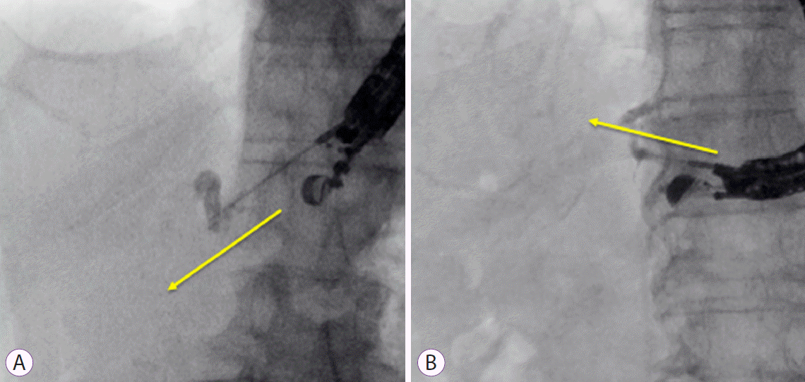
During EUS-HGS, the bile duct of segment 2 (B2) or segment 3 (B3) can be selected as the puncture site, although the more dilated B2 is easier to puncture. However, if B2 is punctured, the esophagus can be used as the entry route. To avoid mediastinitis, many endoscopists consider B3 a more suitable puncture site. To advance the guidewire to the hepatic hilum, the bile duct (which runs from the upper left to the lower right on EUS images) should be punctured. However, when the bile duct is punctured using this strategy, the guidewire can sometimes be advanced into the periphery of the bile duct. It is also important that the shape of the echoendoscope be checked during fluoroscopic imaging prior to bile duct puncture. If the angle between the echoendoscope and the needle is large, the guidewire can be inserted into the periphery of the bile duct. In contrast, if the angle between the echoendoscope and the needle is small, the guidewire can be inserted into the hepatic hilum (Fig. 1). We previously evaluated the influence of the angle between the needle and the echoendoscope on guidewire insertion failure using receiver-operating characteristic (ROC) curves [13]. The area under the ROC curve was 0.86 (95% confidence interval [CI], 0.00–0.76), and an angle of 135° offered 88.0% sensitivity and 82.9% specificity for predicting successful guidewire insertion. In multivariable analysis, an angle >135° between the needle and the echoendoscope was independently associated with successful guidewire insertion (odds ratio, 0.03; 95% CI, 0.01–0.14; p<0.05), whereas sex, puncture site, and diameter of the puncture site were not. Therefore, the shape of the echoendoscope should be checked using fluoroscopic imaging prior to bile duct puncture.
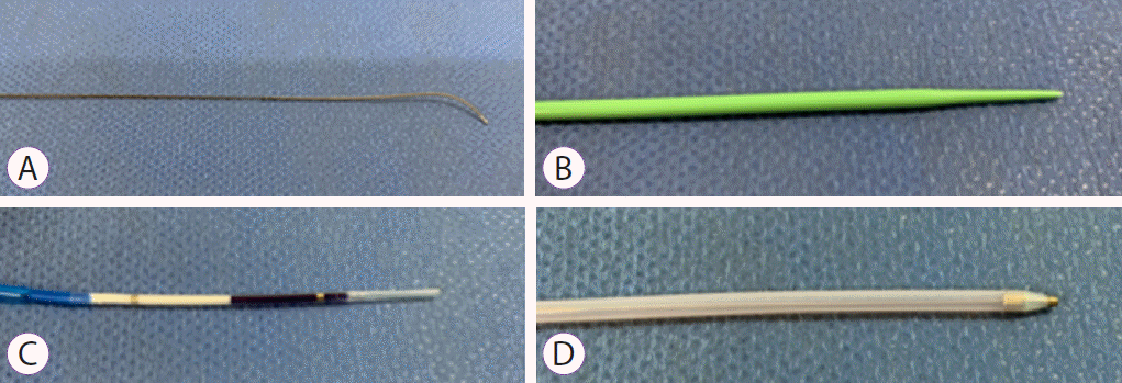
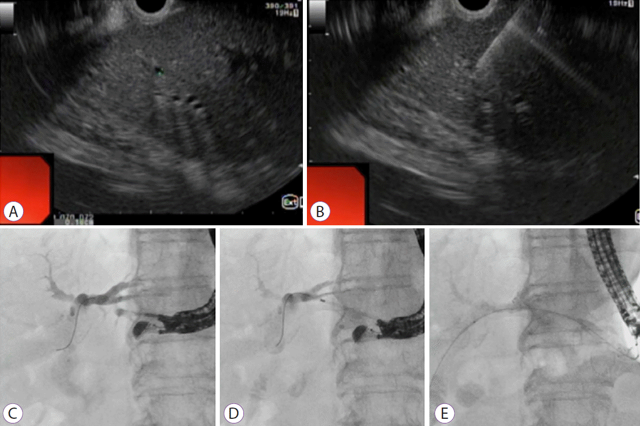
Most endoscopists select a 19 G needle because a stiff guidewire is required for insertion of devices such as a balloon catheter, mechanical dilator, or metallic stent delivery system. However, it is more difficult to manipulate a 19 G needle than a 22 G needle. The difficulty of bile duct puncture may depend on the diameter of the puncture site. Therefore, if the intrahepatic bile duct is not sufficiently dilated, especially in benign biliary disease, bile duct puncture using a 19 G needle can be challenging. A novel 0.018-inch guidewire (Fielder; Olympus Medical, Tokyo, Japan) recently became available in Japan (Fig. 2A). This guidewire has improved manipulation and stiffness compared to a conventional 0.018-inch guidewire. In addition, fine-gauge dilators such as mechanical dilators (ES dilator; Zeon Medical Co., Ltd., Tokyo, Japan) (Fig. 2B), balloon catheters (REN biliary balloon catheter; KANEKA, Osaka, Japan) (Fig. 2C), and fine-gauge electrocautery dilators (Fine 025; Medicos HIRATA, Osaka, Japan) (Fig. 2D) have been approved for use as dilation devices in Japan. Therefore, after bile duct puncture using a 22 G needle, fistula dilation may be less challenging than expected. Fig. 3. shows technical tips for EUS-HGS using a novel 0.018-inch guidewire under 22 G needle guidance. Firstly, the intrahepatic bile duct measuring 1.6 mm in diameter was detected under EUS guidance (Fig. 3A). The intrahepatic bile duct was successfully punctured using a 22 G needle (Fig. 3B). Appropriate bile duct puncture is usually confirmed by aspiration of bile juice through the needle; however, it can be difficult to aspirate bile juice with minimal dilation of the intrahepatic bile duct. In such situations, a small amount of normal saline should be injected. If resistance is felt during injection or if a high echoic area is detected in the hepatic parenchyma during EUS imaging, injection of saline is stopped. If these features are not observed, a cholangiogram should be obtained by injecting a contrast medium. A 0.018-inch guidewire was gently inserted into the intrahepatic bile duct (Fig. 3C) and successful fistula dilation was done using a fine-gauge mechanical dilator was as a dilation device (Fig. 3D). Finally, a self-expandable metal stent was deployed without any adverse events (Fig. 3E).
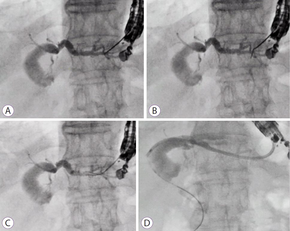
Guidewire insertion and manipulation are the most challenging steps of EUS-HGS. According to a national survey conducted in Spain [2], guidewire manipulation inside the duct was a critically limiting step that caused 28 of 41 failures. Therefore, improvement of this step would lead to the greatest improvement in the overall technical success rate of EUS-HGS. Importantly, the stiff part of the guidewire should be substantially deployed into the biliary tract before inserting the dilation device or stent delivery system. By doing so, the EUS-HGS procedure is performed in a stable manner because the echoendoscope is fixed. To deploy the stiff part of the guidewire, the guidewire should be inserted into the hepatic hilum. As mentioned above, endoscopists should pay attention to the puncture site and the angle of the echoendoscope. However, advancement of the guidewire into the periphery of the bile duct can occur even when these guidelines are followed. If the guidewire is pulled back into the needle after it reaches the periphery of the bile duct, guidewire shearing can occur. We have previously described technical tips for the liver impaction technique [18]. Briefly, if the guidewire is advanced into the periphery of the bile duct, the needle itself must be pulled back into the hepatic parenchyma to prevent shearing of the guidewire. Because the tip of the fine-needle aspiration needle remains in the hepatic parenchyma, guidewire shearing can be avoided. Guidewire selection is also an important factor in successful guidewire insertion. Various types of guidewire can be used. The guidewire might penetrate or injure the bile duct while being inserted via the needle. Therefore, guidewire flexibility is important. In addition, forming loop shape with the guidewire can be a useful technique for preventing duct penetration; however, it should be noted that the ability to create a loop differs among guidewires types [19]. A novel specialized guidewire for creating a loop has recently become available in Japan (0.025-inch, MICHIISUJI; KANEKA). Fig. 4. shows EUS-HGS performed using the loop technique. Guidewire insertion was attempted after puncture of the intrahepatic bile duct (Fig. 4A) but was complicated by penetration of the bile duct (Fig. 4B). Therefore, we exchanged the conventional guidewire for the novel 0.025-inch guidewire. Guidewire advancement was then achieved using the loop technique (Fig. 4C). After tract dilation, a plastic stent was deployed successfully (Fig. 4D). Further experimental and clinical comparison studies are required to confirm this technique.
Ryou et al. recently reported their initial clinical experience using a steerable access device for EUS-BD [20]. This novel device assumes a predetermined curvature (90° or 135°), is fully rotatable, and can theoretically prevent wire shearing because of its blunt tip and the coaxial orientation of the guidewire relative to the tip of the catheter. One patient in their study underwent EUS-HGS, and curved access was successfully performed within the intrahepatic bile duct after intrahepatic bile duct puncture. This device may have an impact on clinical practice if the guidewire is advanced from the periphery of the bile duct, although additional cases and prospective evaluation are necessary to verify its applicability.
After guidewire deployment, the stomach and bile duct wall are commonly dilated before insertion of the stent delivery system. Various dilation devices are available for this purpose. However, there is still insufficient evidence regarding the most suitable dilation device for use during EUS-HGS. According to previous reports, an ERCP or balloon catheter, electrocautery dilator, needle knife, or bougie dilator can be used [1-15]. Novel fine-gauge devices, such as the fine-gauge balloon catheter with a 3 Fr tip (Fig. 2B), have recently become available for this purpose. In our previous evaluation of this balloon catheter as a dilation device during EUS-HGS [16], the stent delivery system was inserted successfully after fistula dilation in nine patients without additional dilation. Stent deployment was performed successfully in all patients, and there were no severe adverse events. In contrast, Honjo et al. [9] performed a retrospective evaluation of EUS-BD using an ultra-tapered mechanical dilator. This dilator is tapered up to 2.5 Fr and is designed for a 0.025-inch guidewire. In that study, 26 patients underwent EUS-HGS. Although additional dilation of the fistula was needed in two patients (technical success rate, 92.3%), the only adverse event was abdominal pain (n=4), which was treated conservatively. Although these devices are clinically useful and safe, the procedure time is relatively long, which may increase the risk of bile peritonitis. Indeed, procedure time was shorter in the electrocautery dilation group than in the mechanical dilation group (19.7±6.1 min vs. 21.5±6.5 min), although the difference was not statistically significant (p=0.09). If the device is difficult to advance due to inflammation or thickening of the bile duct or stomach wall, electrocautery dilation might be useful and help reduce the duration of the procedure. However, the risk of bleeding due to heat is a disadvantage of dilation using electrocautery. The authors of this study [9] reported that electrocautery dilation could burn the hepatic parenchyma, the vessels around the needle tract, and the gastrointestinal lumen, causing unexpected bleeding and inflammation.
With this background in mind, we produced the Fine025 electrocautery dilator (Medicos HIRATA) with a 3 Fr tip and hypothesized that the burning effect would be less with a fine tip than with a conventional electrocautery dilator. Experimental evaluation confirmed that the burning effect was indeed less with the Fine025 dilator compared to a conventional electrocautery dilator (6 Fr, Cysto-Gastro-Set; Endo-Flex GmbH, Voerde, Germany) [17]. In clinical evaluation, EUS-HGS was attempted using this electrocautery dilator in nine patients, all of whom underwent EUS-HGS successfully without any adverse events, including bleeding [14]. Therefore, although additional cases and a prospective evaluation study are needed to confirm these results, the Fine025 dilator has shown promise as a safe and effective tract dilation device.
Stent migration into the abdominal cavity is the most critical adverse event that can occur during EUS-HGS. There are two mechanisms of stent migration. Firstly, the stent can be completely released within the abdominal cavity if release is performed between the hepatic parenchyma and stomach wall. Because covered metal stents are self-expandable, the risk of stent migration can be reduced if stent release is performed across the stomach wall; therefore, stent release should be performed across the stomach wall. Secondly, stent shortening should be considered. If the stent length is short in the luminal portion, stent migration can be a late complication after full stent expansion. These two points are extremely important in preventing stent migration.
Regarding technical tips for stent deployment, the intra- scope channel release technique may be useful for preventing stent migration [21]. When the stent delivery system is inserted into the intrahepatic bile duct, the echoendoscope may be expelled from the hepatic parenchyma due to the pushing force of the stent delivery system (Fig. 5A). In such situations, stent release might occur within the abdominal cavity because of low adhesion between the hepatic parenchyma and the echoendoscope. To avoid this, the stent delivery system should be withdrawn slowly (Fig. 5B) so that adhesion is maintained between the hepatic parenchyma and the echoendoscope. Stent release is then performed from the intrahepatic bile duct to the echoendoscope. During this procedure, the echoendoscope is stabilized until the stent is released up to 2–4 cm inside the echoendoscope because stent deployment across the stomach wall is certain. Finally, the echoendoscope is gradually withdrawn while the stent delivery system is pushed (Fig. 5C). We previously used computed tomography to compare the distance between the hepatic parenchyma and the stomach wall one day after EUS-HGS between the extra-scope (n=20) and intra-scope (n=21) channel release technique groups [20]. This distance was shorter in the intra-scope channel release technique group (0.66±0.97 cm) than in the extra-scope channel release technique group (2.52±0.97 cm) (p<0.05).
Stent length may also be an important factor for preventing stent migration, especially in the late phase, due to full stent expansion. Indeed, recent studies (Table 1) found that several endoscopists select covered metal stents with length of 10 or 12 cm. Nakai et al. evaluated the long-term outcomes of using a long, partially covered metal stent (LP-CMS) for EUS-HGS [11]. That study included 110 patients who underwent EUS-HGS using LP-CMS and reported a technical success rate of 100% and a high clinical success rate (103/110, 94%). The stent length was 10 cm or 12 cm in all but two patients (8 cm). The median stent length in the luminal portion was 54 mm (interquartile range, 46–60 mm). Recurrent biliary obstruction was observed in 36 patients (33%) due to hyperplasia (n=23), sludge (n=7), or other causes (n=4). Notably, no patient experienced stent migration after stent deployment because sufficient length was secured in the luminal portion. Stent length in the luminal portion may be clinically important for obtaining lasting stent patency and preventing stent migration [21]. In our previous evaluation of stent patency, based on stent length in the luminal portion, the median duration of stent patency was significantly shorter for stent a length <3 cm (52 days) than for a stent length of ≥3 cm (195 days; p<0.01). Multivariate analysis for predictive factors of stent patency revealed that stent length in the luminal portion ≥3 cm (hazard ratio [HR] 5.444; p<0.05) and performance of chemotherapy (HR, 4.501; p<0.05) were associated with lasting stent patency. The findings of these studies indicate that long metal stents might be suitable for EUS-HGS, although a randomized comparison study of stent length is needed to confirm this hypothesis.
There have been recent advances in stent design with the aim of preventing stent migration. Cho et al. evaluated longterm outcomes for a newly developed hybrid metal stent (Standard Sci Tech Inc., Seoul, Korea) for EUS-guided biliary drainage [22]. The hybrid metal stent, a partially covered self-expandable metal stent, was used during EUS-BD. The length of the covered distal portion was 3 cm; and the proximal portion, which can have a length of 1.5–6.5 cm, was uncovered. To prevent stent migration, four anchoring flaps were prepared. EUS-HGS using this novel stent was attempted in 21 patients. Stent deployment was successful in all patients (technical success rate, 100%) with a favorable clinical success rate (85.7%). Although pneumoperitoneum (n=2) and bleeding (n=1) were early complications, stent migration did not occur, and there was no stent migration or dislocation observed during the follow-up period (median, 148.5 days). A novel partially covered lumen-apposing metal stent (Spring Stopper; Taewoong Medical, Gimpo, Korea) that also prevents stent migration has been developed in Japan (Fig. 6A). The length of the proximal uncovered site was 1.5–2 cm to prevent stent dislocation and side branch obstruction. Although clinical evaluation is required, this stent may be safer during EUS-HGS for preventing stent migration (Fig. 6B, C).
Because the diameter of these stents is 8.0 Fr or 8.5 Fr, fistula dilation is necessary prior to insertion of the stent delivery system. Leakage of bile juice can easily occur during and after fistula dilation, causing bile peritonitis, which is one of the most frequent complications. A novel covered metal stent with a fine-gauge stent delivery system has recently become available, and several authors have provided technical tips for performing EUS-HGS without fistula dilation. Maehara et al. described EUS-HGS or EUS-guided hepaticojejunostomy without dilation using a stent having a thinner delivery system [23]. A fully covered braided-type metal stent with a 6-Fr diameter delivery system was used as the EUS-HGS stent in six patients (6 mm diameter, Braided 6; S&G Biotech, Seongnam, Korea). After puncturing the intrahepatic bile duct, the guidewire was deployed at an appropriate site. Technical success was defined as insertion of the stent delivery system into the biliary tract without fistula dilation. Technical success was obtained in all patients, and the median procedure time was relatively short (18 min). No early adverse events such as abdominal pain, bile peritonitis, or fever were observed. We have also reported technical tips for one-step EUS-HGS [24]. Compared with the system used by Maehara et al. [23], we used a wider metal stent, which we consider to be more effective for biliary drainage, and a smaller stent delivery system (5.9 Fr) (8×12 cm, HANAROSTENT® Biliary Full Cover BenefitTM; M.I. Tech, Seoul, Korea). Although the procedure time was short (7 min) and no complications occurred, one-step EUS-HGS and conventional EUS-HGS should be compared in a randomized trial to evaluate their clinical usefulness.
Several recent advances in technique and the availability of new devices and stents for EUS-HGS have shown promise for improving the technical success rate of EUS-HGS and reducing the rate of complications. Despite these advances, further improvements are required and endoscopists should be aware of the possibility of severe complications such as stent migration. It is notable that favorable results regarding EUS-HGS have been reported in cases wherein high-level experts performed EUS-HGS. Therefore, further studies, including endoscopists with varying levels of expertise, are needed to verify clinical outcomes following EUS-HGS.
ACKNOWLEDGMENTS
I am grateful to all members of the International Digestive Endoscopy Network 2020 and the Korean Digestive Disease Week 2020 for their assistance despite the COVID-19 crisis. I extend particular thanks to my Korean colleagues with whom I participated in several valuable discussions.
REFERENCES
1. Park DH, Jang JW, Lee SS, Seo DW, Lee SK, Kim MH. EUS-guided biliary drainage with transluminal stenting after failed ERCP: predictors of adverse events and long-term results. Gastrointest Endosc. 2011; 74:1276–1284.
2. Vila JJ, Pérez-Miranda M, Vazquez-Sequeiros E, et al. Initial experience with EUS-guided cholangiopancreatography for biliary and pancreatic duct drainage: a Spanish national survey. Gastrointest Endosc. 2012; 76:1133–1141.
3. Poincloux L, Rouquette O, Buc E, et al. Endoscopic ultrasound-guided biliary drainage after failed ERCP: cumulative experience of 101 procedures at a single center. Endoscopy. 2015; 47:794–801.
4. Khashab MA, Messallam AA, Penas I, et al. International multicenter comparative trial of transluminal EUS-guided biliary drainage via hepatogastrostomy vs. choledochoduodenostomy approaches. Endosc Int Open. 2016; 4:E175–E181.
5. Nakai Y, Isayama H, Yamamoto N, et al. Safety and effectiveness of a long, partially covered metal stent for endoscopic ultrasound-guided hepaticogastrostomy in patients with malignant biliary obstruction. Endoscopy. 2016; 48:1125–1128.
6. Minaga K, Takenaka M, Kitano M, et al. Rescue EUS-guided intrahepatic biliary drainage for malignant hilar biliary stricture after failed transpapillary re-intervention. Surg Endosc. 2017; 31:4764–4772.
7. Sportes A, Camus M, Greget M, et al. Endoscopic ultrasound-guided hepaticogastrostomy versus percutaneous transhepatic drainage for malignant biliary obstruction after failed endoscopic retrograde cholangiopancreatography: a retrospective expertise-based study from two centers. Therap Adv Gastroenterol. 2017; 10:483–493.
8. Oh D, Park DH, Song TJ, et al. Optimal biliary access point and learning curve for endoscopic ultrasound-guided hepaticogastrostomy with transmural stenting. Therap Adv Gastroenterol. 2017; 10:42–53.
9. Honjo M, Itoi T, Tsuchiya T, et al. Safety and efficacy of ultra-tapered mechanical dilator for EUS-guided hepaticogastrostomy and pancreatic duct drainage compared with electrocautery dilator (with video). Endosc Ultrasound. 2018; 7:376–382.
10. Paik WH, Lee TH, Park DH, et al. EUS-guided biliary drainage versus ERCP for the primary palliation of malignant biliary obstruction: a multicenter randomized clinical trial. Am J Gastroenterol. 2018; 113:987–997.
11. Nakai Y, Sato T, Hakuta R, et al. Long-term outcomes of a long, partially covered metal stent for EUS-guided hepaticogastrostomy in patients with malignant biliary obstruction (with video). Gastrointest Endosc. 2020; 92:623–631.e1.
12. Krishnamoorthi R, Dasari CS, Thoguluva Chandrasekar V, et al. Effectiveness and safety of EUS-guided choledochoduodenostomy using lumen-apposing metal stents (LAMS): a systematic review and meta-analysis. Surg Endosc. 2020; 34:2866–2877.
13. Ogura T, Nishioka N, Ueno S, et al. Effect of echoendoscope angle on success of guidewire manipulation during endoscopic ultrasound-guided hepaticogastrostomy. Endoscopy. 2021; 53:369–375.
14. Ma KW, So H, Cho DH, Oh JS, Cheung TT, Park DH. Durability and outcome of endoscopic ultrasound-guided hepaticoduodenostomy using a fully covered metal stent for segregated right intrahepatic duct dilatation. J Gastroenterol Hepatol. 2020; 35:1753–1760.
15. Ogura T, Kitano M, Takenaka M, et al. Multicenter prospective evaluation study of endoscopic ultrasound-guided hepaticogastrostomy combined with antegrade stenting (with video). Dig Endosc. 2018; 30:252–259.
16. Amano M, Ogura T, Onda S, et al. Prospective clinical study of endoscopic ultrasound-guided biliary drainage using novel balloon catheter (with video). J Gastroenterol Hepatol. 2017; 32:716–720.
17. Ogura T, Nakai Y, Iwashita T, Higuchi K, Itoi T. Novel fine gauge electrocautery dilator for endoscopic ultrasound-guided biliary drainage: experimental and clinical evaluation study (with video). Endosc Int Open. 2019; 7:E1652–E1657.
18. Ogura T, Masuda D, Takeuchi T, Fukunishi S, Higuchi K. Liver impaction technique to prevent shearing of the guidewire during endoscopic ultrasound-guided hepaticogastrostomy. Endoscopy. 2015; 47:E583–E584.
19. Kwon CI, Koh DH, Song TJ, Park WS, Lee DH, Jeong S. Technical reports of endoscopic retrograde cholangiopancreatography guidewires on the basis of physical properties. Clin Endosc. 2020; 53:65–72.
20. Ryou M, Benias PC, Kumbhari V. Initial clinical experience of a steerable access device for EUS-guided biliary drainage. Gastrointest Endosc. 2020; 91:178–184.
21. Miyano A, Ogura T, Yamamoto K, Okuda A, Nishioka N, Higuchi K. Clinical impact of the intra-scope channel stent release technique in preventing stent migration during EUS-guided hepaticogastrostomy. J Gastrointest Surg. 2018; 22:1312–1318.
22. Cho DH, Lee SS, Oh D, et al. Long-term outcomes of a newly developed hybrid metal stent for EUS-guided biliary drainage (with videos). Gastrointest Endosc. 2017; 85:1067–1075.
Table 1.
Study | Number of Pt | Technical success rate, % (n) | Clinical success rate, % (n) | Dilation devices | Type of stent | Adverse events |
---|---|---|---|---|---|---|
Park et al. (2011) [1] | 31 | 100 (31/31) | 87 (27/31) | ERCP catheter (4 Fr), dilator (6 and 7 Fr), needle knife | PS (7 Fr, 6–8 cm), FCSEMS (8–10 mm, 4–10 cm) | Pneumoperitoneum (4), bleeding (2) |
Vila et al. (2012) [2] | 34 | 65 (22/34) | N/D | N/D | N/D | Bleeding (3), biloma (3), perforation (2), liver hematoma (2), abscess (1) |
Poincloux et al. (2015) [3] | 66 | 98 (65/66) | 94 (61/65) | Needle-knife, dilator (6 or 7Fr), | Plastic stent (10 Fr), FCSEMS (10 mm, 6–8 cm), PCSEMS (0 mm, 8–10 cm) | Bile leak (5), pneumoperitoneum (2), liver hamatoma (1), severe sepsis and death (2) |
Khashab et al. (2016) [4] | 61 | 92 (52/61) | 89 (50/61) | Balloon, dilator, cautery dilator | Metal stent | None |
Nakai et al. (2016) [5] | 33 | 100 (33/33) | 100 (33/33) | Cautery dilator, bougie dilator (9, 10 Fr) | PCSEMS | Bleeding (1), abscess (1), cholangitis (1) |
Minaga et al. (2017) [6] | 30 | 97 (29/30) | 76 (22/29) | Dilator (6, 7 Fr), balloon (4 mm) | Plastic stent, CSEMS | Bile peritonitis (1) |
Sportes et al. (2017) [7] | 31 | 100 (31/31) | 81 (25/31) | Cystotome | FCSEMS | Severe sepsis (2), bile leak (2), bleeding and death (1) |
Oh et al. (2017) [8] | 129 | 93 (120/129) | 88 (105/120) | Cannula (4 Fr), dilator (6, 7 Fr), needle-knife | Plastic stent (7–10 Fr, 6–10 cm), FCSEMS (6–10 mm, 6–10 cm) | Bacteremia (6), bleeding (5), bile peritonitis (4), pneumoperitoneum (4), intrahepatic stent migration (3) |
Honjo et al. (2018) [9] | 49 | 100 (49/49) | N/D | Tapered mechanical dilator, cystotome, balloon (4 mm) | PCSEMS (6, 8 mm, 10, 12 cm), plastic stent (Type IT) | Abdominal pain (6), bleeding (5) |
Paik et al. (2018) [10] | 32 | 97 (31/32) | 84 (26/31) | None | PCSEMS (DEUS) | Cholangitis (1) |
Nakai et al. (2020) [11] | 110 | 100 (110/110) | 94 (193/110) | Electrocautery, bougie dilator (9 or 10 Fr), or balloon catheter | PCSEMS (8 or 10 mm, 8, 10, or 12 cm) | Transient fever (10), abdominal pain (4), peritonitis (4), cholangitis (3), pseudoaneurysm (1), abscess (1), hemobilia (1), cholecystitis (1) |